
Comprehensive Introduction and Patho-Epidemiology
Definition and Scope of the Deformity
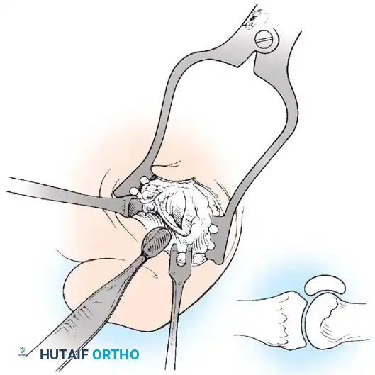
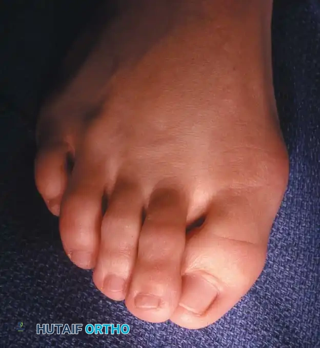
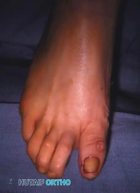
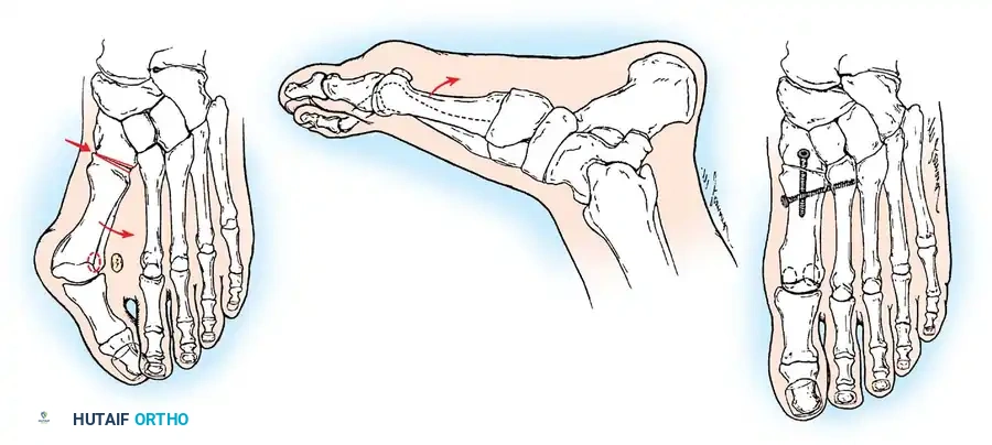
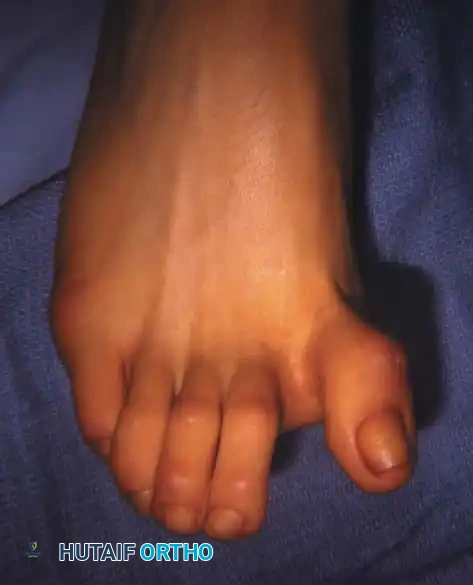
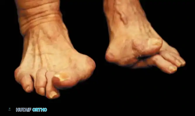
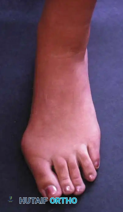
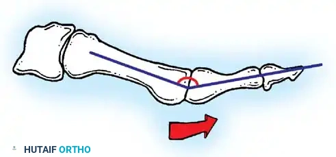
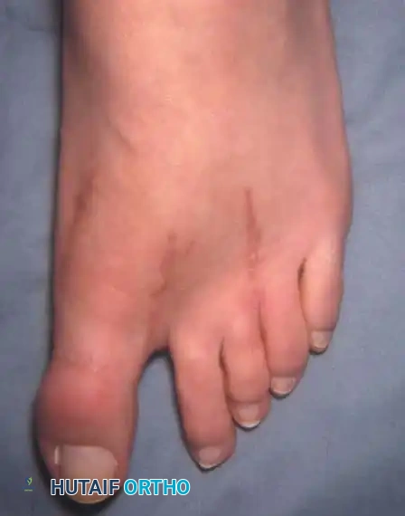
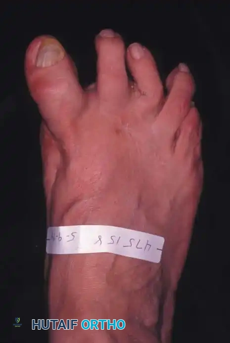
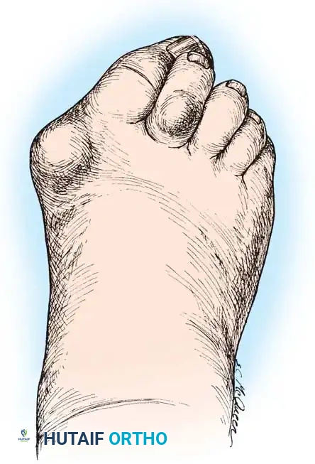
Hallux valgus, colloquially referred to as a bunion, is unequivocally not a singular, isolated osseous disorder as the layman’s terminology might imply. Rather, it represents a highly complex, progressive, three-dimensional deformity of the first ray. This pathological cascade frequently manifests with concomitant deformities and debilitating symptoms in the lesser toes, creating a globally dysfunctional forefoot. The primary architectural derangement involves the lateral deviation and pronation of the great toe, coupled with medial deviation and supination of the first metatarsal. This divergent relationship disrupts the highly synchronized biomechanics of the medial column, leading to significant gait alterations and functional impairment.
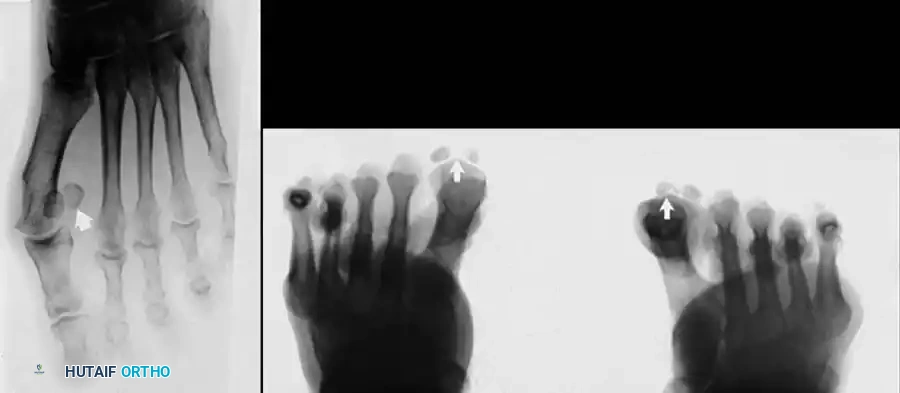
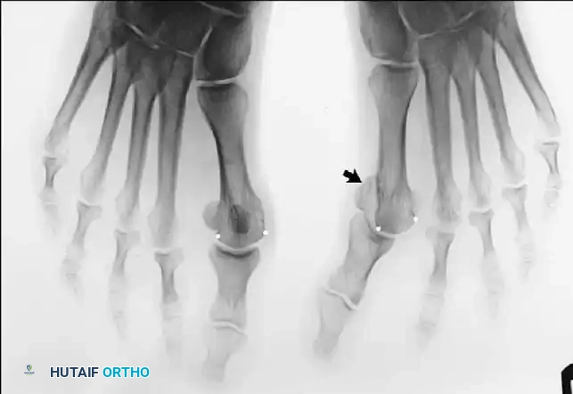
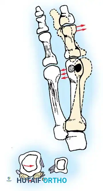
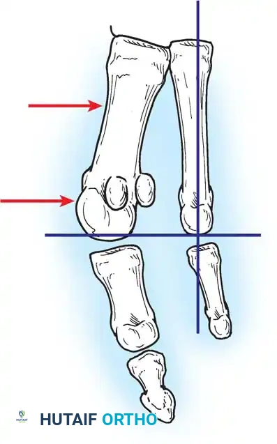
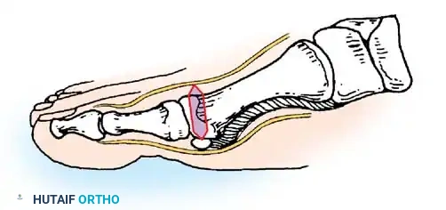
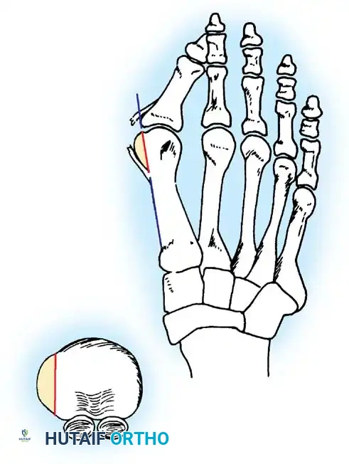
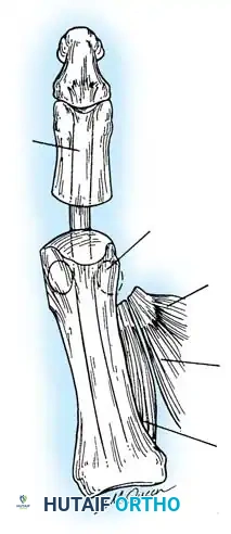
The morphological alterations inherent to hallux valgus extend far beyond simple angular divergence. As the first metatarsal drifts medially, the sesamoid apparatus—anchored by the transverse metatarsal ligament—maintains its spatial relationship with the second metatarsal. Consequently, the first metatarsal head is effectively "pushed" off its sesamoid articulation, leading to uncovering of the fibular sesamoid and erosion of the plantar crista. This subluxation of the first metatarsophalangeal (MTP) joint fundamentally alters the moment arms of the intrinsic and extrinsic musculature traversing the joint, converting dynamic stabilizers into deforming forces.
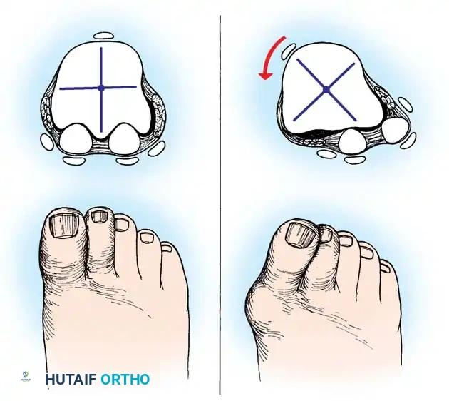
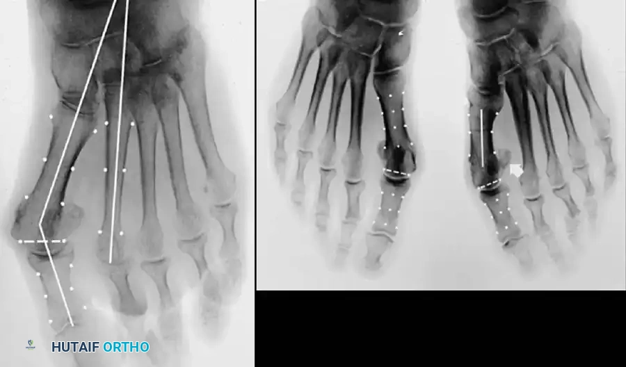
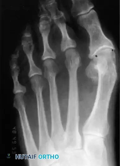
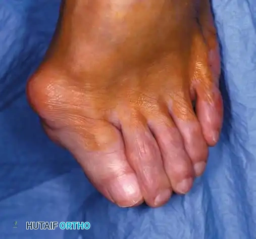
Furthermore, the clinical presentation of the so-called "bunion" is largely an anatomical illusion. Hypertrophy of the medial eminence has been described as a component of hallux valgus since the earliest historical reports; however, rigorous modern investigations, notably those by Thordarson and Krewer, have definitively demonstrated that true bony proliferation is rarely the primary pathoanatomic driver. Comparing radiographic parameters between operative hallux valgus cohorts and normal controls, they found negligible differences in the absolute width of the medial eminence (4.4 mm versus 4.1 mm). The prominent medial eminence is, in reality, the uncovered anteromedial aspect of the first metatarsal head resulting from metatarsus primus varus and lateral phalangeal deviation.
Epidemiological Factors and Etiology
The etiology of hallux valgus is undeniably multifactorial, representing a complex interplay between intrinsic genetic predispositions and extrinsic environmental exacerbators. A robust familial predilection is consistently observed, particularly in juvenile and adolescent hallux valgus presentations. In these younger cohorts, the deformity often manifests prior to significant exposure to constrictive footwear, strongly implicating hereditary structural anomalies such as an oblique first metatarsocuneiform joint, metatarsus primus varus, or a hypermobile first ray. Genetic transmission is frequently described as an autosomal dominant pattern with incomplete penetrance.
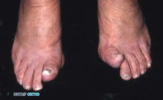
Despite the undeniable genetic component, the role of unphysiologically designed footwear remains the most significant extrinsic catalyst in modern, industrialized societies. While no definitive anthropological study of shod versus unshod populations has implicated footwear as the sole genesis of hallux valgus, the compressive forces exerted by narrow toe boxes and elevated heels unequivocally accelerate the pathomechanical cascade. These shoes force the hallux into a valgus posture while simultaneously plantarflexing the first ray, thereby maximizing the deforming vectors of the extrinsic tendons. The observation of unilateral hallux valgus in patients with otherwise symmetric pedal architecture further underscores the nuanced interplay between individual biomechanical susceptibility and asymmetric environmental stressors.
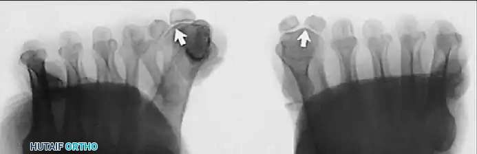
Controversy persists regarding the precise role of first ray hypermobility in the genesis of hallux valgus. Authors such as Coughlin and Shurnas, Myerson and Badekas, and King and Toolan have extensively debated whether hypermobility at the tarsometatarsal (TMT) joint is the primary instigator of metatarsus primus varus, or merely a secondary phenomenon resulting from the loss of the windlass mechanism and lateral displacement of the sesamoids. Regardless of its temporal onset, associated anatomical variants such as pes planovalgus, abnormal insertion of the tibialis posterior tendon, an abnormally long first ray, and incongruous articular surfaces collectively contribute to the destabilization of the medial column, dictating a comprehensive approach to surgical reconstruction.
Pathomechanics of Hallux Valgus Progression
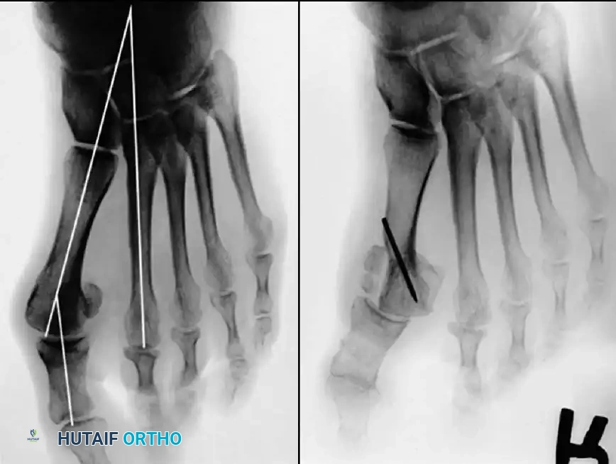
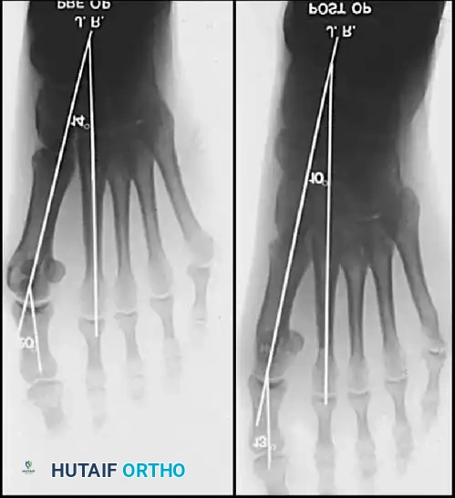
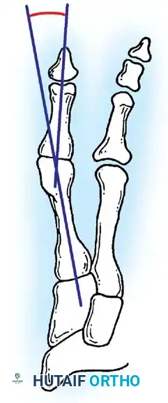
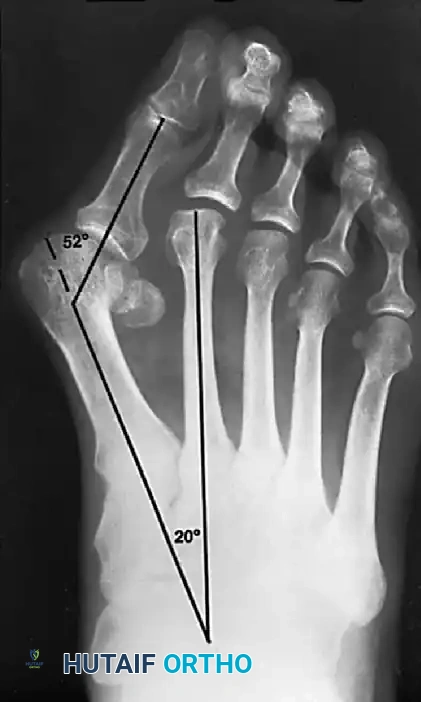
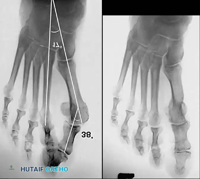
The progression of hallux valgus is driven by a vicious cycle of structural attenuation and muscular imbalance. In the normal physiologic state, the intermetatarsal angle (IMA) between the first and second metatarsals is strictly maintained below 8 to 9 degrees, and the hallux valgus angle (HVA) is contained within 15 to 20 degrees. When the capsuloligamentous restraints fail, and the HVA exceeds 30 to 35 degrees, a critical biomechanical threshold is crossed. At this juncture, the hallux undergoes obligate pronation due to the eccentric pull of the adductor hallucis and the lateralized extensor hallucis longus (EHL).
With this abnormal pronatory rotation, the abductor hallucis—which normally resides plantar to the flexion-extension axis of the first MTP joint, providing a medial stabilizing force—migrates further plantarward. This displacement neutralizes its abductory capacity, leaving the medial capsular ligament (specifically its capsulosesamoid and capsulophalangeal portions) as the sole restraint against valgus drift. Unopposed by the abductor hallucis, the adductor hallucis exerts a relentless lateral vector on the proximal phalanx, severely attenuating the medial capsule and allowing the metatarsal head to subluxate medially off the sesamoid sling.
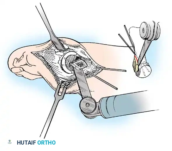
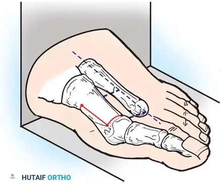
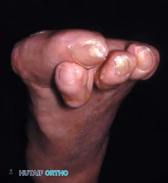
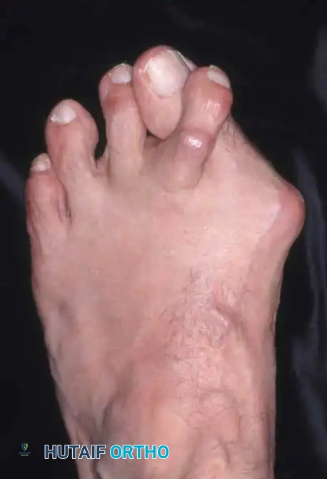
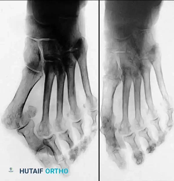
As the structural integrity of the first ray collapses, load-bearing mechanics are drastically altered. The first ray becomes functionally incompetent, shifting the burden of weight-bearing laterally to the lesser metatarsal heads. This phenomenon, known as transfer metatarsalgia, is a hallmark of advanced hallux valgus. The increased pressure beneath the second and third metatarsal heads leads to intractable plantar keratoses, capsulitis, and potential stress fractures. Concurrently, the valgus thrust of the hallux mechanically crowds the second toe, frequently resulting in a rigid crossover hammer toe deformity, which often becomes the patient's primary chief complaint, superseding the pain of the bunion itself.
Detailed Surgical Anatomy and Biomechanics
Osteology and Articular Configuration
The osseous architecture of the first ray is uniquely adapted for both dynamic propulsion and static load distribution. The first metatarsal is the shortest and most robust of the metatarsals, articulating proximally with the medial cuneiform to form the first TMT joint. The geometry of this joint is highly variable; a more oblique or vertically oriented facet inherently predisposes the first ray to medial deviation (metatarsus primus varus) under axial load. Distally, the metatarsal head presents a complex articular surface. The dorsal aspect is smoothly convex to accommodate the proximal phalanx during terminal stance, while the plantar aspect features two distinct longitudinal grooves separated by a prominent bony ridge known as the crista, which guides the sesamoids.
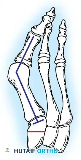
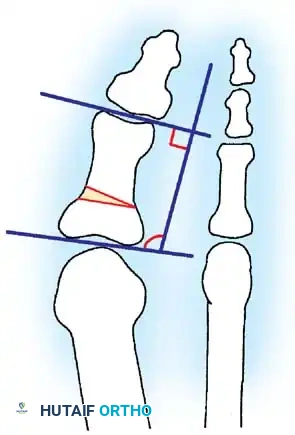
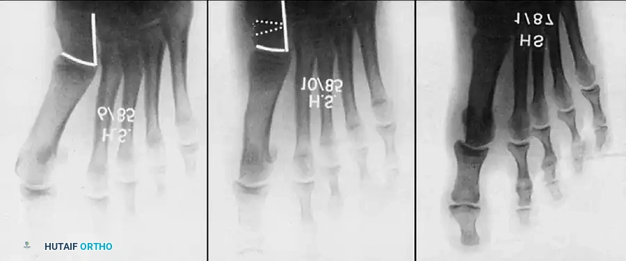
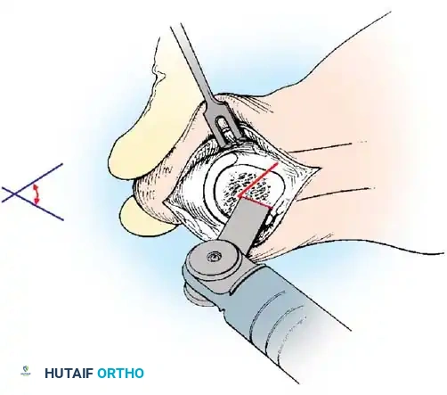
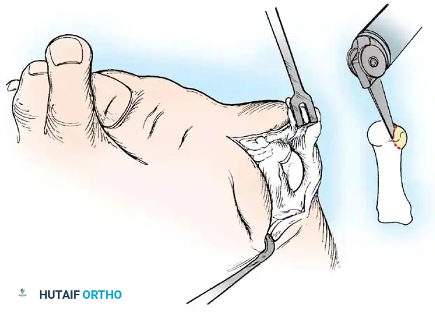
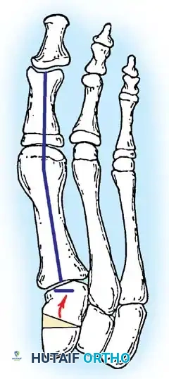
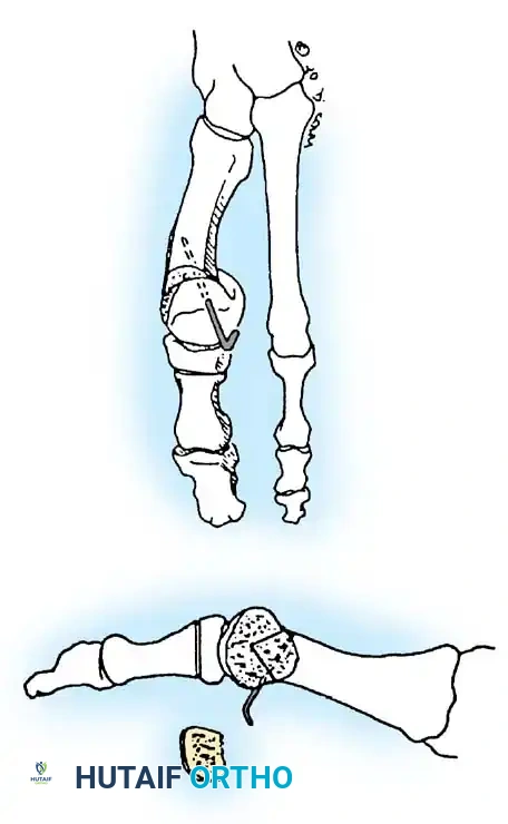
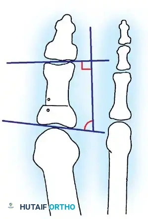
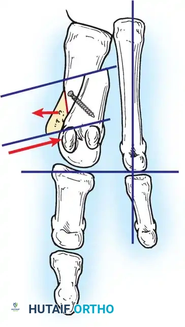
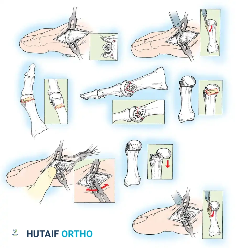
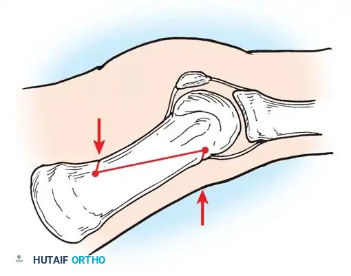
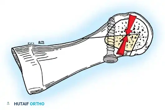
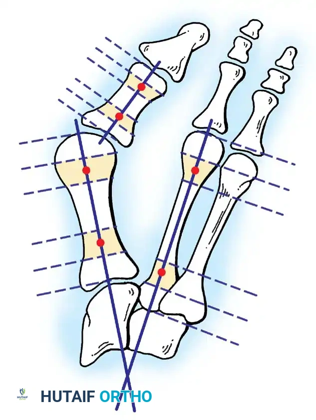
Two critical anatomical variants involving the articular surfaces heavily dictate the surgical algorithm: the Distal Metatarsal Articular Angle (DMAA) and the Phalangeal Articular Angle (PAA). The DMAA represents the orientation of the metatarsal head's articular cartilage relative to the longitudinal axis of the metatarsal shaft. A normal DMAA ranges from 10 to 15 degrees. In certain pathologic variants, the articular surface is significantly laterally deviated—described analogously as a scoop of ice cream sitting obliquely on a cone. Failure to recognize and surgically correct an abnormal DMAA inevitably leads to joint incongruency, restricted range of motion, and rapid recurrence of the deformity.
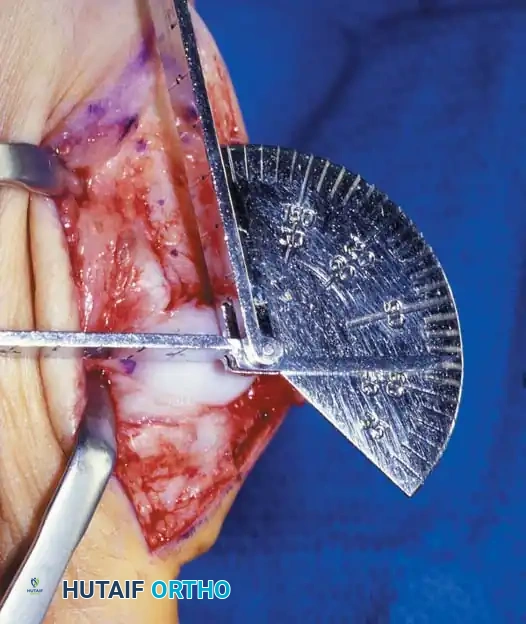
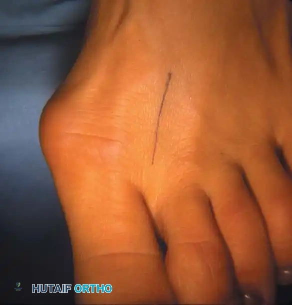
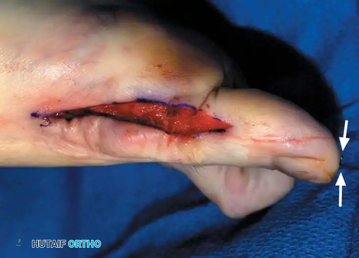
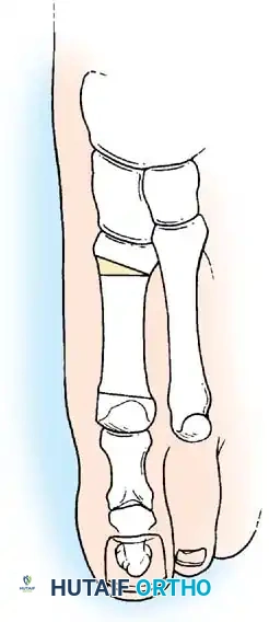
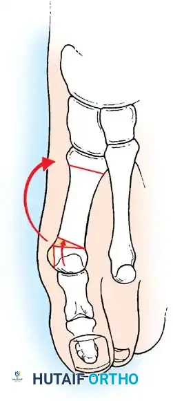
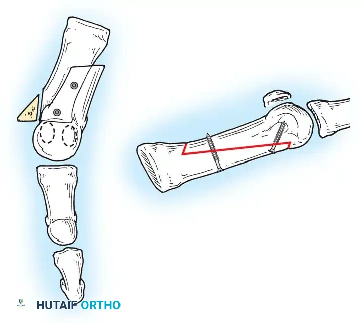
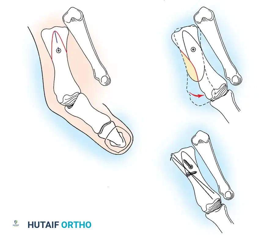
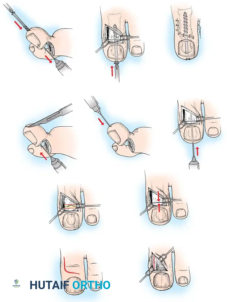
Similarly, the PAA dictates the intrinsic geometry of the proximal phalanx. The normal PAA is generally considered to be 7 to 10 degrees. When the valgus deformity is localized primarily within the proximal phalanx itself (hallux valgus interphalangeus), isolated metatarsal correction will fail to achieve a rectilinear medial border of the foot. In these instances, a proximal phalangeal osteotomy (Akin procedure) is mandated to correct the intrinsic phalangeal valgus. Precise radiographic evaluation of both the DMAA and PAA is paramount; forceful straightening of the hallux via soft-tissue plication alone, while ignoring these osseous angles, will sacrifice joint congruency and precipitate early-onset osteoarthritis.
The Sesamoid Complex and Ligamentous Restraints
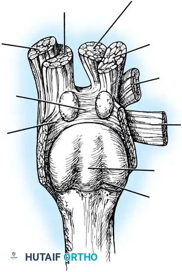
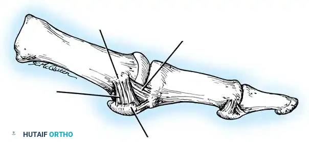
The sesamoid apparatus of the first MTP joint is a biomechanical marvel, functioning as a dynamic pulley system that enhances the mechanical advantage of the flexor hallucis brevis (FHB) while simultaneously protecting the intra-articular flexor hallucis longus (FHL) tendon. The tibial (medial) and fibular (lateral) sesamoids are embedded within the medial and lateral heads of the FHB, respectively. They articulate with the plantar grooves of the first metatarsal head, stabilized centrally by the crista. The sesamoids are interconnected by the robust intersesamoid ligament and are tethered to the proximal phalanx via the plantar plate.
The ligamentous restraints of the first MTP joint are highly complex and intricately balanced. The medial capsular ligament is composed of two distinct functional bands: the capsulosesamoid portion (inserting into the tibial sesamoid) and the capsulophalangeal cord portion (inserting into the base of the proximal phalanx, plantar to the longitudinal axis). During the pathogenesis of hallux valgus, the medial capsulosesamoid ligament undergoes severe plastic deformation. Conversely, the lateral structures—including the lateral collateral ligament, the lateral sesamophalangeal ligament, and the adductor hallucis insertion—become adaptively shortened and contracted.
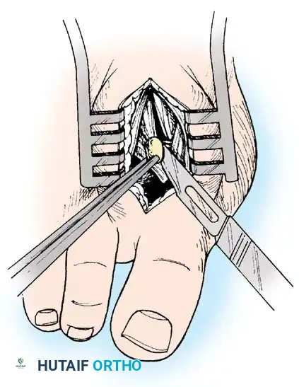
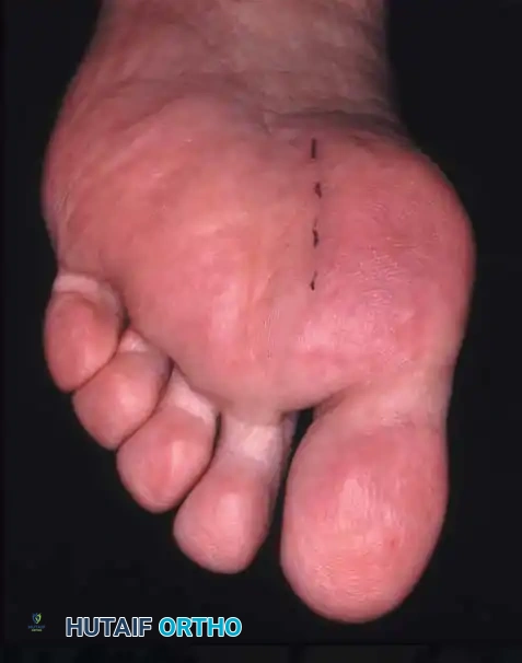
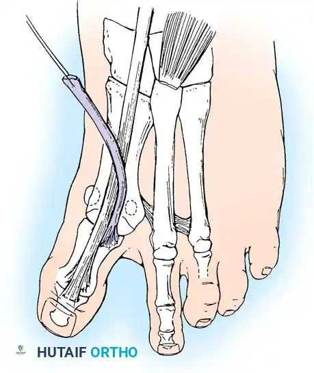
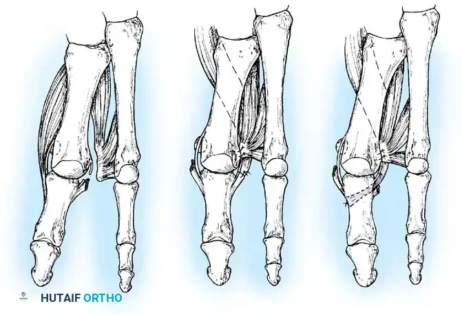
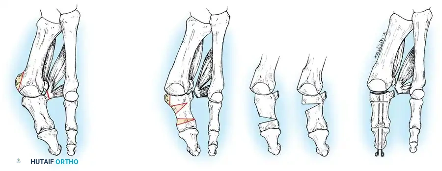
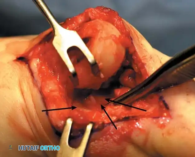
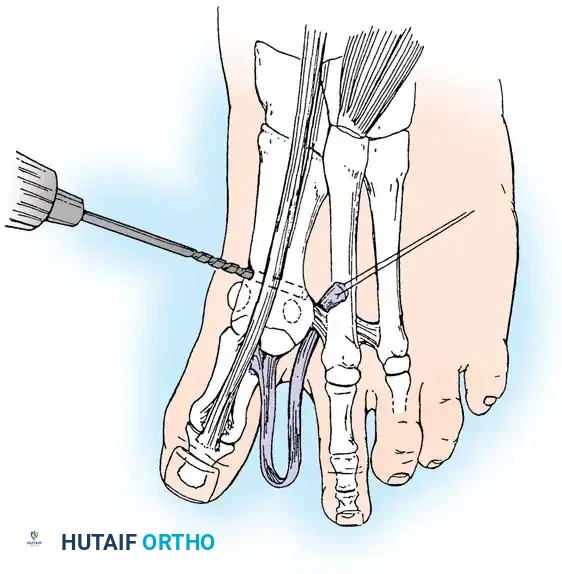
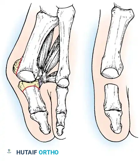
A critical anatomical distinction in the forefoot is the insertion of the deep transverse intermetatarsal ligament. Unlike the lesser metatarsophalangeal joints, where this ligament inserts directly into the adjacent metatarsal heads, at the first interspace, it connects the plantar plate of the first MTP joint to the plantar plate of the second MTP joint. It does not insert into the first metatarsal bone. Consequently, as the first metatarsal deviates medially, the sesamoid apparatus is held firmly in place laterally by the deep transverse intermetatarsal ligament. The first metatarsal head effectively subluxates medially off the sesamoids, causing the crista to flatten due to abnormal abutment against the tibial sesamoid, eventually allowing the fibular sesamoid to displace completely into the first intermetatarsal space.
Intrinsic and Extrinsic Musculature Dynamics
The stability of the first MTP joint relies heavily on the synchronized action of the intrinsic and extrinsic musculature. The extrinsic tendons—the extensor hallucis longus (EHL) and flexor hallucis longus (FHL)—are normally centered over the dorsal and plantar aspects of the joint, respectively. In a rectilinear first ray, their force vectors strictly promote sagittal plane flexion and extension. However, as the hallux deviates laterally into valgus, these long tendons undergo a lateral bowstringing effect. Their line of pull shifts lateral to the joint's axis of rotation, converting them into potent secondary valgus deforming forces that exponentially accelerate the progression of the deformity.
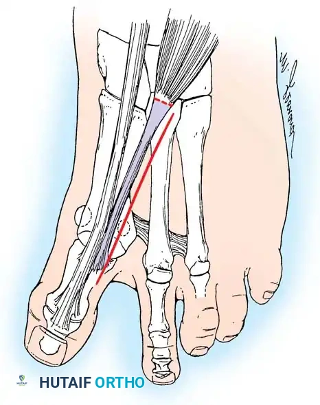
The intrinsic musculature undergoes equally devastating biomechanical alterations. The adductor hallucis, comprising both oblique and transverse heads, inserts firmly into the fibular sesamoid and the lateral base of the proximal phalanx. As the hallux pronates and deviates laterally, the adductor hallucis gains a massive mechanical advantage. Its unopposed contraction pulls the proximal phalanx further into valgus and pronation. Simultaneously, the abductor hallucis, which normally stabilizes the medial aspect of the joint, is displaced plantarward beneath the metatarsal head. In this altered position, it loses its ability to abduct the hallux and instead contributes to the abnormal pronatory moment.
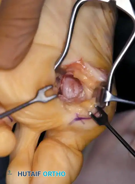
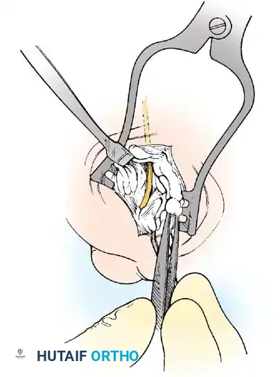
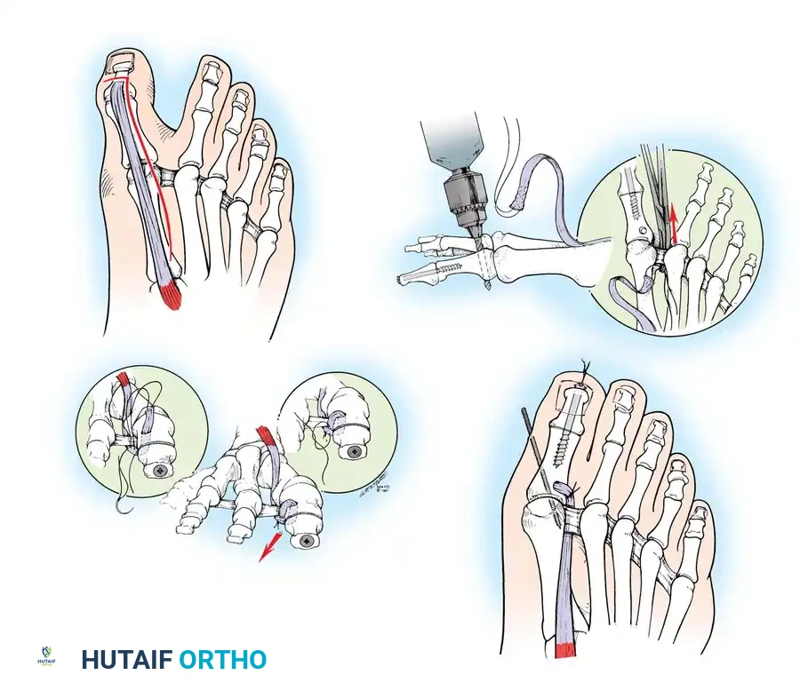
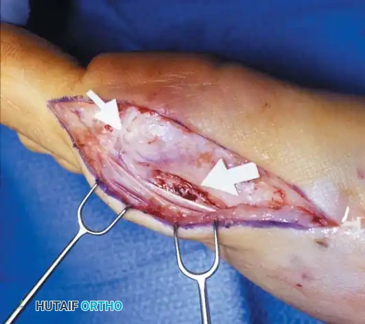
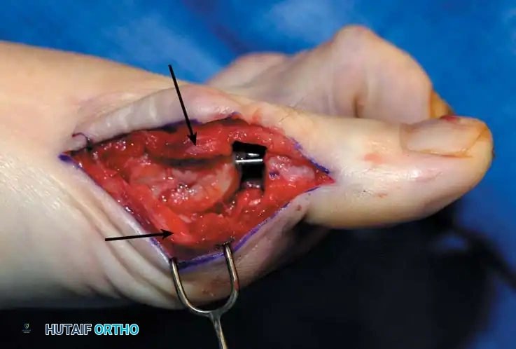
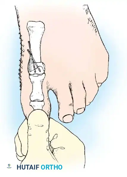
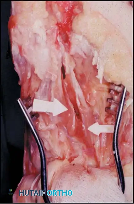
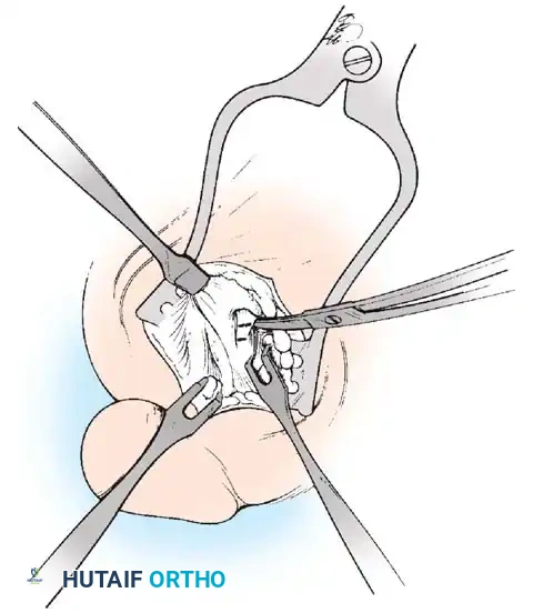
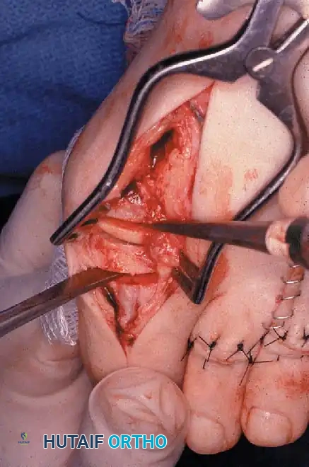
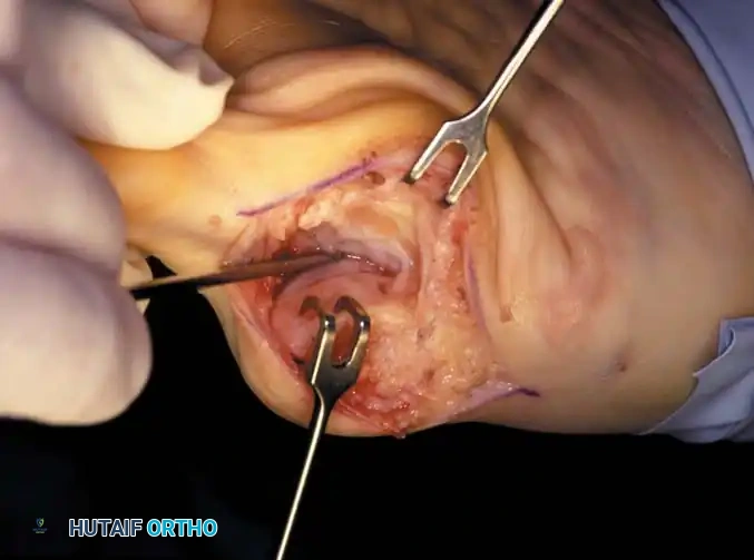
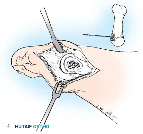
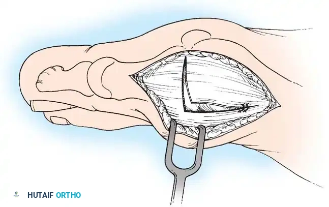
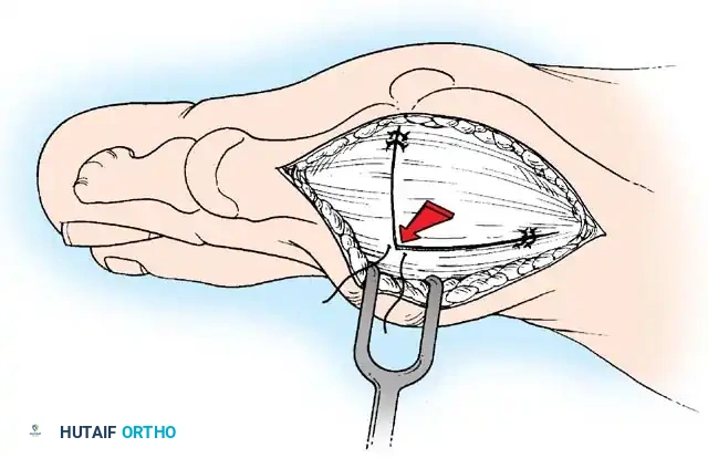
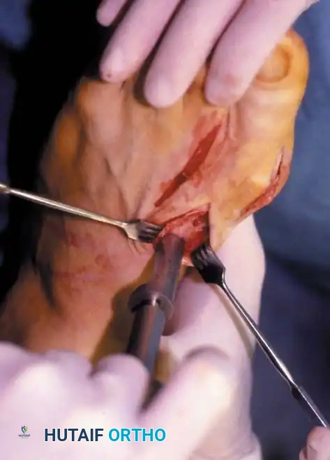
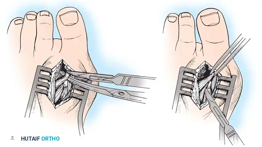
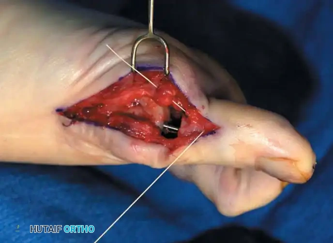
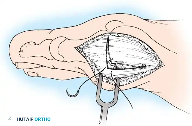
Surgical correction of hallux valgus must, therefore, address these muscular imbalances directly. A lateral soft-tissue release is frequently required to detach the adductor hallucis from the proximal phalanx and release the contracted lateral capsular structures. Concurrently, the medial capsulorrhaphy must not merely tighten the stretched tissues, but actively derotate the sesamoid sling and restore the abductor hallucis to its anatomic, medial position. Failure to restore the central alignment of the extrinsic tendons and balance the intrinsic vectors will inevitably result in rapid recurrence of the deformity, regardless of the osseous correction achieved.
Exhaustive Indications and Contraindications
Clinical Evaluation and Symptomatology
The clinical evaluation of a patient presenting with hallux valgus must be exhaustive, encompassing a detailed history of the deformity's progression, a precise localization of pain, and a comprehensive biomechanical assessment of the entire lower extremity. The primary indication for surgical intervention is intractable pain that significantly limits activities of daily living and is refractory to conservative management, including shoe wear modification, orthoses, and non-steroidal anti-inflammatory drugs (NSAIDs). Cosmetic dissatisfaction, in the absence of pain, is an absolute contraindication to surgical intervention, given the inherent risks of complications and the potential to convert a painless deformity into a painful, stiff joint.
The physical examination must systematically evaluate the entire forefoot, midfoot, and hindfoot. The surgeon must assess the mobility of the first TMT joint in the sagittal plane to rule out gross hypermobility, which may necessitate a Lapidus arthrodesis. The presence of a tight Achilles tendon (equinus contracture) must be identified, as it places excessive load on the forefoot during the terminal stance phase of gait, contributing to both hallux valgus progression and transfer metatarsalgia. The lesser toes must be meticulously examined for crossover deformities, hammer toes, and plantar plate insufficiency, as these frequently require concomitant surgical correction to achieve a balanced, functional forefoot.
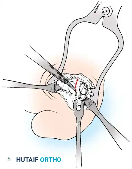
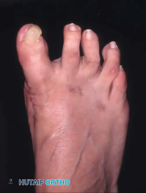
Assessment of the first MTP joint itself must determine the reducibility of the deformity and the presence of crepitus or restricted range of motion. A rigid, non-reducible deformity with significant crepitus suggests advanced degenerative joint disease (hallux rigidus secondary to hallux valgus). In such cases, joint-sparing osteotomies are generally contraindicated, and the surgeon must pivot toward salvage procedures such as an MTP joint arthrodesis or, in low-demand elderly patients, a Keller resection arthroplasty. Furthermore, the neurovascular status of the foot must be rigorously documented; severe peripheral arterial disease or profound peripheral neuropathy (e.g., Charcot neuroarthropathy) represents a strict contraindication to elective reconstructive forefoot surgery.
Radiographic Parameters and Decision Making
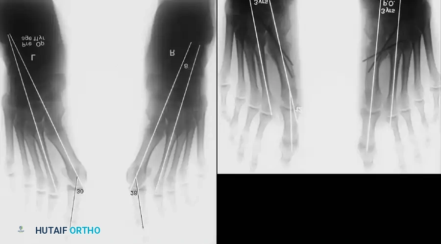
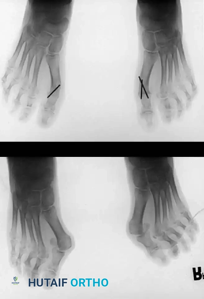
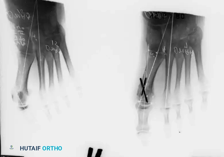
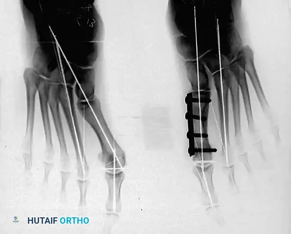
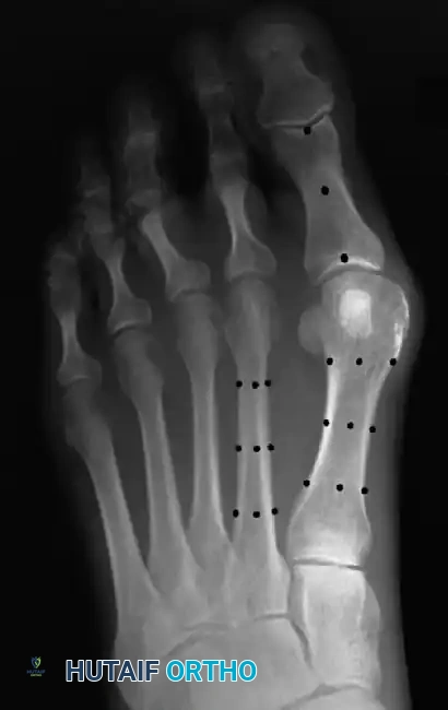
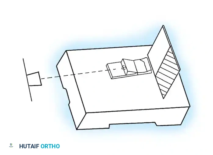
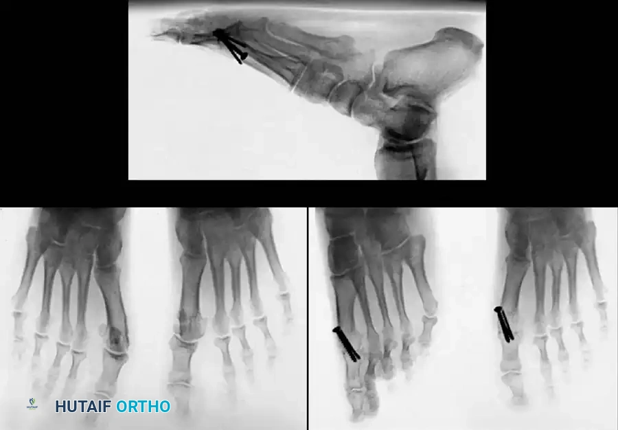
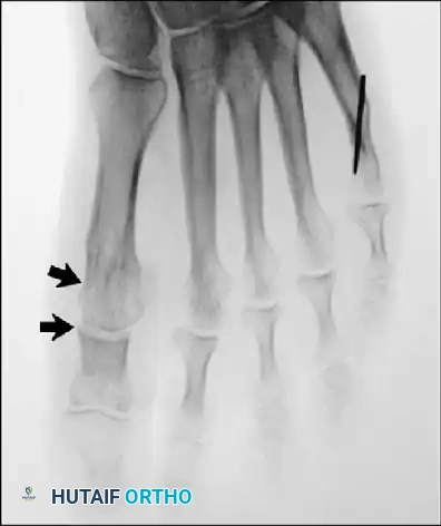
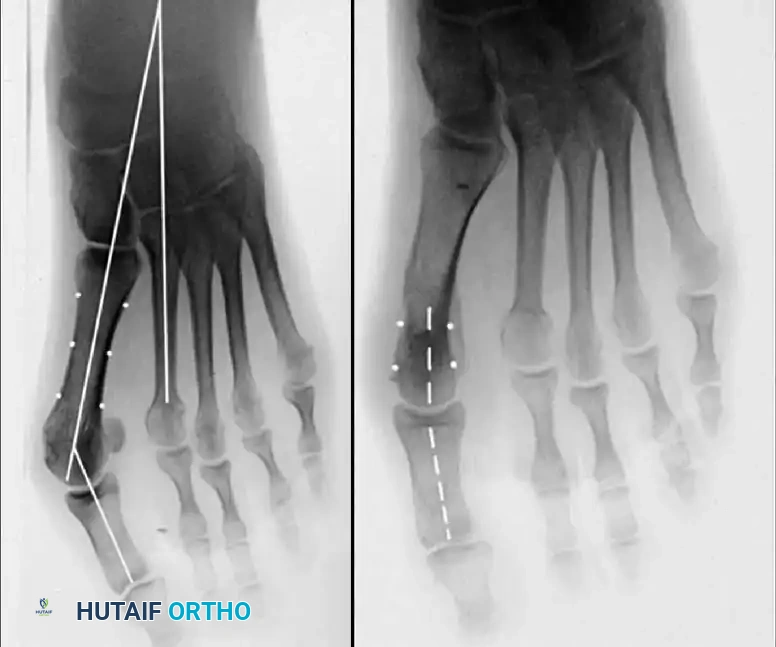
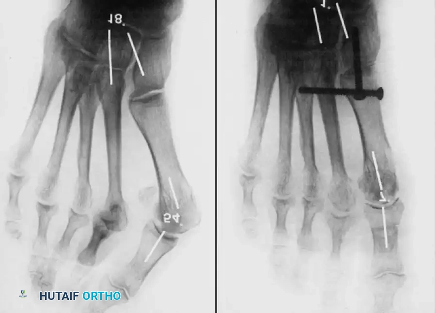
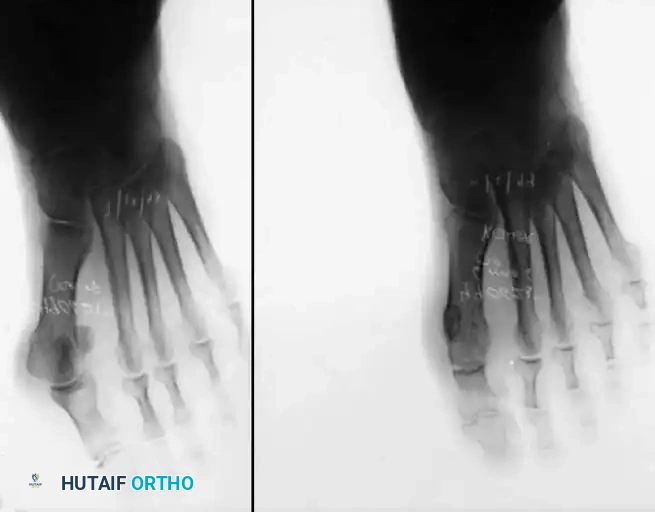
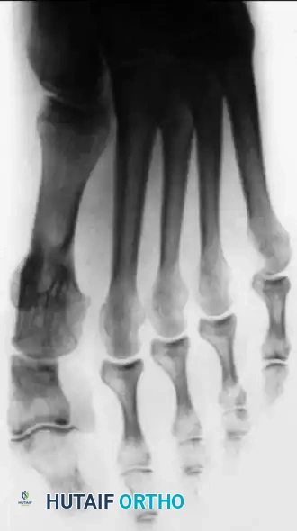
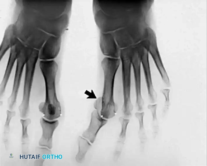
Standardized, weight-bearing radiographs are the cornerstone of preoperative planning for hallux valgus surgery. Non-weight-bearing films are entirely inadequate, as they fail to demonstrate the true dynamic splaying of the forefoot and the functional severity of the deformity. A complete radiographic series must include weight-bearing anteroposterior (AP), lateral, and sesamoid axial views. The AP view is utilized to measure the critical angular parameters: the Hallux Valgus Angle (HVA), the Intermetatarsal Angle (IMA), the Distal Metatarsal Articular Angle (DMAA), and the Phalangeal Articular Angle (PAA).
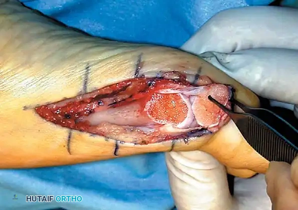
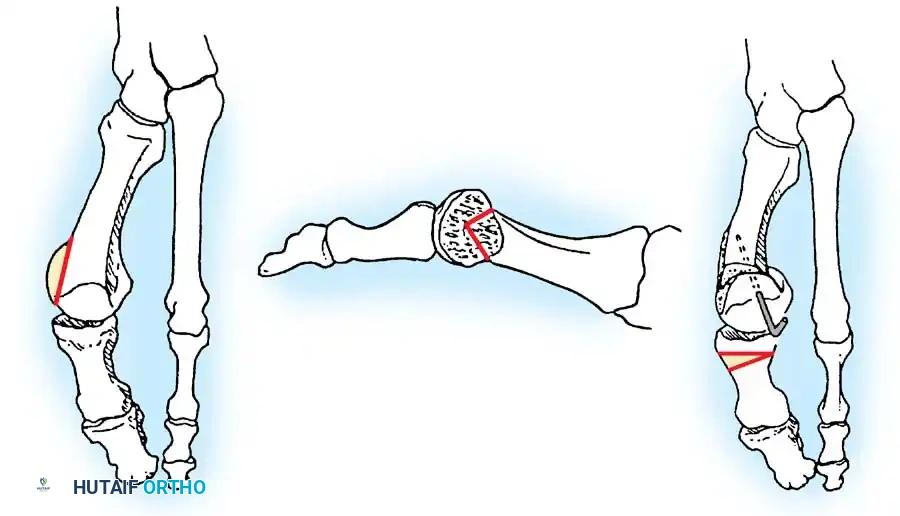
The severity of the deformity is classically categorized based on the HVA and IMA. A mild deformity is characterized by an HVA < 30 degrees and an IMA < 13 degrees. Moderate deformity involves an HVA between 30 and 40 degrees and an IMA between 13 and 20 degrees. Severe deformity is defined by an HVA > 40 degrees and an IMA > 20 degrees. These categorizations directly dictate the surgical algorithm. Mild deformities can often be managed with distal metatarsal osteotomies (e.g., Chevron). Moderate to severe deformities with a high IMA necessitate proximal metatarsal osteotomies (e.g., Crescentic, proximal Chevron) or TMT arthrodesis (Lapidus) to achieve adequate translational and angular correction.
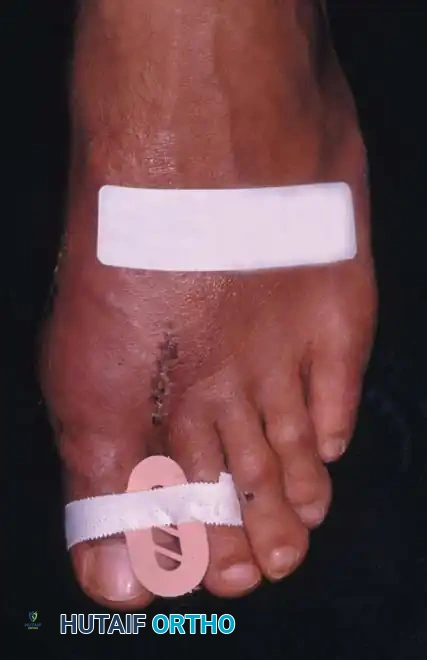
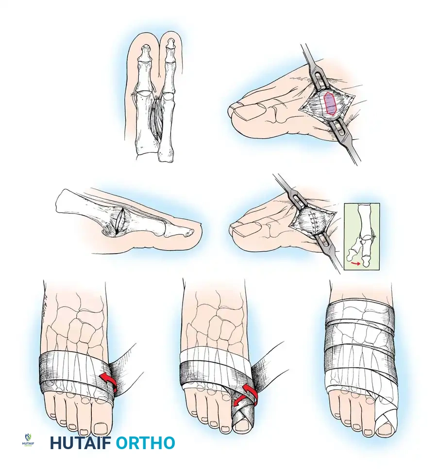
The congruency of the first MTP joint is another critical radiographic determinant. A congruous joint implies that the articular surfaces of the metatarsal head and proximal phalanx are parallel, despite the angular deviation. In a congruous hallux valgus (often seen in juveniles or driven by a high DMAA), soft-tissue releases and standard osteotomies that alter the articular orientation are contraindicated, as they will render the joint incongruous and precipitate arthritis. Instead, extra-articular corrections, such as a double first metatarsal osteotomy or an isolated Akin osteotomy, are required. The sesamoid axial view is indispensable for assessing the degree of lateral subluxation of the fibular sesamoid and evaluating the integrity of the plantar crista.
Surgical Indications Matrix
The selection of the appropriate surgical procedure requires a synthesis of clinical symptoms, physical examination findings, and rigorous radiographic measurements. No single procedure is universally applicable to all variants of hallux valgus. The surgeon must be adept at a diverse armamentarium of techniques, tailoring the approach to the specific pathoanatomy of the individual patient. The following matrix outlines the general indications and contraindications for the most commonly utilized procedures.
| Procedure Category | Specific Technique | Primary Indications | Absolute Contraindications |
|---|---|---|---|
| Soft-Tissue Only | Modified McBride | Mild deformity, HVA < 25°, IMA < 11°, Congruous joint | IMA > 12°, Incongruous joint, Severe pronation |
| Distal Osteotomy | Chevron (Austin) | Mild-to-moderate deformity, HVA < 30°, IMA < 13°, Normal DMAA | IMA > 15°, Advanced MTP arthritis, High DMAA (unless biplanar) |
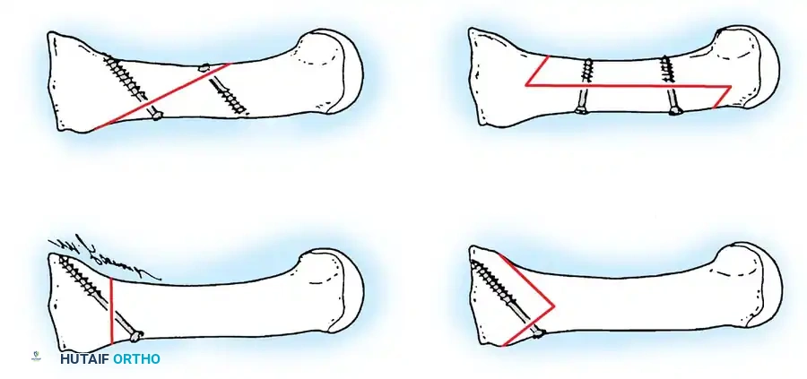
| Diaphyseal Osteotomy | Scarf / Ludloff | Moderate-to-severe deformity, IMA 13-20° | Narrow metatarsal shaft, Severe osteopenia |
| Proximal Osteotomy | Crescentic / Prox. Chevron | Severe deformity, IMA > 15°, HVA > 40° | First TMT hypermobility, MTP arthritis |
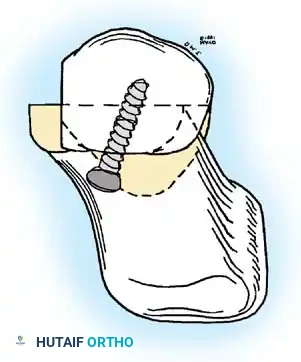
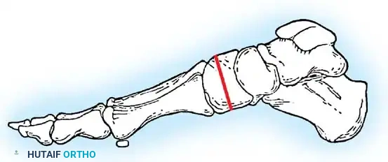
| TMT Arthrodesis | Lapidus Procedure | Severe deformity, First ray hypermobility, Recurrent HV | Open physes (relative), Non-compliant patient |
| MTP Arthrodesis | 1st MTP Fusion | Severe deformity with MTP arthritis, Rheumatoid arthritis, Salvage | Active infection, Severe ipsilateral IP joint arthritis |
| Resection Arthroplasty | Keller Procedure | Low-demand elderly, Severe arthritis, Ulceration salvage | Young/active patients, Significant metatarsus primus varus |
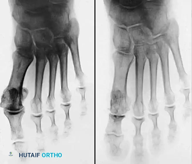
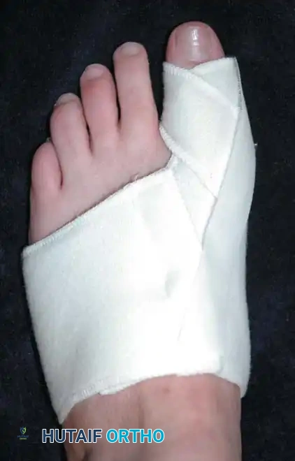
It is imperative to recognize that these indications serve as guidelines rather than immutable laws. Patient-specific factors, such as bone quality, vascular status, occupational demands, and compliance with postoperative rehabilitation, must heavily influence the final surgical decision. For instance, a patient with a moderate deformity (IMA 14 degrees) but profoundly osteopenic bone may be a poor candidate for a diaphyseal Scarf osteotomy due to the risk of troughing and fracture, making a proximal osteotomy or MTP arthrodesis a more prudent choice.
Pre-Operative Planning, Templating, and Patient Positioning
Radiographic Templating and Procedure Selection
Meticulous preoperative radiographic templating is the sine qua non of successful hallux valgus reconstruction. The surgeon must utilize calibrated weight-bearing radiographs to accurately measure the HVA, IMA, DMAA, and PAA. Modern digital templating software allows for precise simulation of various osteotomies to determine the exact degree of translation and angulation required to restore normal biomechanics. The goal of templating is to ensure that the chosen procedure can mathematically achieve an IMA of less than 9 degrees and an H
Clinical & Radiographic Imaging Archive
Detailed Chapters & Topics
Dive deeper into specialized chapters regarding disorders-of-the-hallux