Total Hip Arthroplasty Dislocation: Comprehensive Guide to Causes, Prevention, and Management

Key Takeaway
Total Hip Arthroplasty (THA) dislocation arises from implant malposition, soft tissue imbalance, or specific patient movements (Dorr's classification). Prevention focuses on precise surgical technique, optimal implant selection, soft tissue balancing, and comprehensive patient education on activity restrictions. These measures are crucial for long-term hip stability.
Total Hip Arthroplasty Dislocation: Comprehensive Guide to Causes, Prevention, and Management
Introduction and Epidemiology
Total Hip Arthroplasty (THA) stands as a transformative surgical intervention for end-stage hip pathology, universally recognized as the gold standard for alleviating pain, restoring function, correcting deformity, and profoundly enhancing patient quality of life. Its broad indications encompass a spectrum of conditions, including primary or secondary osteoarthritis, avascular necrosis of the femoral head (etiologies such as alcohol-induced, corticosteroid-induced, or post-traumatic), developmental dysplasia of the hip (DDH), rheumatoid arthritis, ankylosing spondylitis, and complex post-traumatic arthritis. Continuous advancements in biomaterials, prosthetic design, and surgical techniques have propelled THA to achieve exceptionally high overall success rates and commendable patient satisfaction.
Despite these significant strides, postoperative complications remain a paramount concern for orthopedic surgeons. Common sequelae include infection, venous thromboembolism, aseptic loosening, periprosthetic fracture, wear, and dislocation. While the incidence of many complications has markedly decreased, dislocation persists as the second most common complication following aseptic loosening in primary THA, presenting a significant clinical challenge. Reported incidence rates for primary THA dislocation range from 0.5% to 7%, influenced by variables such as surgical approach, patient age, comorbidities, and implant design. In revision THA, owing to compromised soft tissue envelopes, bone stock deficiencies, and the complexities of component repositioning, dislocation rates escalate considerably, reaching 6.6% to 21.2%. Detailed analyses indicate that approximately 70% of dislocation events occur within three months postoperatively, categorized as early dislocations, frequently associated with surgical technique, implant placement, or inadequate early rehabilitation. Late dislocations are often attributed to factors such as prosthetic wear leading to joint space enlargement, soft tissue laxity, osteolysis, suboptimal patient compliance, or spinopelvic sagittal plane imbalance.
Hip dislocation represents a debilitating complication for patients, precipitating acute pain and immediate functional incapacitation. It also carries the potential for secondary injuries, including neurovascular compromise, periprosthetic fractures, prolonged hospitalization, increased rehabilitation demands, and escalating healthcare costs. Recurrent dislocations severely impair daily living activities and psychological well-being, often necessitating multiple revision procedures. Therefore, a thorough understanding of the underlying etiologies, risk factors, precise diagnostic modalities, standardized management strategies, and effective prophylactic measures for THA dislocation is of paramount clinical importance for optimizing patient outcomes and mitigating healthcare burden.
Dorr’s classification system, predicated on the fundamental etiology of THA dislocation, provides a clinically pragmatic framework for diagnosis, individualized treatment planning, and targeted prevention:
-
Positional Dislocation (Type I): In these instances, prosthetic component positioning is typically deemed within acceptable radiographic parameters, and periarticular soft tissue tension and balance are considered adequate. Dislocation primarily results from the patient engaging in inappropriate limb activities in extreme positions, causing impingement and exceeding the mechanical range of motion offered by the prosthetic articulation. This underscores the critical importance of robust postoperative patient education, strict adherence to rehabilitation protocols, and limitations on extreme range of motion activities.
-
Soft Tissue Imbalance Dislocation (Type II): This category of dislocation is directly linked to functional or structural anomalies of the perihilar soft tissues. Manifestations may include non-union or delayed union of the greater trochanteric osteotomy, leading to significant abductor insufficiency and compromised joint stability; insufficient femoral offset due to excessive femoral neck resection, thereby reducing the abductor lever arm and overall soft tissue tension, weakening joint stability; or excessive intraoperative soft tissue disruption, particularly of the posterior capsule and short external rotators, without adequate repair, resulting in intrinsic joint instability.
-
Implant Malposition Dislocation (Type III): This is one of the most prevalent root causes of THA dislocation, directly attributable to inappropriate positioning and orientation of the acetabular cup and/or femoral stem. For example, excessive anteversion or retroversion, or an inclination angle outside the established Lewinnek "safe zone" (conventionally defined as 30-50° inclination and 5-25° anteversion), can significantly constrict the effective range of motion of the articulation, increasing the risk of impingement and subsequent dislocation. Similar issues can arise from excessive femoral stem anteversion or retroversion.
-
Mechanical Impingement Dislocation (Type IV): This type of dislocation is precipitated by mechanical impingement between prosthetic components or between a component and bone, frequently coexisting with Type III (Implant Malposition). Impingement can occur between the femoral neck and the acetabular cup rim, or between the femoral stem and pelvic bone, restricting hip motion within specific ranges and ultimately levering the femoral head out of the acetabular component. This classification highlights the critical interplay between prosthetic design, size matching, and the patient's intrinsic anatomical architecture.
Surgical Anatomy and Biomechanics
Surgical Anatomy
A successful THA fundamentally relies on a precise understanding of complex hip joint anatomy. Intraoperative identification and protection of critical structures, alongside meticulous reconstruction of stabilizing elements, are foundational to dislocation prevention.
-
Bone Anatomy: The key bony structures include the acetabulum of the pelvis and the proximal femur. The acetabulum, a hemispherical socket, is formed by the ilium, ischium, and pubis. Its orientation (acetabular version and inclination) is critical for stability. The proximal femur comprises the femoral head, neck, greater and lesser trochanters, and the femoral shaft. Femoral neck anteversion and offset significantly influence joint mechanics and soft tissue tension. The relationship between the anterior inferior iliac spine (AIIS) and the rectus femoris origin, the ischial tuberosity, and the pubic symphysis provides crucial landmarks.
-
Capsuloligamentous Structures: The hip joint capsule, a robust fibrous structure, attaches proximally to the acetabular rim and transverse acetabular ligament, and distally to the intertrochanteric line anteriorly and the femoral neck posteriorly. Key thickenings form the iliofemoral, pubofemoral, and ischiofemoral ligaments, which provide significant passive stability. The posterior capsule and short external rotators (piriformis, gemelli, obturators internus and externus, quadratus femoris) are particularly vital for posterior stability and are often incised during a posterior approach. Meticulous repair of these structures is paramount.
-
Musculature:
- Abductors: Gluteus medius and minimus, originating from the ilium and inserting on the greater trochanter, are the primary abductors. Their integrity and proper tensioning are essential for maintaining lateral stability and gait. Damage to these muscles (e.g., during an anterolateral approach) or insufficient femoral offset can compromise abductor function.
- Adductors: Pectineus, adductor longus, brevis, magnus, and gracilis provide medial stability but can cause impingement with the femoral component if hypertrophied or if component malposition exists.
- Flexors: Iliopsoas (ilioacus and psoas major) and rectus femoris are powerful flexors. Iliopsoas impingement on an anteriorly proud acetabular component can lead to anterior pain and, rarely, dislocation.
- Extensors: Gluteus maximus and hamstrings are the primary extensors. The gluteus maximus provides significant posterior stability.
- Short External Rotators: As mentioned, these muscles contribute to posterior stability and their repair is critical after a posterior approach.
-
Neurovascular Structures: Proximity of the sciatic nerve (posteriorly), femoral nerve (anteriorly), obturator nerve (medially), and various vessels (femoral, superior and inferior gluteal) necessitates careful surgical dissection to prevent iatrogenic injury.
Biomechanics of Hip Stability
The stability of a THA construct is a complex interplay of inherent factors and surgical modifications:
-
Joint Congruency and Constraint: The articular congruency between the femoral head and acetabular liner is a primary stabilizer. Larger femoral head diameters enhance the "jump distance"—the distance the head must travel before dislocating from the liner—thereby increasing stability. However, this must be balanced against bearing surface wear and material considerations. Constrained liners provide increased mechanical stability but transfer more stress to the bone-prosthesis interface, increasing the risk of aseptic loosening.
`
-
Soft Tissue Tension and Balance: Adequate tension in the periarticular musculature and reconstructed capsule provides dynamic and static stability. Insufficient soft tissue tension, often due to leg length discrepancy or inadequate femoral offset restoration, can result in laxity and increased dislocation risk. Proper restoration of femoral offset directly impacts abductor muscle tension and the efficiency of the abductor lever arm.
-
Component Position and Orientation: This is perhaps the most controllable factor influencing THA stability.
- Acetabular Component: Lewinnek's "safe zone" (inclination 30-50°, anteversion 5-25°) remains a widely cited guideline. However, functional anteversion, which accounts for pelvic tilt in various positions, is increasingly recognized as more critical than static radiographic anteversion. Excessive inclination reduces superior coverage, while excessive anteversion can lead to anterior instability. Excessive retroversion increases posterior instability.
- Femoral Component: Femoral anteversion must be matched to the acetabular component and native anatomy. Proper restoration of femoral offset and leg length is essential for soft tissue tension and abductor mechanics.
- Combined Anteversion: The sum of acetabular and femoral anteversion (often aimed for 30-45°) is a critical parameter to optimize the overall functional arc of motion without impingement.
-
Impingement: Mechanical abutment between prosthetic components (e.g., femoral neck-liner) or between a component and native bone (e.g., femoral stem-pelvic rim) limits range of motion and can lever the head out of the socket. This is often exacerbated by malpositioned components or inadequate implant sizing.
-
Spinopelvic Kinematics: Recent literature emphasizes the dynamic relationship between the lumbar spine and pelvis, particularly in patients with spinal stiffness or deformity. Changes in pelvic tilt (e.g., increased posterior tilt upon sitting) can alter the functional anteversion of the acetabular cup, predisposing to impingement and dislocation even with components placed in the traditional "safe zone." Understanding a patient's spinopelvic parameters (e.g., pelvic incidence, sacral slope, pelvic tilt) preoperatively is increasingly vital.
Indications and Contraindications
Indications for Total Hip Arthroplasty
The primary indication for THA is intractable hip pain and functional limitation recalcitrant to conservative measures, stemming from significant degenerative or destructive hip joint pathology. These pathologies are typically characterized by radiographic evidence of joint space narrowing, osteophytes, subchondral sclerosis, or cyst formation.
- Primary Osteoarthritis: Idiopathic degenerative joint disease.
- Secondary Osteoarthritis: Post-traumatic arthritis, avascular necrosis (AVN) of the femoral head (alcohol-induced, corticosteroid-induced, idiopathic, post-fracture), developmental dysplasia of the hip (DDH), Legg-Calvé-Perthes disease sequelae, slipped capital femoral epiphysis (SCFE) sequelae, acetabular protrusio.
- Inflammatory Arthritis: Rheumatoid arthritis, ankylosing spondylitis, psoriatic arthritis.
- Other Conditions: Certain complex hip fractures in elderly patients (e.g., displaced femoral neck fractures unsuitable for osteosynthesis), tumors of the proximal femur or acetabulum requiring reconstruction, Paget's disease of bone with secondary arthritis.
Contraindications for Total Hip Arthroplasty
Contraindications are categorized as absolute or relative, reflecting the balance between surgical risk and potential benefit.
-
Absolute Contraindications:
- Active systemic infection or local infection in the hip joint.
- Sepsis.
- Distant uncontrolled infection (e.g., urinary tract infection, dental abscess) that could hematogenously seed the implant.
- Skeletally immature patients (relative, but generally avoided).
- Rapidly progressive neurological disorder that would preclude rehabilitation or lead to recurrent dislocation (e.g., uncontrolled spasticity, severe Parkinson's disease).
- Charcot arthropathy of the hip (relative, but high failure rates).
-
Relative Contraindications:
- Severe morbid obesity (increased infection, dislocation, DVT risk; challenging surgical exposure).
- Significant medical comorbidities posing unacceptably high anesthetic or surgical risk (e.g., severe cardiopulmonary disease).
- Non-ambulatory status, inability to cooperate with rehabilitation.
- Neuropathic joint disease without adequate control.
- Compromised skin integrity over the operative site.
- Arterial insufficiency or venous stasis in the affected limb.
- Severe bone loss or poor bone quality that compromises implant fixation (e.g., severe osteopenia, prior radiation).
- Recurrent hip dislocation in a patient with uncorrectable neuromuscular impairment.
Table: Indications for Total Hip Arthroplasty
| Category | Operative Indications | Non-Operative Considerations (Prior to Surgery) |
|---|---|---|
| Primary OA | Persistent, severe pain and functional limitation refractory to conservative measures, radiographic evidence of severe arthritis. | Physical therapy, NSAIDs, intra-articular injections, activity modification, weight loss. |
| Secondary OA | Same as primary OA, arising from identified underlying pathology (AVN, DDH, trauma, etc.). | Addressing underlying cause where possible, symptom management as above. |
| Inflammatory Arthritis | Symptomatic, radiographic evidence of joint destruction despite optimal medical management (DMARDs, biologics). | Disease-modifying anti-rheumatic drugs, corticosteroids, physical therapy. |
| Acute Trauma | Displaced femoral neck fractures in elderly, active patients where osteosynthesis is unlikely to succeed. | Internal fixation for younger patients or less active elderly patients, conservative management for stable fractures. |
| Tumors | Reconstruction following oncologic resection of proximal femur or acetabular tumors. | Chemotherapy, radiation therapy (depending on tumor type and stage). |
| Revision THA | Aseptic loosening, osteolysis, recurrent dislocation, chronic infection, periprosthetic fracture. | Non-operative management for minor symptoms or if surgical risk is prohibitive. |
Pre Operative Planning and Patient Positioning
Effective preoperative planning is paramount to minimize THA dislocation risk, addressing patient-specific factors, implant selection, and surgical technique.
Preoperative Assessment
-
Patient History and Physical Examination:
- Detailed history focusing on pain, functional limitations, prior hip surgeries, and medical comorbidities.
- Identify risk factors for dislocation: neuromuscular disorders (e.g., Parkinson's disease, stroke, cerebral palsy, spinal cord injury), cognitive impairment, obesity, alcohol abuse, prior hip surgery (especially open reduction for DDH), spinal deformities (e.g., kyphosis, scoliosis, previous lumbar fusion).
- Assess hip range of motion, muscle strength, gait pattern, and leg length discrepancy. Document any fixed hip deformities (e.g., flexion contracture, adduction contracture).
- Evaluate skin integrity over the surgical site.
-
Radiographic Assessment:
- Standard Hip Series: AP pelvis with both hips (including entire hemipelvis), lateral hip, and potentially frog-leg lateral. Assess joint space, osteophytes, bone defects, and implant positioning from prior surgeries if revision.
- Templating: Crucial for planning implant size (acetabular cup diameter, femoral stem size and offset), anticipated leg length change, and prosthetic head size. Use digital templating software to accurately estimate component placement and identify potential challenges (e.g., protrusio, cavitary defects).
- Spinopelvic Radiographs: For patients with spinal pathology or suspected spinopelvic imbalance, standing lateral lumbar spine radiographs and sitting lateral hip radiographs are invaluable. These help quantify pelvic tilt, sacral slope, and pelvic incidence, allowing for estimation of dynamic changes in acetabular orientation with posture. For instance, a stiff spine with reduced lumbar lordosis and resultant posterior pelvic tilt in sitting can significantly retrovert the functional acetabular cup, predisposing to anterior impingement and posterior dislocation.
- CT Scan: Reserved for complex cases (e.g., severe acetabular bone loss, protrusio, previous trauma, revision surgery) to accurately define bone stock, quantify acetabular version and inclination, and assess femoral torsion. 3D reconstruction can aid in understanding complex deformities.
Risk Stratification for Dislocation
Synthesize clinical and radiographic findings to identify high-risk patients. These patients may benefit from specific prophylactic measures, such as larger head options, dual mobility constructs, or highly constrained liners, and more intensive postoperative rehabilitation and activity restrictions.
Surgical Planning for Dislocation Prevention
- Component Selection:
- Femoral Head Size: Larger femoral heads (≥32mm, increasingly 36mm or even 40+mm) significantly increase the "jump distance" required for dislocation, offering enhanced stability.
- Dual Mobility (DM) Articulations: These systems feature a small femoral head articulating within a larger polyethylene liner, which then articulates with the metal shell. This provides a very large jump distance and greater stability, particularly beneficial in high-risk patients, revision cases, or those with neuromuscular disorders.
- Constrained Liners: For highly unstable situations or recurrent dislocations unresponsive to other measures, constrained liners mechanically capture the femoral head within the acetabular cup. However, they transfer more stress to the shell-bone interface, increasing the risk of aseptic loosening and mechanical failure.
- Target Component Positioning:
- Acetabular Cup: Aim for appropriate inclination (30-45°) and anteversion (15-25°) relative to the anterior pelvic plane, adjusting for spinopelvic kinematics. Consider an "anatomic" or "functional" safe zone rather than a static one, especially in patients with spinal stiffness.
- Femoral Stem: Restore appropriate femoral anteversion and offset to optimize soft tissue tension and avoid impingement. Correct leg length discrepancy within 5-10mm to avoid gait disturbances and optimize soft tissue tension.
- Approach Selection: The chosen surgical approach significantly impacts soft tissue integrity and subsequent dislocation risk. This decision should be individualized based on surgeon experience, patient anatomy, and risk factors.
Patient Positioning
The choice of patient positioning (lateral decubitus vs. supine) and the specific surgical approach are intimately linked.
-
Lateral Decubitus Position:
- Typically used for posterior and anterolateral approaches.
- Ensures adequate exposure of the posterior and lateral hip.
- Careful padding is essential to prevent nerve compression (peroneal, ulnar) and skin breakdown.
- Requires secure stabilization of the pelvis (e.g., with anterior and posterior posts) to prevent intraoperative rotation that can mislead orientation of acetabular component placement.
- Disadvantage: Can be challenging to accurately assess leg length and offset compared to supine, requiring experience or intraoperative imaging.
-
Supine Position:
- Typically used for direct anterior and sometimes anterolateral approaches.
- Allows for easier intraoperative leg length and offset assessment, and use of fluoroscopy.
- Provides access to both hips if bilateral THA is planned.
- Requires traction table for direct anterior approach, which can have its own risks (femoral nerve neurapraxia).
- Disadvantage: May limit posterior exposure and maneuverability for certain revisions.
Regardless of position, ensure proper draping to allow for intraoperative range of motion assessment and reduction trials.
Detailed Surgical Approach and Technique
The choice of surgical approach, meticulous soft tissue handling, and precise component placement are critical for minimizing the risk of THA dislocation.
Choice of Surgical Approach
The "ideal" surgical approach is contentious, with proponents for each emphasizing advantages in dislocation rates, recovery, and specific patient populations.
-
Posterior Approach (PA):
- Internervous Plane: Between gluteus maximus (innervated by inferior gluteal nerve) and gluteus medius/minimus (innervated by superior gluteal nerve) for skin incision, then through the short external rotators and posterior capsule.
- Advantages: Excellent exposure of the acetabulum and femoral canal, relatively straightforward for revision surgery, good for complex deformities.
- Dislocation Risk: Traditionally associated with a higher posterior dislocation rate if the posterior capsule and short external rotators are not meticulously repaired. Modern techniques with robust soft tissue repair have significantly reduced this risk.
- Technique Considerations:
- Skin incision centered over the greater trochanter, directed proximally and distally.
- Gluteus maximus fascia incised, fibers split bluntly.
- Short external rotators (piriformis, gemelli, obturator internus) are identified, tagged, and carefully detached from their femoral insertions. The posterior capsule is incised.
- After component implantation, meticulous repair of the short external rotators and capsule back to the posterior aspect of the greater trochanter or drill holes in the femur is crucial. This step is non-negotiable for stability.
-
Direct Lateral (Hardinge) and Anterolateral Approaches (ALA):
- Internervous Plane: Direct lateral approach splits the gluteus medius and minimus, while the anterolateral approach utilizes the interval between tensor fascia latae and gluteus medius.
- Advantages: Lower perceived posterior dislocation rate due to preservation of posterior structures.
- Dislocation Risk: Can be associated with abductor weakness or limp if the abductor muscles are extensively damaged or inadequately repaired.
- Technique Considerations (Direct Lateral):
- Incision over the greater trochanter, extending proximally and distally.
- Fascia lata incised, then gluteus medius and minimus are split longitudinally at their anterior one-third.
- Can lead to persistent limp or heterotopic ossification.
-
Direct Anterior Approach (DAA):
- Internervous Plane: Between the tensor fascia latae (superior gluteal nerve) and the sartorius/rectus femoris (femoral nerve).
- Advantages: Muscle-sparing, potentially faster initial recovery, theoretically lower dislocation rates (especially posterior) due to preservation of all posterior structures, easier intraoperative fluoroscopy use for component positioning.
- Dislocation Risk: Low posterior dislocation risk. However, anterior dislocation can occur if component anteversion is excessive, or with specific extreme motions. Femoral preparation can be challenging, particularly in obese or muscular patients, increasing risk of femoral fracture or malposition.
- Technique Considerations:
- Patient typically supine, often on a traction table.
- Skin incision anterior to the greater trochanter.
- Tensor fascia latae and sartorius/rectus femoris are retracted to expose the hip capsule.
- Capsulotomy is performed, and care is taken to protect the lateral femoral cutaneous nerve.
- Femoral elevation and preparation require specific instrumentation and maneuvers (e.g., external rotation, extension, adduction of the leg to expose the femoral neck).
- No posterior soft tissue repair is typically performed as these structures are undisturbed.
Acetabular Preparation and Component Placement
- Exposure and Reaming: After capsulectomy and femoral head dislocation/osteotomy, the acetabulum is exposed. Sequential reaming is performed to remove articular cartilage and subchondral bone, creating a hemispherical socket for the acetabular cup. Reaming should restore the anatomical center of rotation.
- Cup Orientation: This is a critical step for preventing dislocation.
- Inclination (Abduction Angle): Aim for 30-45° relative to the transverse acetabular ligament or the interischial line. Excessive inclination reduces superior coverage and increases superior dislocation risk. Insufficient inclination may restrict range of motion and cause impingement.
- Anteversion: Aim for 15-25° relative to the anterior pelvic plane. Excessive anteversion can lead to anterior instability and posterior impingement. Insufficient anteversion (retroversion) increases posterior instability and anterior impingement.
- Functional Anteversion: Consider intraoperative fluoroscopy or navigation systems, especially in patients with spinal deformities, to assess component orientation in dynamic positions. This helps account for changes in pelvic tilt.
- Fixation: Press-fit fixation is common for uncemented cups. Supplemental screw fixation may be used to enhance stability in cases of poor bone quality or large defects. Ensure screws are not excessively long or directed into neurovascular structures.
Femoral Preparation and Component Placement
- Femoral Osteotomy: The femoral neck is osteotomized at a predetermined level to allow for proper leg length and offset restoration.
- Femoral Canal Preparation: Sequential reaming and broaching of the femoral canal are performed to prepare for the femoral stem. The final broach should provide a snug fit, indicating optimal implant-bone contact for primary stability.
- Stem Orientation:
- Anteversion: The femoral stem should be implanted with appropriate anteversion, typically matching the native femoral anteversion, or adjusted to complement the acetabular cup anteversion for optimal combined anteversion (30-45°). Excessive or insufficient femoral anteversion can contribute to impingement and dislocation.
- Offset: Restoration of femoral offset (the horizontal distance from the center of rotation to the femoral shaft axis) is crucial for tensioning the abductor muscles and optimizing their lever arm, thereby enhancing dynamic stability and reducing limping. Modular neck options can assist in fine-tuning offset and version.
- Trial Reduction and Stability Testing:
- Before definitive component implantation, a trial reduction is performed with trial components of appropriate sizes.
- Range of Motion Assessment: Systematically test the hip through its full range of motion (flexion, extension, abduction, adduction, internal, external rotation) to identify any impingement points or instability.
- Stability Assessment: Assess stability in positions that commonly cause dislocation for the chosen approach (e.g., flexion, adduction, internal rotation for posterior approach). Look for smooth, unrestricted motion without subluxation or clunking.
- Leg Length and Offset Assessment: Compare intraoperatively to preoperative planning and the contralateral limb. Adjust trial components (e.g., neck length, head size) until optimal parameters are achieved.
- Definitive Implantation: Once stability, range of motion, leg length, and offset are optimized with trial components, the definitive femoral head is placed, and the hip is reduced.
Soft Tissue Repair
- Posterior Approach: Meticulous repair of the posterior capsule and short external rotators (e.g., using transosseous sutures through the greater trochanter or drill holes in the femur) is essential to reconstruct the posterolateral soft tissue sleeve, significantly reducing posterior dislocation risk.
- Other Approaches: While not as extensive, primary capsular repair is beneficial across all approaches to enhance stability.
Complications and Management
THA dislocation, while extensively preventable, remains a significant complication requiring prompt and systematic management.
Diagnosis of Dislocation
- Clinical Presentation: Patients typically present with acute, severe hip pain and inability to bear weight. The affected limb is usually held in a characteristic position:
- Posterior Dislocation (most common): Flexion, adduction, and internal rotation.
- Anterior Dislocation (less common): Flexion, abduction, and external rotation.
- Superior Dislocation: Less common, typically secondary to severe abductor deficiency.
- Radiographic Confirmation: Antero-posterior (AP) pelvis and lateral hip radiographs are mandatory. These confirm the dislocation, identify the direction, and rule out periprosthetic fracture. Post-reduction radiographs are essential to confirm concentric reduction and rule out retained intra-articular fragments or recurrent subluxation.
Acute Dislocation Management
- Closed Reduction: This is the immediate goal. It should be performed as soon as possible to minimize soft tissue swelling, pain, and potential neurovascular compromise.
- Technique: Typically performed under conscious sedation or general anesthesia in the emergency department or operating room. Various maneuvers are described, often involving traction, flexion, and then rotation and abduction (for posterior dislocation) or adduction (for anterior dislocation). The patient should be monitored for vital signs and neurovascular status.
- Post-Reduction Assessment: After reduction, the hip should be carefully examined for stability through a controlled range of motion. Post-reduction radiographs confirm concentric reduction.
- Post-Reduction Care: Depending on the suspected cause and stability, the limb may be immobilized in a brace (e.g., abduction brace) for 4-6 weeks, particularly for early, traumatic dislocations without obvious component malposition. Progressive weight-bearing and activity restrictions are implemented.
Recurrent Dislocation Etiology and Workup
Recurrent dislocation necessitates a thorough investigation to identify the underlying cause, often aligning with Dorr's classification.
- Comprehensive Workup:
- Patient History: Detailed inquiry into activity levels, specific positions leading to dislocation, compliance with precautions, history of neuromuscular disorders or cognitive impairment.
- Physical Examination: Assess for leg length discrepancy, abductor strength, range of motion, and signs of neuromuscular deficit.
- Radiographic Assessment:
- Standard Hip Series: AP pelvis, lateral hip. Evaluate component position (inclination, anteversion), head size, femoral offset, and look for signs of osteolysis or loosening.
- CT Scan: Gold standard for precisely measuring acetabular and femoral component version, detecting subtle impingement points, and assessing for subtle bone defects. It can also identify soft tissue interposition.
- Spinopelvic Radiographs: Standing lateral lumbar spine and sitting lateral hip views are crucial to evaluate dynamic pelvic tilt and its impact on functional acetabular orientation, especially in patients with spinal pathology.
- MRI: Less commonly used for dislocation but can assess soft tissue integrity, differentiate fluid collections, or evaluate for abductor tendon pathology.
- Exclude Infection: ESR, CRP, aspiration with cell count and culture if suspicion of infection.
Management of Recurrent Dislocation
Management strategy hinges on the identified etiology.
-
Non-Operative Management: For patients with positional dislocations, severe comorbidities precluding surgery, or those with neuromuscular disorders where surgical solutions are unlikely to be durable. This includes:
- Activity Modification: Strict avoidance of high-risk positions.
- Bracing: Use of abduction braces or orthoses to limit extreme ranges of motion.
- Physical Therapy: Strengthening exercises, gait training, balance improvement, if feasible.
-
Revision Surgery: Indicated when a correctable mechanical cause is identified and non-operative measures fail, or for multiple dislocations. The goal is to address the underlying mechanism of instability.
-
Address Component Malposition:
- Acetabular Revision: Repositioning the cup into a more favorable inclination and anteversion. This may involve removing and reimplanting the cup, or using a constrained liner if minor malposition exists.
- Femoral Revision: Repositioning the stem to correct femoral anteversion or altering femoral offset with a different stem or modular neck.
-
Increase Head-Neck Ratio and Jump Distance:
- Larger Femoral Heads: If the existing acetabular liner allows, upgrading to a larger femoral head (e.g., 36mm, 40mm) significantly increases stability.
- Dual Mobility (DM) Constructs: Conversion to a dual mobility system is a highly effective strategy for recurrent dislocation, particularly in high-risk patients, revision settings, and cases with soft tissue deficiency.
- Constrained Liners: For recalcitrant instability where other measures have failed, or in patients with severe neuromuscular compromise. These provide mechanical capture but require careful consideration due to higher stresses on the implant-bone interface.
-
Address Soft Tissue Deficiency/Imbalance (Dorr Type II):
- Trochanteric Advancement: For severe abductor insufficiency or non-union of a prior trochanteric osteotomy, the greater trochanter can be osteotomized and advanced distally and laterally to restore abductor tension.
- Abductor Repair/Reconstruction: Direct repair of torn abductor tendons or reconstruction using allograft/autograft.
- Soft Tissue Release: In rare cases of severe soft tissue contracture causing impingement, judicious release may be considered.
-
Address Impingement (Dorr Type IV):
- Component Repositioning: As above, correct malpositioned components.
- Osteophyte Resection: Remove any impinging osteophytes around the acetabular rim or femoral neck.
- Bone Resection: In rare cases, minor bone resection (e.g., of a proud anterior inferior iliac spine) may be considered, but with caution to avoid weakening the bone.
-
Address Spinopelvic Imbalance: In patients with significant fixed spinal deformity, specific adjustments to acetabular cup orientation (e.g., targeting less anteversion or higher inclination for patients with severe posterior pelvic tilt in sitting) may be necessary to accommodate the altered functional acetabular position. This requires careful preoperative assessment and intraoperative confirmation.
-
Table: Common THA Complications, Incidence, and Salvage Strategies
| Complication | Incidence (Primary THA) | Salvage Strategies | Specific Dislocation Link |
|---|---|---|---|
| Dislocation | 0.5% - 7% | Closed reduction, activity restriction, bracing, revision THA (component repositioning, larger head, dual mobility, constrained liner, soft tissue repair). | Directly related to all aspects of the current discussion. |
| Aseptic Loosening | 2-5% at 10 years | Revision THA (component exchange, bone grafting). | Increased stress from constrained liners can accelerate loosening. |
| Periprosthetic Joint Infection | 0.5% - 2% | Debridement and implant retention (DAIR), single-stage or two-stage revision, suppressive antibiotics. | N/A |
| Periprosthetic Fracture | 0.1% - 1.5% | ORIF with or without revision components, stem exchange. | Dislocation maneuvers can cause or exacerbate fractures. |
| Neurovascular Injury | 0.1% - 1% | Conservative management (nerve palsy), surgical exploration/repair (vascular injury). | Dislocation event or reduction maneuver can cause injury. |
| Venous Thromboembolism | 0.5% - 5% (symptomatic) | Anticoagulation, IVC filter. | N/A |
| Leg Length Discrepancy | 10% - 20% (minor) | Shoe lift, revision THA (component exchange). | Can alter soft tissue tension and contribute to instability. |
| Heterotopic Ossification | 5% - 20% (mild) | NSAIDs, radiation prophylaxis, surgical excision. | Impingement from HO can lead to reduced ROM and dislocation. |
Post Operative Rehabilitation Protocols
Postoperative rehabilitation is an integral component of dislocation prevention and successful THA outcome, reinforcing surgical stability and restoring function. Protocols must be individualized, considering the surgical approach, patient-specific risk factors for dislocation, and the stability achieved intraoperatively.
Early Postoperative Period (Days 0-6 Weeks)
- Weight-Bearing: Typically immediate weight-bearing as tolerated for uncemented components with good primary stability, or weight-bearing as tolerated for cemented components. Protected weight-bearing may be prescribed in cases of fragile bone or specific osteotomies.
- Activity Restrictions (Approach-Specific Dislocation Precautions):
- Posterior Approach: Emphasize avoiding hip flexion beyond 90 degrees, adduction past midline, and internal rotation. Use an abduction wedge in bed and avoid crossing legs.
- Direct Anterior Approach: Emphasize avoiding hip extension and external rotation, especially combined with adduction. Avoid "figure-4" position.
- General Precautions: Avoid low chairs, squatting, and twisting movements.
- Early Mobilization:
- Initiate gentle range of motion exercises, guided by precautions, to prevent stiffness and promote circulation.
- Early ambulation with assistive devices (walker, crutches) is encouraged to reduce DVT risk and restore functional mobility.
- Isometric strengthening of quadriceps, hamstrings, and gluteal muscles (within limits of precautions).
- Patient Education: Crucial for preventing positional dislocations. Patients and caregivers must be thoroughly educated on activity restrictions, safe transfer techniques, and warning signs of dislocation. Provide written and pictorial instructions.
Intermediate Postoperative Period (6 Weeks - 3 Months)
- Gradual Increase in Activity: As soft tissue healing progresses and pain subsides, activity restrictions are gradually relaxed, typically around 6 weeks, based on surgeon assessment and intraoperative stability.
- Progressive Strengthening: Focus on strengthening hip abductors, adductors, flexors, and extensors. Closed kinetic chain exercises are generally preferred.
- Balance and Proprioception Training: Initiate exercises to improve balance and coordination, especially important for reducing fall risk.
- Gait Training: Progress from assistive devices to independent ambulation, emphasizing a normal heel-toe gait pattern.
- Stair Climbing: Practice stair negotiation, leading with the unaffected leg when ascending ("good to heaven") and the affected leg when descending ("bad to hell").
Late Postoperative Period (3 Months Onwards)
- Return to Activities: Most patients can gradually return to low-impact activities like walking, swimming, cycling. High-impact sports (running, jumping) or activities involving extreme hip motion (e.g., deep squatting, certain yoga poses) are generally discouraged.
- Lifelong Precautions: While most surgical approaches allow for near-normal range of motion over time, lifelong vigilance regarding extreme movements that might provoke impingement or exceed the prosthetic range of motion is prudent, especially in high-risk patients.
- Maintenance Strengthening: Encourage ongoing exercises to maintain hip and core strength.
- Weight Management: Encourage weight control to reduce stress on the hip joint.
Summary of Key Literature and Guidelines
The literature on THA dislocation is extensive, reflecting its prevalence and clinical significance. Evolving surgical techniques, implant designs, and a deeper understanding of patient-specific risk factors continue to shape best practices.
-
Surgical Approach and Dislocation Risk:
- Meta-analyses and large registry studies have historically suggested a higher dislocation rate with the posterior approach compared to anterolateral or direct anterior approaches. However, this distinction has become less pronounced with contemporary posterior approaches that include meticulous posterior capsule and short external rotator repair.
- The direct anterior approach has gained popularity, often cited for its muscle-sparing nature and low posterior dislocation rates. However, its anterior dislocation risk, while lower, still exists, and it carries specific risks of femoral fracture or nerve injury.
- Ultimately, surgeon experience and adherence to sound surgical principles are often more important than the specific approach itself.
-
Component Positioning:
- The "Lewinnek Safe Zone" (acetabular inclination 30-50°, anteversion 5-25°) remains a foundational concept. However, this static radiographic zone has been challenged by the recognition of dynamic spinopelvic kinematics.
- Studies by Lazennec et al. and Parratte et al. highlighted the impact of spinal sagittal balance and pelvic tilt on functional acetabular anteversion, demonstrating that patients with stiff spines or severe spinal deformity may be prone to dislocation even within the "safe zone" due to changes in pelvic orientation during sitting or standing. This has led to the concept of individualized target zones for component positioning.
- Combined anteversion (acetabular + femoral) is increasingly recognized as a key parameter, with a target range of 30-45° to optimize overall hip stability.
-
Implant Design:
- Femoral Head Size: Numerous studies consistently demonstrate that larger femoral head diameters (≥32mm, preferably ≥36mm or 40mm) significantly reduce the risk of dislocation by increasing the "jump distance" and impingement-free range of motion. This benefit is a major driver of the shift away from smaller heads.
- Dual Mobility (DM) Articulations: DM implants have shown robust evidence of reducing dislocation rates, particularly in high-risk patients (e.g., neuromuscular disorders, revision THA, prior dislocation, elderly patients). Their widespread adoption is supported by their biomechanical advantages.
- Constrained Liners: While effective for recalcitrant instability, the literature indicates a higher risk of aseptic loosening and mechanical failure compared to standard or DM bearings. Their use is generally reserved for situations where other options have failed.
-
Patient Factors:
- Neuromuscular disorders (Parkinson's, stroke, cerebral palsy), cognitive impairment, obesity, prior hip surgery, and alcohol abuse are well-established patient-related risk factors for dislocation. Preoperative identification of these factors allows for targeted counseling and consideration of enhanced stability implants.
-
Rehabilitation and Patient Education:
- Strict adherence to postoperative precautions, tailored to the surgical approach, is vital in the early postoperative period.
- Comprehensive patient education, as demonstrated by studies from the American Academy of Orthopaedic Surgeons (AAOS) and other societies, significantly improves patient compliance and reduces positional dislocations.
-
Guidelines:
- Professional organizations like the AAOS and the European Hip Society (EHS) regularly update guidelines and consensus statements on best practices for THA, often addressing dislocation prevention and management. These guidelines typically emphasize meticulous preoperative planning, intraoperative stability assessment, appropriate component selection, and comprehensive postoperative care.
- The management algorithm for recurrent dislocation typically progresses from addressing identifiable mechanical causes (malposition, impingement) to increasing prosthetic stability (larger heads, DM, constrained liners), and finally considering soft tissue augmentation.
In conclusion, THA dislocation remains a multifactorial complication. A thorough understanding of anatomical nuances, biomechanical principles, meticulous surgical technique, careful component selection, and comprehensive postoperative rehabilitation, all informed by a robust literature base and individualized patient assessment, are essential for minimizing its incidence and effectively managing its sequelae. The ongoing evolution of implant technology and surgical understanding, particularly regarding spinopelvic dynamics, continues to refine strategies for optimizing THA stability.
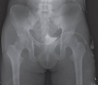
Clinical & Radiographic Imaging
You Might Also Like