Principles of Operative Arthroscopy: Indications, Techniques, and the Mastery of Triangulation
Key Takeaway
Operative arthroscopy demands exceptional psychomotor skills, spatial awareness, and mastery of triangulation. This comprehensive guide details the fundamental principles of arthroscopic surgery, including precise indications, absolute and relative contraindications, and the biomechanics of instrument manipulation within confined intra-articular spaces. Designed for orthopedic residents and consultants, it provides a step-by-step framework for overcoming the steep learning curve associated with advanced arthroscopic techniques.
INTRODUCTION TO OPERATIVE ARTHROSCOPY
The advent of operative arthroscopy represents one of the most profound paradigm shifts in the history of orthopedic surgery. Transitioning from extensive, morbidity-inducing arthrotomies to minimally invasive, visually magnified intra-articular interventions has revolutionized patient care. However, the transition from open surgeon to master arthroscopist requires a fundamental rewiring of psychomotor skills, spatial proprioception, and tactile feedback. This comprehensive academic review delineates the foundational principles, inherent disadvantages, precise indications, and the critical biomechanics of triangulation required for proficiency in operative arthroscopy.
DISADVANTAGES AND INHERENT LIMITATIONS
While the benefits of arthroscopy—decreased postoperative pain, accelerated rehabilitation, and diminished soft-tissue morbidity—are universally recognized, the disadvantages are non-trivial and must be acknowledged by the operating surgeon.
Psychomotor and Cognitive Demands
Not every surgeon possesses the innate temperament or spatial awareness required to excel in arthroscopic surgery. The procedure demands operating through restrictive portals using delicate, elongated instruments. The surgeon must translate a two-dimensional, monocular video feed into a three-dimensional mental map of the joint. This loss of binocular convergence eliminates natural depth perception, forcing the surgeon to rely on secondary visual cues (e.g., relative size, motion parallax, and shadow casting) and altered tactile feedback.
Risk of Iatrogenic Injury
The necessity to maneuver rigid steel instruments within the tight confines of the intra-articular space—often with clearances measured in millimeters—poses a significant risk of iatrogenic injury.
Surgical Warning: Inexperienced surgeons frequently use the articular cartilage as a fulcrum to lever instruments. This produces significant scuffing, scoring, and irreversible iatrogenic chondral damage. Instruments must always be manipulated with the portal acting as the fulcrum, never the articular surface.
Equipment and Time Constraints
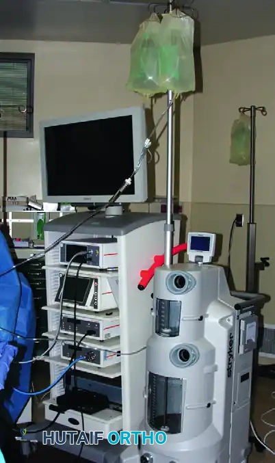
Early in a surgeon's learning curve, arthroscopic procedures can be exceptionally time-consuming. Prolonged operative times increase the risk of fluid extravasation, tourniquet-related complications, and infection. Furthermore, the specialized equipment required—high-definition camera systems, fluid management pumps, radiofrequency ablators, and motorized shavers—is extensive, fragile, and capital-intensive.
INDICATIONS AND CONTRAINDICATIONS
Historically reserved for the "problem joint" that defied clinical diagnosis, arthroscopy is now the gold standard for a vast array of intra-articular pathologies. However, rigorous patient selection remains the cornerstone of successful outcomes.
Diagnostic Indications
Diagnostic arthroscopy is indicated for the preoperative evaluation and definitive confirmation of clinical diagnoses prior to complex arthroscopic or open surgical reconstruction.
* Pathophysiological Mapping: It provides an unparalleled, magnified in vivo method for evaluating the pathophysiological mechanisms of synovial diseases, chondral wear patterns, and ligamentous kinematics.
* Medicolegal and Documentation: It is highly indicated for the precise documentation of specific lesions when medicolegal issues, worker’s compensation claims, or secondary gain factors necessitate irrefutable objective evidence.
Therapeutic Indications
Therapeutic indications span all major joints and include, but are not limited to:
* Meniscal resection and repair.
* Ligamentous reconstruction (e.g., ACL, PCL).
* Rotator cuff repair and labral stabilization.
* Chondroplasty and osteochondral grafting.
* Synovectomy and loose body removal.
Absolute Contraindications
- Active Local Infection: Arthroscopy is strictly contraindicated when there is a risk of joint sepsis from an overlying cellulitis, infected abrasion, or local skin condition.
- Remote Seeding: The presence of a remote, active systemic infection that may seed the operative site via bacteremia.
- Conservative Management: Arthroscopy should never be utilized in a minimally deranged joint that has not undergone an exhaustive trial of conservative, non-operative management.
Relative Contraindications
- Capsular and Ligamentous Disruption: Major collateral ligamentous and capsular disruptions (e.g., acute knee dislocation) permit massive, uncontrolled extravasation of irrigation fluid into the surrounding soft tissues. This can rapidly precipitate compartment syndrome. In such scenarios, the capsule must be allowed to heal ("stick down") or be repaired primarily before arthroscopy is attempted.
- Severe Ankylosis: Partial or complete ankylosis severely restricts joint volume, making instrument insertion dangerous. However, advanced arthroscopists may selectively utilize arthroscopy for the lysis of adhesions (arthroscopic arthrolysis) in the knee, shoulder, or elbow.
Clinical Pearl: Never proceed to arthroscopy without a meticulous history, a thorough physical examination, and appropriate standard non-invasive diagnostic imaging (radiographs, MRI). Arthroscopy is an adjunct to, not a replacement for, clinical acumen.
BASIC ARTHROSCOPIC TECHNIQUES AND SKILL ACQUISITION
Proficiency in arthroscopy requires immense patience, persistence, and the mastery of a surgical skill set entirely distinct from open orthopedics.
The Learning Curve
Everything in the arthroscopic field is magnified. Because the arthroscope is monocular, depth perception is a learned cognitive adaptation rather than a direct observation. While artificial models (dry labs) and cadaveric specimens (wet labs) are invaluable for initial orientation, they rarely provide sufficient realism or pathological variability to achieve true mastery.
The optimal method for skill acquisition is a graduated, mentored approach:
1. Observation: Extensive observation of highly skilled arthroscopists.
2. Diagnostic Mastery: Perfecting the 14-point diagnostic sweep of the knee or the 15-point sweep of the shoulder before attempting therapeutic intervention.
3. Hybrid Procedures: Performing procedures that combine arthroscopic and open techniques (e.g., arthroscopically assisted ACL reconstruction or mini-open rotator cuff repair). Opening the joint allows the surgeon to directly evaluate the efficacy of the arthroscopic portion of the procedure.
Managing Patient Expectations and Surgical Judgment
Modern patients possess high expectations regarding minimally invasive surgery, placing tremendous pressure on practicing orthopedic surgeons.
Pitfall: A surgeon must never be coerced by patient pressure into performing a complex arthroscopic procedure for which they have not yet developed sufficient psychomotor skills.
If an arthroscopic procedure is not progressing safely or efficiently, the surgeon must exercise mature clinical judgment. Aborting the arthroscopic approach and converting to an open arthrotomy that guarantees a safe, effective outcome is a hallmark of surgical wisdom, not a failure. A skillfully performed open procedure is infinitely preferable to a poorly executed, iatrogenically damaging arthroscopic attempt.
THE PRINCIPLE OF TRIANGULATION
Triangulation is the fundamental biomechanical and optical principle upon which all operative arthroscopy is based. It involves the precise spatial coordination of one or more instruments inserted through separate portals, brought into the optical field of the arthroscope.
Biomechanics and Geometry of Triangulation
In a perfectly triangulated setup, the tip of the arthroscope and the tip of the operating instrument form the apex of a triangle, with the distance between the two skin portals forming the base.
* Separation of Axes: Triangulation separates the viewing axis from the operating axis. This allows the viewing arthroscope to be magnified and the field of view to be optimized independently of the instrument's trajectory.
* Independent Movement: The most significant advantage of triangulation is that it permits independent, uncoupled movement of the arthroscope and the surgical instrument.
* The Fulcrum Effect: The skin portal and the joint capsule act as a fulcrum. The surgeon's hand movements outside the joint are translated into opposite movements inside the joint (e.g., moving the hand superiorly drives the instrument tip inferiorly).
Step-by-Step Triangulation Technique
Mastering triangulation requires overcoming the psychomotor challenge of bringing two objects together in a confined, three-dimensional space using only two-dimensional, monocular vision.
- Establish the Visual Field: Begin with the arthroscope retracted slightly away from the target pathology. This provides a wide-angle, panoramic field of vision. A common mistake among novices is driving the scope too close to the target, resulting in a "red-out" or "white-out" and a total loss of spatial orientation.
- Identify Anatomical Landmarks: Utilize constant intra-articular landmarks for orientation. In the knee, recognize the intercondylar notch, the anterior cruciate ligament, or divide the menisci into anatomical thirds. Direct the instrument toward these known structures.
- Instrument Insertion: Insert the accessory instrument through the working portal. Aim the instrument toward the light source of the arthroscope visible through the skin (transillumination), or aim toward the known anatomical target.
- Convergence: Once the instrument enters the wide field of view, advance both the arthroscope and the instrument simultaneously toward the intended pathology. This reduces the field of vision while increasing magnification for precise surgical work.
Overcoming Spatial Disorientation
If the surgeon becomes disoriented and cannot locate the instrument within the visual field, they must avoid blindly sweeping the instrument, which causes chondral damage.
Clinical Pearl (The Rescue Technique): If triangulation is lost, withdraw the accessory instrument slightly. Bring the instrument into the joint until it physically contacts the metallic sheath of the arthroscope. By gently sliding the instrument down the shaft of the sheath to the arthroscope tip, the instrument will seamlessly enter the center of the visual field.
The Role of Lens Angulation
The angle of the arthroscope's fore-lens dictates the difficulty of triangulation.
* 0-Degree Lens: A straight-ahead lens makes learning triangulation significantly easier, as the visual axis perfectly matches the mechanical axis of the scope.
* 30-Degree Lens: The standard in modern arthroscopy. It provides a wider field of view by rotating the light cable, allowing the surgeon to "look around corners," but requires a higher degree of spatial awareness.
* 70-Degree Lens: Used for complex visualization (e.g., posterior compartments of the knee or inferior capsule of the shoulder). Triangulation with a 70-degree lens is highly advanced, as the visual field is severely offset from the mechanical axis.
PATIENT POSITIONING AND OPERATING ROOM SETUP
Optimal triangulation is impossible without meticulous patient positioning and operating room setup. The setup must allow unimpeded manipulation of instruments and fluid management.
Knee Arthroscopy Positioning
- Supine Position: The patient is positioned supine. A lateral post or a circumferential leg holder is applied to the proximal thigh.
- Biomechanics of Setup: The leg holder acts as a fulcrum against which a valgus or varus force can be applied to open the medial or lateral compartments, respectively. This joint distraction is critical; without it, the tight confines of the tibiofemoral joint will not permit safe instrument triangulation, leading to iatrogenic cartilage scuffing.
- Tourniquet: A pneumatic tourniquet is placed proximally but is ideally left uninflated unless visualization is compromised by bleeding, thereby minimizing ischemic complications.
Shoulder Arthroscopy Positioning
- Lateral Decubitus: The patient is placed in the lateral decubitus position with the operative arm suspended in 10 to 15 pounds of traction at 45 degrees of abduction and 15 degrees of forward flexion. This maximizes the subacromial and glenohumeral spaces, facilitating easier triangulation.
- Beach Chair Position: The patient is seated upright at 45 to 60 degrees. This provides a more anatomical orientation of the shoulder, making the transition from arthroscopic to open procedures seamless, though it requires careful management of cerebral perfusion pressure.
POSTOPERATIVE PROTOCOLS AND REHABILITATION
The minimally invasive nature of arthroscopy allows for accelerated, yet carefully phased, postoperative rehabilitation. Protocols must be tailored to the specific therapeutic intervention performed.
Phase I: Immediate Postoperative (Days 1-7)
- Edema and Pain Control: Strict adherence to cryotherapy, elevation, and compression. Intra-articular local anesthetics (e.g., bupivacaine) and multimodal oral analgesia are utilized.
- Weight-Bearing Status: For simple diagnostic procedures or partial meniscectomies, weight-bearing as tolerated (WBAT) is permitted immediately. For meniscal repairs or osteochondral procedures, strict non-weight-bearing (NWB) or restricted weight-bearing is enforced to protect the repair.
- Early Range of Motion (ROM): Immediate initiation of passive and active-assisted ROM to prevent intra-articular adhesions and stimulate chondrocyte nutrition via synovial fluid diffusion.
Phase II: Intermediate Rehabilitation (Weeks 2-6)
- Restoration of Kinematics: Focus on restoring full, symmetrical ROM.
- Muscle Activation: Initiation of closed-kinetic-chain exercises to stimulate proprioception and rebuild quadriceps/rotator cuff strength without placing undue shear stress on the joint surfaces.
Phase III: Return to Function (Weeks 6+)
- Sport-Specific Training: Progression to plyometrics, cutting maneuvers, and sport-specific biomechanical training.
- Clearance: Return to play is dictated by the resolution of effusions, restoration of 90% contralateral limb strength, and the biological healing timeline of any repaired tissues.
CONCLUSION
Operative arthroscopy is an exacting discipline that marries advanced technology with refined psychomotor skills. The transition from a novice to a master arthroscopist is defined by a deep respect for the inherent disadvantages of the modality, strict adherence to indications, and the relentless pursuit of perfection in triangulation. By mastering the spatial geometry of the joint and respecting the biomechanical limitations of the instruments, the orthopedic surgeon can consistently deliver the profound benefits of minimally invasive surgery while mitigating the risks of iatrogenic harm.
📚 Medical References
- Operative arthroscopy of the knee, Instr Course Lect 30:357, 1981.
- Michot M, Conen D, Holtz D, et al: Prevention of deep-vein thrombosis in ambulatory arthroscopic knee surgery: a randomized trial of prophylaxis with low–molecular weight heparin, Arthroscopy 18:257, 2002.
- Miller DV, O’Brien SJ, Kelly AM, et al: The use of the contact Nd : YAG laser in arthroscopic surgery. Presented at the annual meeting of the Arthroscopy Association of North America, Seattle, Wash, Apr 13-16, 1989.
- Miller GK, Dickason JM, Blazina ME, et al: The use of electrosurgery for arthroscopic subcutaneous lateral release, Orthopedics 5:309, 1982.
- Miskulin M, Maldini B: Outpatient arthroscopic knee surgery under multimodal analgesic regimens, Arthroscopy 22:978, 2006.
- Mohr M, Henche HR: The morbidity associated with lost or irretrievable resected meniscal fragments, Arthroscopy 9:94, 1992.
- Muellner T, Menth-Chiari WA, Reihsner R, et al: Accuracy of pressure and fl ow capacities of four arthroscopic fl uid management systems, Arthroscopy 17:760, 2001.
- Mulhollan JS: Complications of arthroscopic surgery. Paper presented at the International Arthroscopy Association meeting, Philadelphia, Oct 4, 1980.
- Nelson WE, Henderson RC, Almekinders LC, et al: An evaluation of preand postoperative nonsteroidal antiinfl ammatory drugs in patients undergoing knee arthroscopy: a prospective, randomized, double-blinded study, Am J Sports Med 21:510, 1993.
- Noonan TJ, Tokish JM, Briggs KK, et al: Laser-assisted thermal capsulorrhaphy, Arthroscopy 19:815, 2003.
- Noyes FR, Spievack ES: Extraarticular fl uid dissection in tissues during arthroscopy: a report of clinical cases and a study of intraarticular and thigh pressures in cadavers, Am J Sports Med 10:346, 1982.
- Oberlander MA, Shalvoy RM, Hughston JC: The accuracy of the clinical knee examination documented by arthroscopy: a prospective study, Am J Sports Med 21:773, 1993.
- O’Connor RL: Arthroscopy in the diagnosis and treatment of acute ligament injuries of the knee, J Bone Joint Surg 56A:333, 1974.
- O’Connor RL: Arthroscopy , Philadelphia, 1977, JB Lippincott. Olszewski AD, Jones R, Farrell R, et al: The effects of dilute epinephrine saline irrigation on the need for tourniquet use in routine arthroscopic knee surgery, Am J Sports Med 27:354, 1999.
- Patel D, Guhl JF: The use of bovine knees in operative arthroscopy, Orthopedics 6:1119, 1983.
- Polousky JD, Hedman TP, Vangsness CT Jr: Electrosurgical methods for arthroscopic meniscectomy: a review of the literature, Arthroscopy 16:813, 2000.
- Poulsen KA, Borris LC, Lassen MR: Thromboembolic complications after arthroscopy of the knee, Arthroscopy 9:570, 1993.
- Price AJ, Jones NAG, Webb PJ, et al: Do tourniquets prevent deep vein thrombosis? J Bone Joint Surg 62B:529, 1980.
- Rand JA, Gaffey TA: Effect of electrocautery on fresh human articular cartilage, Arthroscopy 1:242, 1985.
- Rasmussen S, Thomsen S, Madsen SN, et al: The clinical effect of naproxen sodium after arthroscopy of the knee: a randomized, double-blind, prospective study, Arthroscopy 9:375, 1993.
- Reagan BF, McInerny VK, Treadwell BV, et al: Irrigating solutions for arthroscopy: a metabolic study, J Bone Joint Surg 65A:629, 1983.
- Reigstad O, Grimsgaard C: Complications in knee arthroscopy, Knee Surg Sports Traumatol Arthrosc 14:473, 2006.
- Ritt MJPF, TeSlaa RL, Koning J, et al: Popliteal pseudoaneurysm after arthroscopic meniscectomy: a report of two cases, Clin Orthop Relat Res 295:198, 1993.
- Rodeo SA, Forster RA, Weiland AJ: Current concepts review. Neurological complications due to arthroscopy, J Bone Joint Surg 75A:917, 1993.
- Schippinger G, Wirnsberger GH, Obernosterer A, et al: Thromboembolic complications after arthroscopic knee surgery: incidence and risk factors in 101 patients, Acta Orthop Scand 69:144, 1998.
- Schonholtz GJ: The use of closed circuit television in arthroscopy, Orthopedics 7:342, 1984.
- Schonsheim PM, Caspari RB: Evaluation of electrosurgical meniscectomy in rabbits, Arthroscopy 2:71, 1986.
- Shahriaree H: O’Connor’s textbook of arthroscopic surgery , Philadelphia, 1984, JB Lippincott. Shinjo H, Nakata K, Shino K, et al: Effect of irrigation solution for arthroscopic surgery on intraarticular tissue: comparison in human meniscus–derived primary cell culture between lactate Ringer’s solution and saline solution, J Orthop Res 20:1305, 2002.
- Shoji H, Gutierrez MM, Aldridge KE: The use of 2 glutaraldehyde as a disinfectant for arthroscopes used in septic joints, Orthopedics 7:241, 1984.
- Sisto DJ, Blazina ME, Hirsch LC: The synovial response after CO 2 laser arthroscopy of the knee, Arthroscopy 9:574, 1993.
- Skyhar MJ, Altcheck DW, Warren RF, et al: Shoulder arthroscopy with the patient in the beach-chair position, Arthroscopy 4:256, 1988.
- Small NC: Complications in arthroscopy: the knee and other joints, Committee on Complications of Arthroscopy Association of North America, Arthroscopy 2:253, 1986.
- Small NC: Complications in arthroscopic surgery performed by experienced arthroscopists, Arthroscopy 4:215, 1988.
- Smith JB: Laser energy for arthroscopic meniscus surgery: a preliminary report. Paper presented at the annual meeting of the Arthroscopy Association of North America, Coronado, Calif, Jan 1983.
- Stringer MD, Steadman CA, Hedges AR, et al: Deep vein thrombosis after elective knee surgery: an incidence study in 312 patients, J Bone Joint Surg 71B:492, 1989.
- Takagi K: The classic arthroscope, Clin Orthop Relat Res 167:6, 1982.
- Takahashi T, Tanaka M, Ikeuchi M, et al: Pain in arthroscopic knee surgery under local anesthesia, Acta Orthop Scand 75:580, 2004.
- Tawes RL, Etheredge SN, Webb RL, et al: Popliteal artery injury complicating arthroscopic meniscectomy, Am J Surg 156:136, 1988.
- Vangsness CT, Thordarson DB, Park K: A disposable fi beroptic arthroscope: a cadaver study, Foot Ankle 15:502, 1994.
- Watanabe M, Takeda S: The number 21 arthroscope, J Jpn Orthop Assoc 34:1041, 1960.
- Watanabe M, Takeda S, Ikeuchi H: Atlas of arthroscopy , Tokyo, 1969, Igaku Shoin. Weber SC, Abrams JS, Nottage WM: Complications associated with arthroscopic shoulder surgery, Arthroscopy 18(suppl 1):88, 2002.
- Whipple TL, Caspari RB, Meyers JF: Synovial response to laserinduced carbon ash residue, Lasers Surg Med 3:291, 1984.
- Whipple TL, Caspari RB, Meyers JF: Arthroscopic laser meniscectomy in a gas medium, Arthroscopy 1:2, 1985.
- Williams R, Laurencin CT, Warren RF, et al: Septic arthritis after arthroscopic anterior cruciate reconstruction, Am J Sports Med 25:261, 1997.
- Wredmark T, Lundh R: Arthroscopy under local anesthesia using controlled pressure-irrigation with prilocaine, J Bone Joint Surg 64B:583, 1982.
- Yang CY, Cheng SC, Shen CL: Effect of irrigation fl uids on the articular cartilage: a scanning electron microscope study, Arthroscopy 9:425, 1993.
- Yoshiya S, Kurosaka M, Hirohata K, et al: Knee arthroscopy using local anesthetic, Arthroscopy 4:86, 1988.
You Might Also Like