Complex OTA/AO 43-C3 Pilon Fracture: A Detailed Orthopedic Case Study
Key Takeaway
A pilon fracture, specifically an OTA/AO 43-C3, involves a comminuted intra-articular fracture of the distal tibia due to high-energy axial load. Diagnosis relies on plain radiographs and detailed CT scans to assess articular involvement, fragment displacement, and comminution. This guides pre-operative planning, crucial for anatomical reduction and successful management of severe ankle trauma.
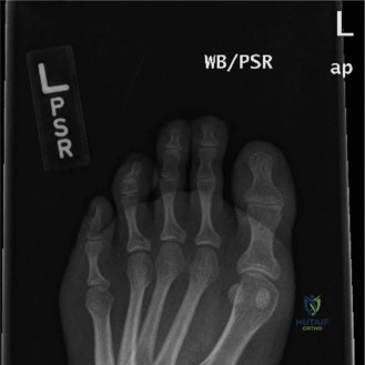
A 42-year-old male presents with this ankle injury after a fall from 4 meters. Describe the key clinical findings and the immediate management priorities for this limb.
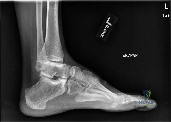
Candidate: The patient has a high-energy pilon fracture. I see significant comminution, articular depression, and lateral subluxation of the talus. Clinically, I would perform a thorough neurovascular assessment, document the soft tissue status (Tscherne classification), and look for signs of compartment syndrome. Immediate management involves neurovascular stabilization, splinting, and likely a staged protocol: spanning external fixation to allow for soft tissue recovery before definitive ORIF.
Focusing only on the fracture and jumping to internal fixation. Failing to mention the "soft tissue envelope" as the primary driver of decision-making, or forgetting to mention that this is a systemic high-energy injury requiring a trauma survey (ATLS) to rule out associated calcaneal, tibial plateau, or spinal fractures.
Structure the response: 1. Assessment: Neurovascular status, ATLS (rule out high-energy associates), and detailed Soft Tissue assessment (Tscherne/wrinkle sign). 2. Management: "Span, Scan, and Plan." Apply a spanning external fixator for damage control, perform a post-fixator CT for surgical planning, and counsel the patient on the high risk of soft tissue complications and long-term post-traumatic arthritis.
The patient has been stabilized with a fixator and is now 14 days post-injury. We are planning definitive fixation. Based on this CT reconstruction, what are the primary surgical objectives and how would you approach the reconstruction?
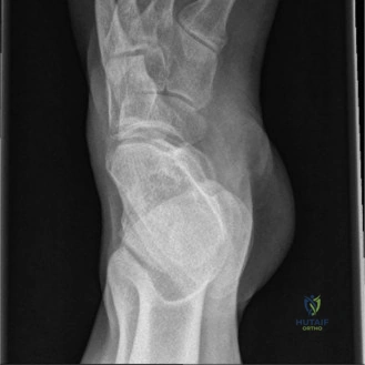
Candidate: The goal is anatomic reconstruction of the articular surface. I would use the 'outside-in' technique. I'd perform an ORIF of the fibula first to establish length. Then, I would utilize a dual-incision approach: an anterolateral approach to elevate the central die-punch fragments and the Chaput fragment, and a posteromedial approach to buttress the medial column and prevent varus collapse, as this is a high-risk area for failure.
Suggesting a single approach (e.g., just anterior) which risks leaving the posterior (Volkmann) fragment malreduced, or failing to address the "void" created by the elevated fragments, which will lead to secondary articular subsidence.
Emphasize: 1. Fibular fixation for length. 2. Anterolateral approach for the central plafond (the "die-punch"). 3. Posteromedial approach for medial/posterior stability. 4. Bone grafting the metaphyseal void to support the reconstructed articular block. 5. Locking plate construct to provide 'rafting' support to the articular surface.
Look at the final construct. What are the specific biomechanical reasons for choosing this dual-plate configuration, and what is your plan for postoperative weight-bearing?
Candidate: The dual-plate construct provides stability against the axial loading and rotational forces that caused the injury. The lateral plate supports the lateral column and provides rafting screws for the articular surface. The medial plate is a buttress against varus deformity. Regarding weight-bearing, I would maintain strict non-weight-bearing for 6 weeks, then progress to touch-down weight-bearing based on radiographic healing at the 6-week follow-up.
Ignoring the biology. Encouraging early weight-bearing without evidence of radiographic healing, or ignoring the need for active early range of motion to prevent stiffness, which is a major morbidity factor in these injuries.
State clearly: "Stability is provided by the dual construct to neutralize varus and compressive forces." Crucially, mention that while weight-bearing is delayed to protect the articular reconstruction, early non-weight-bearing active range of motion is essential to prevent arthrofibrosis and to facilitate synovial fluid movement across the cartilage.
