Decoding Pilon Fractures: Anterolateral and Medial Fragments Revealed
Key Takeaway
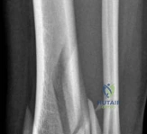
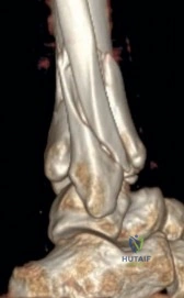
Your ultimate guide to Decoding Pilon Fractures: Anterolateral and Medial Fragments Revealed starts here. A metaphyseal fracture with joint involvement is a displaced, multifragmentary distal tibia fracture with articular involvement, often seen with an associated fibular fracture and tibiotalar joint dislocation. Imaging reveals displaced posterior, anterolateral, and medial fragments, causing intraarticular step-off and gap, which compromises the tibiotalar joint.
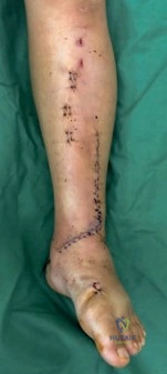
A 38-year-old female presents following a high-energy motor vehicle collision with a gross deformity of the left ankle. Radiographs show an AO/OTA 43C3.3 distal tibial fracture. What are your immediate priorities, and what is your rationale for the management strategy?
Candidate: My priority is to assess the neurovascular status and the soft tissue envelope. Since this is a high-energy injury with potential swelling, I would perform a closed reduction to restore length and apply a temporary ankle-spanning external fixator. This avoids early surgery on compromised skin to prevent infection.
Failure to mention the "span, scan, and plan" philosophy. Candidates often forget to mention the immediate neurovascular assessment or fail to justify why definitive ORIF is avoided acutely (the "swelling/blister" risk).
The candidate should state: "My immediate priority is the ATLS assessment of the polytrauma patient. Specifically for the ankle, I perform a neurovascular exam and assess the soft tissue for tension or open wounds. I advocate for the 'span, scan, and plan' protocol: emergent closed reduction and application of an ankle-spanning external fixator to neutralize deforming forces, restore length, and allow the soft tissue to recover. I would then obtain a CT scan to map the articular fragments before planning definitive fixation once the 'wrinkle sign' is present, typically at 10–21 days."
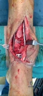
You have decided to proceed with definitive fixation. How do you plan your surgical approaches while respecting the local vascularity and the 7 cm skin bridge rule?
Candidate: I use the anterolateral approach for the Chaput fragment and the anteromedial approach for the medial column. I keep a 7 cm skin bridge between them to protect the angiosomes of the distal tibia.
Ignoring the danger of crossing angiosomes or failing to mention the preservation of the saphenous nerve/vein (medial) and the superficial peroneal nerve (lateral).
Structure the answer by anatomy: 1. Acknowledge the vascular supply (angiosomes of the anterior tibial artery). 2. Define the approaches: The anterolateral incision (lateral to peroneus tertius) to address the Chaput fragment and fibula; the anteromedial incision (lateral to the tibial crest) to address the medial column. 3. Emphasize the 7 cm rule to prevent necrosis of the skin bridge. 4. Mention "no-touch" soft tissue technique, avoiding excessive traction or crushing forceps.
During the procedure, you encounter a "die-punch" fragment. How do you manage this central impaction, and what is its biomechanical significance?
Candidate: I would elevate the impacted fragment back to the articular level using a bone tamp. Because there is often a metaphyseal void underneath, I would fill this with bone graft to prevent subsidence.
Failing to mention the critical need for a stable "plate-buttress" or locking construct after elevation, leading to re-impaction post-operatively.
The candidate must define the die-punch fragment as a central, non-articular-tethered fragment that, if left depressed, creates a massive incongruity. The answer should include: 1. Elevation of the fragment using an intra-articular tamp. 2. Bone grafting (autograft/substitute) to support the subchondral bone. 3. Fixation: Use of a raft of subchondral screws or a periarticular locking plate to act as a buttress, ensuring the articular surface remains supported during early mobilization to prevent post-traumatic osteoarthritis.
