Acute Dislocations: Indications for Open Reduction and Joint-Specific Management
Key Takeaway
Open reduction of acute dislocations is mandated when closed reduction fails due to interposed soft tissues, when the reduction is inherently unstable, or when accompanied by progressive neurological deficits or persistent vascular ischemia. This comprehensive review details the absolute indications for surgical intervention, alongside joint-specific operative strategies for complex ankle dislocations and acute patellofemoral instability, providing evidence-based protocols for orthopedic surgeons.
INTRODUCTION TO ACUTE DISLOCATIONS
The management of acute joint dislocations represents a fundamental pillar of emergency orthopedic surgery. While the vast majority of acute dislocations can and should be managed with prompt, gentle closed reduction under appropriate analgesia or conscious sedation, a distinct subset of these injuries defies conservative measures. Failure to recognize the indications for open reduction can lead to catastrophic outcomes, including irreversible chondral damage, chronic instability, avascular necrosis, and permanent neurovascular compromise.
This comprehensive guide delineates the absolute and relative indications for the open reduction of acute dislocations, expanding upon the foundational principles of operative orthopedics. Furthermore, it provides an in-depth, evidence-based analysis of the pathoanatomy, biomechanics, and surgical management of two highly specific and complex entities: acute ankle dislocations and acute patellofemoral dislocations.
INDICATIONS FOR OPEN REDUCTION
The decision to transition from closed management to operative intervention must be guided by strict clinical and radiographic criteria. Open reduction of an acute dislocation is definitively indicated under the following five circumstances:
1. Irreducibility by Closed Techniques
If an anatomical, concentric reduction cannot be achieved by gentle, closed techniques with the patient under general anesthesia or deep sedation, open reduction is mandatory. Repeated, forceful attempts at closed reduction are contraindicated, as they exponentially increase the risk of iatrogenic cartilage damage, neurovascular traction injuries, and fracture propagation.
Irreducibility is most commonly caused by:
* Soft Tissue Interposition: Tendons, ligaments, or joint capsule may "buttonhole" around the dislocated osseous structures. Classic examples include the interposition of the posterior tibial tendon in subtalar dislocations or the long head of the biceps tendon in shoulder dislocations.
* Osteochondral Fragments: Intra-articular fracture fragments can become wedged within the joint space, physically blocking concentric reduction.
Clinical Pearl: A widened joint space on post-reduction radiographs, even if the joint appears grossly aligned, is highly suspicious for interposed soft tissue or osteochondral debris and warrants advanced imaging (CT or MRI) or immediate surgical exploration.
2. Inability to Maintain a Stable Reduction
A joint that easily reduces but spontaneously redislocates upon the release of traction is inherently unstable. This profound instability is frequently associated with massive capsuloligamentous avulsions or significant articular shear fractures. Articular fractures often compromise the osseous buttress of the joint (e.g., posterior wall fractures of the acetabulum, coronoid fractures of the ulna). These must be anatomically reduced and rigidly fixed to restore the geometric stability of the articulation.
3. Progressive Neurological Deficit
Careful, documented evaluation of motor and sensory function prior to any reduction attempt is medicolegally and clinically imperative. If pre-reduction neurological function is normal, but a definite, complete motor and sensory nerve deficit becomes evident after closed reduction, the nerve may be entrapped within the joint or stretched across a fracture fragment. This scenario demands immediate surgical exploration, nerve decompression, and open reduction.
4. Documented Circulatory Impairment
If circulatory impairment distal to the injury (e.g., absent pulses, delayed capillary refill, abnormal Ankle-Brachial Index) is well documented before reduction and persists after a radiographically confirmed reduction, an arterial injury must be presumed. Further assessment of the circulation is essential and should immediately include CT angiography or on-table arteriography.
5. Persistent Ischemia
Persistent ischemia is an absolute surgical emergency. Surgical exploration with appropriate management of the vascular injury—often requiring a multidisciplinary approach with vascular surgery—is indicated. The orthopedic surgeon must rapidly stabilize the skeletal structures (often via external fixation or rapid internal fixation) to protect the subsequent vascular repair.
Surgical Warning: In cases of combined orthopedic and vascular injuries, the sequence of repair is critical. If the limb is profoundly ischemic, a temporary vascular shunt should be placed first, followed by rapid skeletal stabilization, and finally, definitive vascular bypass or repair.
ACUTE ANKLE DISLOCATIONS
Pathoanatomy and Biomechanics
Pure dislocations of the ankle—defined as a dislocation without an associated fracture of the medial malleolus, lateral malleolus, or the anterior/posterior lip of the distal tibial articular surface—are exceedingly rare. The inherent stability of the ankle mortise is provided by its highly congruent osseous architecture and the robust syndesmotic and collateral ligamentous complexes.
When dislocations do occur, they are typically fracture-dislocations. However, in the rare event of a purely ligamentous dislocation, ruptures of the deltoid ligament, the anterior tibiotalar ligament, and the anterior and posterior talofibular ligaments (ATFL and PTFL) occur either in isolation or in massive combination.
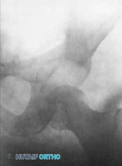
The Bosworth Lesion
A specific entity that frequently necessitates open reduction is the posterior dislocation of the fibula behind the tibia, known as a Bosworth fracture-dislocation. In this severe injury, the proximal fragment of the fractured fibula becomes entrapped behind the posterior tubercle of the distal tibia. The intact interosseous membrane acts as a tether, making closed reduction virtually impossible.
Indications for Surgery in Ankle Dislocations
Usually, acute ankle dislocations (even those with fractures) can be provisionally reduced in the emergency department using closed methods (traction and counter-traction). However, operative intervention is required when:
* The dislocation is irreducible (e.g., Bosworth lesion, interposed deltoid ligament).
* There is persistent syndesmotic diastasis or mortise widening after reduction.
* Associated unstable malleolar fractures require Open Reduction and Internal Fixation (ORIF).
Surgical Approach and Technique
When open reduction is mandated for an irreducible ankle dislocation, a systematic approach is required:
- Positioning: The patient is placed supine with a bump under the ipsilateral hip to internally rotate the leg, providing excellent access to the lateral malleolus and syndesmosis.
- Incision: A standard lateral approach to the fibula is utilized. If the fibula is entrapped behind the tibia (Bosworth lesion), the incision may need to be extended proximally.
- Reduction Maneuver: Soft tissue interposition (often the torn syndesmotic ligaments or joint capsule) is cleared. For a Bosworth lesion, a bone hook or periosteal elevator is used to lever the fibula anteriorly out from behind the posterior tibial tubercle.
- Ligamentous Management: Controversy exists regarding the acute repair of the deltoid ligament in the absence of a medial malleolar fracture. Current evidence-based consensus suggests that good-to-excellent results are achievable without acute direct repair of the deltoid ligament, provided the lateral malleolus is anatomically reduced and the syndesmosis is rigidly stabilized.
- Syndesmotic Stabilization: If syndesmosis and mortise widening persist, it must be treated operatively. This is typically achieved using one or two syndesmotic screws (3.5 mm or 4.5 mm) placed parallel to the joint line, or via dynamic suture-button constructs.
Pitfall: Over-compression of the syndesmosis can lead to restricted dorsiflexion and chronic pain. The syndesmosis should be held in an anatomically reduced position (often with a large pointed reduction clamp) while the ankle is held in neutral dorsiflexion during fixation.
Postoperative Protocol
Postoperatively, the ankle is immobilized in a well-padded short leg splint in neutral dorsiflexion. Patients are typically kept non-weight-bearing for 6 weeks. If syndesmotic screws are used, institutional protocols vary regarding routine removal at 3 to 4 months versus retention unless symptomatic.
ACUTE DISLOCATIONS OF THE PATELLA
Pathoanatomy and Biomechanics
Acute lateral dislocation of the patella is a common injury, particularly among young, active individuals and adolescents. The stability of the patellofemoral joint relies on a complex interplay between osseous geometry (trochlear depth) and soft tissue restraints.
The Medial Patellofemoral Ligament (MPFL) is the primary passive restraint to lateral patellar translation, providing 50% to 60% of the restraining force at 0 to 20 degrees of knee flexion. An acute lateral patellar dislocation almost universally results in a rupture or severe sprain of the MPFL.
Recent biomechanical and MRI studies have demonstrated that the site of MPFL injury varies significantly:
* Femoral Origin: The most common site of injury (near Schöttle's point).
* Patellar Insertion: Less common, but can involve bony avulsions.
* Midsubstance: Often associated with interstitial tearing.
* Combined Tears: Involving multiple zones of the ligament.
Furthermore, underlying anatomic predispositions play a massive role. The Tibial Tuberosity-Trochlear Groove (TT-TG) distance is a critical measurement. A TT-TG distance greater than 20 mm is considered highly abnormal and strongly predisposes the patient to recurrent instability. Studies indicate that patients with MPFL injuries at the patellar origin often exhibit significantly greater TT-TG distances compared to those with femoral origin or midsubstance tears.
Indications for Surgical Intervention
The management of primary (first-time) acute patellar dislocations remains highly controversial. Sufficient high-level evidence does not currently exist to advocate for routine surgical intervention in all primary patellar dislocations.
Non-Operative Management:
In most patients, the long-term subjective and functional results after non-operative management of an acute primary patellar dislocation are satisfactory. Good or excellent results have been reported in up to 75% of non-operatively treated knees.
Operative Management Indications:
Surgical intervention in the acute setting is strictly indicated in the following scenarios:
1. Osteochondral Fracture: The presence of a large, displaced osteochondral fragment (usually from the medial patellar facet or the lateral femoral condyle) that requires excision or internal fixation.
2. Massive Medial Sided Avulsion: A palpable defect with a massive bony avulsion of the MPFL that leaves the patella grossly unstable even in extension.
3. Recurrent Dislocation: After a second dislocation, the redislocation rate skyrockets to nearly 50%. At this juncture, surgical intervention (MPFL reconstruction) is strongly considered.
Clinical Pearl: Routine repair of the torn medial stabilizing soft tissues in acute patellar dislocation is not recommended in children or adolescents with open physes, as initial operative repair combined with lateral release has not been shown to improve long-term outcomes compared to conservative management.
Surgical Techniques: Repair vs. Reconstruction
Because the sites of injury of the MPFL differ, treating the specific pathology is critical.
Direct Repair:
Direct repair may be adequate for a clean avulsion lesion at the femoral or patellar origin. However, direct repair is only satisfactory if the intrinsic quality of the ligament is otherwise excellent. We prefer the open method for direct repair of the medial patellar retinaculum and MPFL if acute repair is indicated, utilizing suture anchors at the anatomic footprint. Arthroscopic techniques have been described but often fail to adequately tension the primary stabilizing fibers.
MPFL Reconstruction:
If the ligament quality is poor, if there is a combined/midsubstance tear, or if the patient has a history of recurrent instability, an anatomic MPFL reconstruction is far more appropriate than a direct repair.
* Graft Choice: Autograft (gracilis or semitendinosus) or allograft can be utilized.
* Femoral Fixation: Identifying the exact anatomic femoral origin (Schöttle's point) fluoroscopically is the most critical step. Non-anatomic femoral placement (especially too proximal or anterior) will result in severe over-constraint of the patellofemoral joint in flexion, leading to rapid chondrolysis and loss of motion.
* Patellar Fixation: The graft is secured to the proximal half of the medial patellar border, taking care not to fracture the patella with overly large drill holes.
Concomitant Procedures:
If the TT-TG distance exceeds 20 mm, an MPFL reconstruction alone will likely fail due to excessive lateralizing vectors. In these cases, a tibial tubercle osteotomy (TTO)—specifically an anteromedialization (Fulkerson osteotomy)—must be performed concomitantly to normalize the extensor mechanism mechanics.
Postoperative Protocol and Outcomes
Following MPFL repair or reconstruction, the knee is typically braced in extension for weight-bearing to protect the healing soft tissues. Early, protected range of motion (0 to 90 degrees) is initiated within the first two weeks to prevent arthrofibrosis.
Despite optimal treatment, the orthopedic surgeon must counsel the patient extensively. Recurrent dislocation occurs in approximately 71% of non-operatively treated knees and up to 67% of operatively treated knees in some long-term cohorts, with 52% of patients experiencing their first redislocation within 2 years after the primary injury. The patient must be warned of the distinct possibility of future episodes of recurrent patellar subluxation, dislocation, and the eventual development of patellofemoral osteoarthritis.
CONCLUSION
The management of acute dislocations requires a profound understanding of joint kinematics, soft tissue restraints, and strict adherence to surgical indications. While closed reduction remains the first line of treatment, the orthopedic surgeon must be vigilant in identifying irreducibility, instability, and neurovascular compromise that mandate open reduction. Whether addressing a complex Bosworth fracture-dislocation of the ankle or navigating the controversies of acute patellofemoral instability, a meticulous, evidence-based surgical approach is paramount to restoring joint congruity and optimizing long-term patient outcomes.
You Might Also Like