Uncover Ankle Stability: The Critical Surface of the Talus
Key Takeaway
Discover the latest medical recommendations for Uncover Ankle Stability: The Critical Surface of the Talus. The ankle joint is a hinge allowing dorsiflexion and plantarflexion, with load-bearing primarily on the upper articular surface of the talus and tibia. Its inherent stability is reinforced by ligaments and bony congruency. The articular surface of the talus is wider anteriorly, which, during dorsiflexion, firmly grips between the malleoli, preventing displacement and contributing significantly to the joint's stability.
Introduction and Epidemiology
Chronic ankle instability and the associated pathology of the talar articular surface represent a complex biomechanical challenge in orthopedic surgery. The ankle joint, frequently subjected to high-velocity inversion and rotational forces, relies on a delicate interplay between osseous congruency and ligamentous integrity. Lateral ankle sprains are among the most ubiquitous musculoskeletal injuries, with an estimated incidence of 2.15 per 1,000 person-years in the general population and significantly higher rates in athletic cohorts. While the majority of acute capsuloligamentous injuries resolve with functional rehabilitation, up to 20% to 30% of patients progress to chronic ankle instability, characterized by recurrent episodes of giving way, persistent pain, and mechanoreceptor deficit.


The critical surface of the talus plays a paramount role in the pathogenesis of long-term morbidity following ankle instability. Recurrent microtrauma and altered kinematics lead to asymmetric load-bearing stresses, predominantly affecting the medial and lateral borders of the talar dome. This abnormal sheer force frequently culminates in osteochondral lesions of the talus. Epidemiological data suggest that osteochondral lesions are present in up to 73% of patients undergoing surgical intervention for chronic lateral ankle instability. Understanding the intricate relationship between the ligamentous restraints and the articular surface geometry is essential for the orthopedic surgeon to execute precise anatomical reconstructions and mitigate the progression to post-traumatic osteoarthritis.
The socioeconomic burden of chronic ankle instability and concomitant talar surface pathology is substantial, encompassing direct healthcare expenditures and indirect costs related to lost productivity and premature cessation of athletic participation. The pathogenesis of osteochondral lesions in the setting of chronic instability is multifactorial, involving repetitive shear stress, altered joint contact mechanics, and localized ischemic events within the subchondral bone. As the lateral ligamentous complex attenuates, the talus undergoes abnormal anterior translation and internal rotation during the stance phase of gait. This kinematic aberration shifts the center of pressure anteriorly and medially, overloading the relatively constrained medial talar dome and subjecting the lateral dome to recurrent impaction against the fibula.
Furthermore, the mechanoreceptor deficit inherent to chronic ankle instability perpetuates a cycle of delayed peroneal muscle latency and diminished proprioceptive acuity. This neuromuscular uncoupling exacerbates the frequency and severity of inversion events, accelerating the degradation of the talar articular cartilage. The resultant osteochondral lesions are often characterized by a zone of necrotic subchondral bone overlying intact but mechanically compromised hyaline cartilage, eventually leading to delamination, loose body formation, and global joint degeneration if left untreated.


Surgical Anatomy and Biomechanics
The ankle joint functions as a highly modified hinge joint. While its primary arc of motion occurs in the sagittal plane permitting dorsiflexion and plantarflexion, the joint kinematics also accommodate up to 18 degrees of axial rotation of the talus within the tibial mortise. This complex motion is dictated by the unique osteology of the talocrural articulation and its robust ligamentous envelopes.
Osteology of the Tibiotalar Articulation
Load-bearing stresses are transmitted almost exclusively through the superior articular surface of the talus and the tibial plafond. The fibula, while critical for lateral mortise stability, does not participate in axial load transmission. The inherent osseous stability of the ankle is derived from the mortise configuration. Medial translation of the talus is restricted by the robust medial malleolus, while lateral translation is blocked by the lateral malleolus.


A critical anatomical feature of the talar dome is its trapezoidal geometry in the axial plane; the articular surface is approximately 2.5 mm wider anteriorly than posteriorly. Biomechanically, this mandates that as the ankle is brought into maximum dorsiflexion, the wider anterior segment of the talus wedges into the mortise, conferring maximal osseous stability. Conversely, in plantarflexion, the narrower posterior portion of the talus occupies the mortise, rendering the joint reliant primarily on ligamentous restraints for stability. This fundamental geometric property explains why the majority of lateral ankle sprains occur with the foot in a plantarflexed and inverted position.
The sagittal profile of the talar dome is characterized by a variable radius of curvature. The medial border has a larger radius of curvature compared to the lateral border, creating a subtle conical shape with the apex directed laterally. This morphology dictates that during dorsiflexion, the talus undergoes obligate external rotation, while plantarflexion is coupled with internal rotation. Disruptions to the lateral ligamentous complex alter this coupled motion, leading to abnormal sheer forces across the articular cartilage, particularly at the anterolateral and posteromedial aspects of the talar dome.
Ligamentous Anatomy and Restraints
The lateral collateral ligament complex is composed of three distinct structures: the anterior talofibular ligament (ATFL), the calcaneofibular ligament (CFL), and the posterior talofibular ligament (PTFL). The ATFL is the primary restraint to anterior translation of the talus and is the most frequently injured ligament in the human body. It originates from the anterior margin of the distal fibula and inserts onto the talar body just anterior to the lateral articular facet. Anatomical studies reveal that the ATFL is often composed of two distinct bands (superior and inferior), separated by vascular branches from the peroneal artery. The inferior band shares a common fibular footprint with the CFL, a relationship that is surgically relevant during anatomical reconstruction procedures.
The CFL is an extra-articular structure that spans both the tibiotalar and subtalar joints, originating from the anterior border of the distal fibula and inserting onto the lateral calcaneal tubercle. It acts as the primary restraint to inversion when the ankle is in a neutral or dorsiflexed position. The PTFL, the strongest component of the lateral complex, originates from the medial surface of the lateral malleolus and inserts onto the lateral tubercle of the posterior talar process. It is rarely injured in isolation and serves to limit posterior translation and external rotation of the talus.
On the medial aspect, the deltoid ligament complex provides robust stabilization against valgus and external rotation stresses. It is divided into superficial and deep components. The superficial deltoid crosses two joints and includes the tibionavicular, tibiocalcaneal, and superficial tibiotalar ligaments. The deep deltoid, primarily the deep anterior and posterior tibiotalar ligaments, is the primary medial stabilizer of the talus within the mortise. Chronic lateral ankle instability can lead to secondary attenuation of the deltoid ligament due to chronic rotational malalignment, a phenomenon that must be evaluated during preoperative planning.
Vascular Supply to the Talus
The vascular anatomy of the talus is highly relevant to the pathogenesis and treatment of osteochondral lesions. Approximately 60% of the talar surface is covered by articular cartilage, leaving limited areas for vascular penetration. The blood supply is derived from three main sources: the artery of the tarsal canal (a branch of the posterior tibial artery), the artery of the sinus tarsi (formed by anastomoses from the anterior lateral malleolar and perforating peroneal arteries), and branches from the dorsal pedis artery. The delicate intraosseous anastomotic network is highly susceptible to disruption from trauma or chronic microvascular sheer, contributing to the development of subchondral cysts and avascular necrosis associated with advanced osteochondral lesions.


Indications and Contraindications
The decision-making paradigm for surgical intervention in the setting of chronic ankle instability and talar surface pathology relies on a comprehensive assessment of symptom duration, failure of conservative measures, morphological characteristics of the osteochondral lesion, and the presence of concomitant pathology. Surgical management is generally indicated for patients who experience recurrent instability, persistent pain, and functional limitation despite a minimum of three to six months of dedicated physical therapy emphasizing peroneal strengthening and proprioceptive retraining.
When evaluating the critical surface of the talus, the size, depth, and containment of the osteochondral lesion dictate the surgical algorithm. Lesions smaller than 1.5 cm² are typically amenable to arthroscopic debridement and bone marrow stimulation (microfracture), which aims to replace the defect with fibrocartilage. Lesions exceeding 1.5 cm², or those associated with large subchondral cysts, generally require structural restoration via osteochondral autograft transfer (OATS), structural allografting, or matrix-induced autologous chondrocyte implantation (MACI).
Operative Versus Non Operative Management Parameters
| Clinical Parameter | Non-Operative Indications | Operative Indications |
|---|---|---|
| Duration of Symptoms | Acute sprains; < 3-6 months of chronic symptoms | > 6 months of persistent instability/pain despite PT |
| Ligamentous Status | Grade I/II sprains; functional instability without gross laxity | Grade III tears; mechanical laxity (+ anterior drawer/talar tilt) |
| Osteochondral Lesion Size | Asymptomatic incidental findings; purely cartilaginous contusions | Symptomatic OLTs; failed prior microfracture; cystic changes |
| Lesion Morphology | Intact cartilage over stable subchondral bone (Berndt & Harty Stage I/II) | Detached, displaced, or cystic lesions (Berndt & Harty Stage III/IV) |
| Patient Demographics | Low-demand patients; significant medical comorbidities | High-demand athletes; active individuals failing conservative care |
| Alignment | Normal hindfoot alignment | Correctable varus/valgus malalignment (requires concurrent osteotomy) |
Absolute and Relative Contraindications
Absolute contraindications to surgical reconstruction of the lateral ligaments and talar surface include active soft tissue or intra-articular infection, severe peripheral vascular disease precluding wound healing, and advanced neuroarthropathy (Charcot joint).
Relative contraindications include generalized hyperlaxity syndromes (e.g., Ehlers-Danlos syndrome), which may predispose to premature graft or repair attenuation, and advanced tibiotalar osteoarthritis (Kellgren-Lawrence Grade III or IV), where ligamentous reconstruction or localized cartilage repair will not address the global joint degeneration. In cases of advanced osteoarthritis, arthrodesis or total ankle arthroplasty are more appropriate considerations. Furthermore, uncorrected hindfoot varus is a critical relative contraindication; performing a lateral ligament reconstruction without addressing the osseous malalignment (e.g., via a lateralizing calcaneal osteotomy) will subject the repair to excessive tensile forces, leading to inevitable failure.


Pre Operative Planning and Patient Positioning
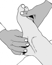
Thorough preoperative planning is paramount to executing a successful reconstruction of the unstable ankle and addressing talar surface defects. The clinical examination must quantify mechanical laxity through the anterior drawer and talar tilt tests, comparing the affected side to the contralateral asymptomatic ankle. Palpation of the anterior joint line may reveal osteophytes indicative of anterior impingement, while tenderness over the peroneal tendons necessitates evaluation for tendinopathy or subluxation, which frequently coexist with lateral instability.
Radiographic and Advanced Imaging Protocols
Standard weight-bearing radiographs, including anteroposterior, lateral, and mortise views, are mandatory. These views allow for the assessment of global joint congruency, the presence of loose bodies, and the identification of subtle varus or valgus malalignment. Stress radiography, utilizing either manual or mechanized techniques, can objectively quantify instability. An anterior translation of the talus exceeding 10 mm (or >3 mm compared to the contralateral side) suggests ATFL incompetence, while a talar tilt angle greater than 10 degrees (or >3 degrees compared to the contralateral side) indicates combined ATFL and CFL disruption.
Magnetic Resonance Imaging (MRI) is the gold standard for evaluating the critical surface of the talus and the integrity of the ligamentous complexes. High-resolution MRI allows for precise morphological assessment of osteochondral lesions, including the integrity of the overlying cartilage, the depth of subchondral bone involvement, and the presence of underlying cystic changes. Fluid signal deep to the osteochondral fragment on T2-weighted sequences is highly indicative of instability and detachment. Additionally, MRI facilitates the evaluation of concomitant soft tissue pathology, such as peroneal tendon tears, syndesmotic injury, and sinus tarsi syndrome. In cases of large, cystic osteochondral lesions where structural grafting is contemplated, a preoperative Computed Tomography (CT) scan is highly recommended to precisely define the osseous architecture and plan the trajectory of the osteotomy or graft harvest.
Patient Positioning and Surgical Preparation
The optimal patient position depends on the planned interventions. For isolated arthroscopy and lateral ligament reconstruction (e.g., modified Broström-Gould procedure), the patient is typically positioned supine with a bump placed under the ipsilateral hip to internally rotate the leg, bringing the lateral malleolus anteriorly and parallel to the floor. A thigh tourniquet is applied to provide a bloodless field.
If a concomitant medial malleolar osteotomy is required to access a posteromedial osteochondral lesion, the patient may be positioned supine with a smaller bump, allowing the leg to rest in external rotation for medial access, while still permitting internal rotation for lateral work. For complex cases requiring extensive posterior access or concomitant Achilles tendon pathology, a prone or lateral decubitus position may be indicated.
Intraoperative fluoroscopy must be readily available and positioned to allow for orthogonal views of the ankle without compromising the sterile field. Non-invasive joint distraction using a standardized strap technique is often employed during the arthroscopic phase to maximize visualization of the talar dome, particularly the central and posterior aspects, without causing iatrogenic scuffing of the articular cartilage.


Detailed Surgical Approach and Technique
The surgical management of chronic ankle instability with concomitant talar surface pathology typically proceeds in a staged manner during the same anesthetic event: diagnostic and therapeutic arthroscopy followed by open or minimally invasive ligamentous reconstruction.
Ankle Arthroscopy and Management of the Talar Surface
The procedure begins with the establishment of standard anteromedial and anterolateral arthroscopic portals. The anteromedial portal is created just medial to the tibialis anterior tendon at the level of the joint line, taking care to avoid the saphenous vein and nerve. The anterolateral portal is established lateral to the peroneus tertius tendon, with meticulous attention paid to the superficial peroneal nerve, which can often be transilluminated or palpated prior to incision.
A systematic 21-point arthroscopic examination is performed, evaluating the articular surfaces, the medial and lateral gutters, and the syndesmosis. Hypertrophic synovitis and scar tissue in the lateral gutter, common sequelae of chronic instability, are debrided using a motorized shaver and radiofrequency ablation.
For the management of osteochondral lesions, the unstable cartilage is excised to create stable, vertical margins. The necrotic subchondral bone is curetted to a bleeding base. If the lesion is less than 1.5 cm², bone marrow stimulation is performed. Using a specialized awl or a small-diameter drill, microfracture holes are created 2 to 3 mm apart and to a depth of 2 to 4 mm, ensuring penetration into the subchondral marrow spaces. The egress of fat droplets upon tourniquet deflation confirms adequate depth. This technique relies on the influx of mesenchymal stem cells to form a fibrocartilaginous repair tissue, predominantly composed of Type I collagen, which, while mechanically inferior to native hyaline cartilage, provides satisfactory clinical outcomes in appropriately selected lesions.
Structural Grafting for Large Osteochondral Lesions
For lesions exceeding 1.5 cm² or those with substantial subchondral cysts, structural restoration is required. Autologous osteochondral transplantation (OATS) involves harvesting a cylindrical graft from a non-weight-bearing portion of the ipsilateral knee (e.g., the lateral periphery of the lateral femoral condyle) and press-fitting it into a prepared recipient socket in the talus.
Accessing posteromedial lesions often necessitates a medial malleolar osteotomy. A chevron or step-cut osteotomy is preferred to maximize rotational stability and surface area for healing. Prior to completing the osteotomy, the malleolus is pre-drilled and tapped for two 4.0 mm partially threaded cancellous screws to ensure perfect anatomic reduction during closure. The osteotomy is directed to exit the tibial plafond precisely at the medial border of the talar dome, avoiding injury to the weight-bearing cartilage. Once the osteochondral graft is impacted flush with the surrounding native cartilage, the malleolus is reduced and secured.
Lateral Ligament Reconstruction Technique
Following the intra-articular work, attention is directed to the lateral ligamentous complex. The modified Broström-Gould procedure remains the gold standard for anatomical repair. A curvilinear incision is made over the anterior margin of the distal
Clinical & Radiographic Imaging


You Might Also Like
