Solving the Pain Syndrome Complex: Modern Insights for CRPS

Key Takeaway
In this comprehensive guide, we discuss everything you need to know about Solving the Pain Syndrome Complex: Modern Insights for CRPS. Complex Regional Pain Syndrome (CRPS) is a modern pain syndrome complex characterized by abnormal pain, swelling, and dysfunction in the vasomotor and sudomotor systems, contracture, and osteoporosis, often following an injury. Historically, it was recognized as causalgia by Silas Weir Mitchell or as a posttraumatic pain syndrome with edema and trophic changes by Paul Südeck.
Introduction and Epidemiology
During the American Civil War, Silas Weir Mitchell described a syndrome that occurred in patients who had suffered gunshot injuries to major nerves. Noting that a leading feature was burning pain, he called the condition causalgia. At the beginning of the 20th century, Paul Südeck, a clinician in Hamburg, Germany, used the newly invented technique of roentgenology to investigate patients with severe pain after injury. He described a posttraumatic pain syndrome with edema, trophic changes, and osteoporosis. In 1979, the AO group advocated open reduction and rigid internal fixation to prevent fracture disease, which was defined as a combination of circulatory disturbance, inflammation, and pain as a result of dysfunction of joints and muscles. In an intriguing vignette, Channon and Lloyd noted that finger stiffness after Colles fracture could be either simple or associated with swelling and changes in hand temperature. In the latter case, it did not respond well to physiotherapy.
The modern term for the syndrome described in different circumstances by these early researchers is complex regional pain syndrome, usually abbreviated as CRPS. CRPS consists of abnormal pain, swelling, vasomotor and sudomotor dysfunction, contracture, and osteoporosis. It used to be considered a rare, devastating complication of injury, caused by abnormalities in the sympathetic nervous system and seen mainly in psychologically abnormal patients. Modern research is altering this view radically. This review will specifically examine CRPS within the context of orthopaedic trauma surgery. For this reason, the emphasis, descriptions, and concepts differ slightly from those routinely found in publications from the International Association for the Study of Pain. It is important to appreciate that these apparent differences are merely counterpoints. The theme is identical.
Definitions of Abnormal Pain Perception
A cardinal feature of CRPS is abnormalities of pain perception, which are mainly foreign to orthopaedic surgeons. They have been codified by Merskey and Bogduk and are fundamental to understanding the clinical presentation.
Allodynia literally means other pain and represents a painful perception of a stimulus that should not usually be painful. Thus, a patient will find gentle stroking of the affected part painful. Allodynia differs from referred pain, but allodynic pain can occur in areas other than the one stimulated. There are several forms of allodynia. Mechanical or tactile allodynia implies pain in response to touch. It may be further subdivided into static mechanical allodynia, implying pain in response to light touch or pressure, and dynamic mechanical allodynia, where the pain occurs as a result of brushing. In thermal hot or cold allodynia, the pain is caused by mild changes in skin temperature in the affected area.
Hyperalgesia is an increased sensitivity to pain, which may be caused by damage to nociceptors or peripheral nerves. Thus, the patient finds gentle touching with a pin unbearably painful. Hyperalgesia is usually experienced in focal, discrete areas, typically associated with injury. Focal hyperalgesia may be divided into two subtypes. Primary hyperalgesia describes pain sensitivity that occurs directly in the damaged tissues. Secondary hyperalgesia describes pain sensitivity that occurs in surrounding undamaged tissues. Rarely, hyperalgesia is seen in a more diffuse, bodywide form.
Hyperpathia is a temporal and spatial summation of an allodynic or hyperalgesic response. Thus, the patient finds gentle touching painful, but repetitive touching either on the same spot or on another part of the affected limb becomes increasingly unbearable and the pain continues for a period after the stimulus has been removed.
Incidence and Demographic Factors
CRPS is subdivided into Type I, which occurs without a definable nerve lesion formerly reflex sympathetic dystrophy, and Type II, which occurs in the presence of a definable nerve lesion formerly causalgia. In orthopaedic trauma, CRPS Type I is most frequently observed following distal radius fractures, tibial plateau fractures, and foot and ankle trauma. Historically, the incidence of CRPS following distal radius fractures was reported to be as high as thirty percent. However, with the adoption of the stringent Budapest Criteria, the true incidence is estimated to be between four and seven percent. Females are affected three to four times more frequently than males, and the peak incidence occurs in the fifth to seventh decades of life.
Surgical Anatomy and Biomechanics
Understanding the pathophysiology of CRPS requires a thorough grasp of the peripheral nervous system, the autonomic nervous system, and the biomechanical consequences of prolonged sympathetic overdrive on the musculoskeletal system.
Neuroanatomy of the Sympathetic and Nociceptive Pathways
The sympathetic nervous system plays a central role in the vasomotor and sudomotor manifestations of CRPS. Sympathetic preganglionic neurons originate in the intermediolateral horn of the spinal cord from T1 to L2. For the upper extremity, these fibers ascend the sympathetic chain to synapse in the cervical ganglia, predominantly the stellate ganglion cervicothoracic ganglion, located anterior to the transverse process of C7 and the neck of the first rib. Postganglionic fibers then join the brachial plexus to provide sympathetic innervation to the upper limb. For the lower extremity, preganglionic fibers descend to the lumbar sympathetic ganglia L2 to L4 before joining the lumbosacral plexus.
In CRPS, peripheral nociceptors unmyelinated C-fibers and thinly myelinated A-delta fibers become sensitized. Tissue trauma induces the release of pro-inflammatory neuropeptides such as substance P and calcitonin gene-related peptide from the peripheral terminals of these afferent fibers, a process known as neurogenic inflammation. This leads to profound vasodilation, protein extravasation, and the characteristic edema seen in acute CRPS.
Central Sensitization and Ephaptic Transmission
Prolonged peripheral nociceptive input leads to central sensitization within the dorsal horn of the spinal cord. Wide dynamic range neurons in the dorsal horn exhibit wind-up phenomenon, mediated by the activation of N-methyl-D-aspartate receptors. This central amplification is responsible for secondary hyperalgesia and allodynia. Furthermore, in CRPS Type II, damaged peripheral nerves can develop abnormal connections between sympathetic efferent fibers and sensory afferent fibers, known as ephaptic transmission or short-circuiting. This sympathetic-afferent coupling means that normal sympathetic efferent activity triggered by stress or cold directly activates nociceptive pathways, resulting in sympathetically maintained pain.
Biomechanical Consequences of CRPS
The biomechanical implications of CRPS are profound and directly impact orthopaedic management. Chronic neurogenic inflammation and disuse lead to rapid periarticular fibrosis. The joint capsule thickens, and the synovial recesses become obliterated, leading to severe contractures.
Concurrently, regional osteoclastic overactivity, driven by local hypoxia, acidosis, and inflammatory cytokines, results in profound localized osteoporosis, classically described as Südeck's atrophy. Biomechanically, this trabecular bone loss drastically reduces the pull-out strength of orthopaedic implants. If surgical intervention such as hardware removal or corrective osteotomy is attempted during the active phase of CRPS, the structurally compromised bone is highly susceptible to iatrogenic fracture and catastrophic fixation failure.
Indications and Contraindications
Surgical intervention in the setting of CRPS is highly controversial and must be approached with extreme caution. Surgery can exacerbate the condition, triggering a massive inflammatory cascade and sympathetic storm. However, certain structural lesions driving the CRPS must be addressed.
| Intervention Category | Operative Indications | Non-Operative Indications |
|---|---|---|
| Primary Fracture Management | Unstable fractures requiring rigid fixation to allow early mobilization; Compartment syndrome. | Stable fractures amenable to functional bracing; Non-displaced fractures. |
| Nerve Decompression | Clinically and electrodiagnostically proven compressive neuropathy driving CRPS Type II. | Diffuse pain without a localizable nerve entrapment; Acute inflammatory phase of CRPS. |
| Hardware Removal | Prominent, painful implants directly irritating a peripheral nerve; Intra-articular hardware. | Asymptomatic hardware; Active florid CRPS with severe osteopenia. |
| Neuromodulation | Refractory CRPS failing 6 months of comprehensive medical and physical therapy. | Untreated psychiatric comorbidities; Active systemic infection; Coagulopathy. |
| Contracture Release | Severe, fixed joint contractures after the active inflammatory phase has completely burned out. | Active CRPS with ongoing edema, vasomotor instability, and hyperalgesia. |
Pre Operative Planning and Patient Positioning
When surgery is unavoidable in a patient with established CRPS, or when operating on a patient at high risk for developing CRPS, meticulous preoperative planning is mandatory. The goal is to blunt the sympathetic nervous system response and minimize surgical trauma.
Pharmacological Optimization
Preoperative pharmacological optimization requires a multidisciplinary approach involving pain management specialists. The perioperative administration of Vitamin C ascorbic acid has been widely debated but remains a low-risk intervention. Based on the initial trials by Zollinger, a dose of 500 milligrams daily for fifty days post-injury or post-surgery is frequently utilized to mitigate oxidative stress and reduce the incidence of CRPS.
Gabapentinoids such as gabapentin or pregabalin should be initiated preoperatively to dampen central sensitization by binding to the alpha-2-delta subunit of voltage-gated calcium channels. In severe cases, preoperative intravenous ketamine infusions subanesthetic doses may be utilized to block N-methyl-D-aspartate receptors and reset the central nervous system wind-up.
Anesthetic Considerations
Regional anesthesia is strongly preferred over general anesthesia alone. Continuous peripheral nerve blocks or epidural anesthesia provide profound sympathectomy and preemptive analgesia, blocking the afferent nociceptive barrage from reaching the dorsal horn. For upper extremity surgery, an axillary or supraclavicular brachial plexus block with a continuous catheter can be maintained for several days postoperatively. The catheter allows for aggressive early physical therapy while the limb is sympathectomized.
Patient Positioning and Tourniquet Management
Positioning must be executed with extreme care to avoid traction neuropraxia, which can trigger or worsen CRPS Type II. All pressure points must be heavily padded.
Tourniquet use is highly controversial in the CRPS limb. Tourniquet-induced ischemia and subsequent reperfusion injury exacerbate neurogenic inflammation and generate reactive oxygen species. If a tourniquet is absolutely necessary for visualization, the inflation time must be kept to an absolute minimum, and lower inflation pressures utilizing a wide cuff are recommended. Exsanguination should be performed gently with elevation rather than tight Esmarch wrapping, which can provoke mechanical allodynia.
Detailed Surgical Approach and Technique
Surgical intervention in a patient with CRPS demands flawless, atraumatic technique. The following section outlines the principles of orthopaedic surgery in the CRPS limb, followed by the specific techniques utilized for neuromodulation when conservative measures fail.

Principles of Atraumatic Orthopaedic Dissection
When performing hardware removal or nerve decompression in a CRPS limb, the surgical approach must respect the already inflamed and sensitized soft tissues.
- Incision and Exposure: Skin incisions should utilize previous scars if possible, but avoid crossing flexion creases perpendicularly to prevent hypertrophic scarring, which can become a nidus for hyperalgesia. Skin flaps must be full-thickness.
- Internervous Planes: Strict adherence to internervous planes is mandatory. Retraction must be static and gentle. Self-retaining retractors should be released periodically to prevent local tissue ischemia.
- Hemostasis: Meticulous hemostasis is critical. Postoperative hematoma formation increases tissue tension, exacerbates pain, and provides a scaffold for further fibrosis. Bipolar electrocautery is preferred near peripheral nerves to prevent thermal injury to the vasa nervorum.
- Handling of Nerves: If neurolysis is performed for CRPS Type II, the nerve must be handled exclusively by the epineurium using non-toothed micro-forceps. Circumferential external neurolysis should be performed only to the extent necessary to relieve compression, preserving the segmental blood supply. Internal neurolysis is strictly contraindicated as it violates the blood-nerve barrier and worsens intraneural fibrosis.
- Wound Closure: Closure should be tension-free. Subcutaneous tissues are approximated with absorbable sutures, and the skin is closed with non-reactive monofilament sutures or staples. Tight dressings and circumferential casts must be avoided to prevent iatrogenic compression.
Surgical Interventions for Refractory CRPS
When orthopaedic pathology has been addressed or ruled out, and the patient suffers from refractory CRPS, interventional pain techniques become the primary surgical focus.
Stellate Ganglion Block
For upper extremity CRPS, a stellate ganglion block is utilized both diagnostically and therapeutically. The procedure is performed under fluoroscopic or ultrasound guidance. The patient is positioned supine with the neck slightly extended. The target is the anterior tubercle of the C6 transverse process Chassaignac tubercle. A spinal needle is advanced to the bone, withdrawn slightly to avoid periosteal injection, and negative aspiration is confirmed to avoid intravascular vertebral artery or intrathecal injection. A local anesthetic is injected, and successful blockade is confirmed by the development of an ipsilateral Horner syndrome ptosis, miosis, anhidrosis and an increase in limb temperature.
Spinal Cord Stimulation
Spinal cord stimulation is indicated for chronic, refractory CRPS. The technique involves the percutaneous or open surgical placement of epidural electrodes over the dorsal columns of the spinal cord. The electrodes are connected to an implantable pulse generator. The electrical impulses activate large-diameter A-beta mechanoreceptive fibers. According to the Gate Control Theory of Pain, this activation stimulates inhibitory interneurons in the substantia gelatinosa of the dorsal horn, effectively closing the gate to nociceptive signals traveling via C-fibers and A-delta fibers. Furthermore, spinal cord stimulation has been shown to induce orthodromic sympathetic blockade, improving peripheral microcirculation.
Complications and Management
Operating on a patient with CRPS, or managing the complications of the syndrome itself, presents significant challenges. The most devastating complication is the exacerbation or spread of the disease process.
| Complication | Estimated Incidence | Pathophysiology and Clinical Presentation | Prevention and Salvage Strategies |
|---|---|---|---|
| CRPS Flare or Spread | 20% to 40% post-op | Surgical trauma triggers massive neuropeptide release and sympathetic storm. May spread contralaterally. | Preoperative regional blockade; Ketamine infusions; Atraumatic surgical technique; Avoid tourniquet ischemia. |
| Hardware Failure | 10% to 15% | Profound localized osteopenia (Südeck's atrophy) leads to loss of screw purchase and implant pull-out. | Utilize locking plate technology; Augment fixation with bone cement; Delay elective osteotomies until bone density recovers. |
| Severe Joint Contracture | 30% to 50% | Periarticular fibrosis, synovial obliteration, and muscle shortening due to protective posturing. | Aggressive early motion under regional anesthesia; Dynamic splinting; Serial casting; Late surgical arthrolysis. |
| Infection of SCS Implant | 2% to 5% | Bacterial colonization of the implantable pulse generator pocket or epidural leads. | Strict aseptic technique during implantation; Preoperative systemic antibiotics; Explantation required if deep infection occurs. |
| Post-Traumatic Neuroma | 5% to 10% | Iatrogenic nerve injury or inadequate resection of a damaged nerve in CRPS Type II. | Meticulous nerve handling; Transposition of nerve endings into healthy muscle or bone; Targeted muscle reinnervation. |
Post Operative Rehabilitation Protocols
Rehabilitation is the cornerstone of CRPS management and must begin immediately postoperatively. The overarching goal is to restore function and normalize somatosensory processing. Immobilization is the enemy of the CRPS limb.
Early Mobilization and Desensitization
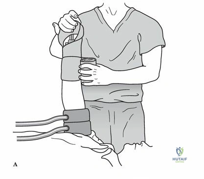
Active range of motion exercises are initiated on postoperative day one. Passive range of motion is generally avoided initially, as it can provoke mechanical allodynia and trigger muscle guarding. Desensitization therapy is employed to normalize tactile processing. This involves progressive tactile stimulation of the hyperalgesic skin, starting with soft textures like silk and progressing to rougher textures like Velcro, combined with contrast baths to habituate the thermal receptors.
Graded Motor Imagery
For patients with severe central sensitization and allodynia who cannot tolerate physical touch, Graded Motor Imagery is a highly evidenced, sequential rehabilitation protocol designed to retrain the brain and eliminate the pain neurotag. It consists of three distinct phases.
- Laterality Reconstruction: Patients view images of left and right limbs in various postures and must quickly identify them. This restores the disrupted cortical body schema without activating motor or pain pathways.
- Motor Imagery: Patients imagine moving their affected limb into various positions without actually contracting the muscles. This activates the premotor cortex and motor networks without triggering nociceptive feedback from the periphery.
- Mirror Visual Feedback: The patient places the affected limb behind a mirror and watches the reflection of the unaffected, moving limb. This creates a visual illusion that the affected limb is moving painlessly, resolving the sensorimotor incongruence that drives central sensitization.
Summary of Key Literature and Guidelines
The diagnosis and management of CRPS have been standardized by the International Association for the Study of Pain. Familiarity with these guidelines is essential for the orthopaedic surgeon to ensure accurate diagnosis and avoid unnecessary surgical interventions.
The Budapest Criteria
The clinical diagnosis of CRPS is currently based on the Budapest Criteria, which offer high sensitivity and specificity. To meet the criteria, the patient must exhibit continuing pain that is disproportionate to any inciting event. Additionally, the patient must report at least one symptom in three of the four following categories, and display at least one sign at the time of evaluation in two or more of the following categories.
- Sensory: Reports of hyperalgesia or allodynia.
- Vasomotor: Reports of temperature asymmetry or skin color changes and asymmetry.
- Sudomotor and Edema: Reports of edema, sweating changes, or sweating asymmetry.
- Motor and Trophic: Reports of decreased range of motion, motor dysfunction such as weakness, tremor, or dystonia, or trophic changes in hair, nails, or skin.
Furthermore, there must be no other diagnosis that better explains the signs and symptoms. Orthopaedic surgeons must rigorously rule out deep infection, compartment syndrome, deep vein thrombosis, and acute hardware failure before diagnosing CRPS.
Evidence Based Prophylaxis and Treatment
The orthopaedic literature provides specific guidance on the prevention of CRPS. The seminal randomized controlled trials by Zollinger demonstrated that Vitamin C supplementation significantly reduces the risk of CRPS following distal radius fractures. While subsequent meta-analyses have debated the absolute efficacy, the American Academy of Orthopaedic Surgeons recognizes Vitamin C as a low-cost, low-risk prophylactic measure.
For treatment, guidelines emphasize a stepwise approach. First-line therapy includes physical rehabilitation and psychological support. Second-line therapy introduces pharmacological agents such as tricyclic antidepressants, gabapentinoids, and short courses of oral corticosteroids for acute inflammatory CRPS. Third-line interventions encompass regional anesthetic blocks and sympathetic ganglion blocks. Finally, fourth-line therapies, reserved for refractory cases, include neuromodulation via spinal cord stimulation or dorsal root ganglion stimulation. Surgical sympathectomy is rarely indicated today due to the high risk of compensatory hyperhidrosis and post-sympathectomy neuralgia.
By adhering to these modern insights and guidelines, the orthopaedic surgeon can navigate the complex pain syndrome, avoiding surgical pitfalls and facilitating optimal functional recovery for the patient.
You Might Also Like