Decompression of the Retrocalcaneal: Endoscopic Relief for Haglund's
Key Takeaway
This article provides essential research regarding Decompression of the Retrocalcaneal: Endoscopic Relief for Haglund's. Decompression of the retrocalcaneal refers to a surgical technique, often endoscopic, used to treat Haglund syndrome. This involves removing the calcaneal prominence and inflamed retrocalcaneal bursa, which cause posterior heel pain, Achilles tendinopathy, and mechanical irritation. This outpatient procedure offers low morbidity, high satisfaction, and a short recovery time after conservative treatments have failed.
Introduction and Epidemiology
Patrick Haglund first described an enlarged posterior border of the os calcis in 1928, establishing the foundational understanding of posterior heel pain related to osseous morphology. This specific anatomical variant, widely known as a Haglund deformity, assumes critical clinical importance when external compressive forces—such as rigid shoe heel counters—combine with repeated hyperdorsiflexion of the ankle joint. This dynamic mechanism forces pathological contact between the anterior aspect of the Achilles tendon, the posterior vertical surface of the calcaneus, and the interposed retrocalcaneal bursa.
Haglund syndrome represents a distinct clinical triad comprising insertional Achilles tendinopathy, retrocalcaneal bursitis, and the presence of a Haglund deformity. The syndrome is fundamentally characterized by an inflammatory cascade within the retrocalcaneal or superficial adventitial bursa, which frequently progresses to secondary insertional Achilles tendinopathy. The posterior heel pain and focal swelling pathognomonic of Haglund syndrome are the direct result of chronic mechanical irritation exerted by the calcaneal prominence on the surrounding soft tissue envelope and the anterior paratenon of the Achilles tendon.
Epidemiologically, this condition exhibits a bimodal distribution, frequently affecting young, active individuals (particularly runners and jumping athletes) as well as middle-aged patients experiencing degenerative tendinopathic changes. There is a documented predilection for females, historically attributed to the rigid heel counters of restrictive footwear, which led to the colloquial and historical designation of the resulting superficial bursa as a "pump bump." However, the true etiology is multifactorial, involving intrinsic foot biomechanics, such as a rigid cavus foot structure, which inherently increases the pitch of the calcaneus and exacerbates the prominence of the posterosuperior tuberosity.

When exhaustive conservative and nonoperative measures fail to provide symptomatic relief, and advanced imaging delineates significant pathology, surgical intervention becomes warranted. Endoscopic decompression of the retrocalcaneal space—encompassing retrocalcaneal bursectomy, debridement of the anterior Achilles paratenon, and resection of the posterosuperior calcaneal prominence (calcaneoplasty)—has emerged as a highly efficacious, minimally invasive alternative to traditional open procedures. This endoscopic technique is typically performed as an outpatient procedure and is associated with reduced soft tissue morbidity, high patient satisfaction, and an accelerated return to pre-injury activity levels.
Surgical Anatomy and Biomechanics
A profound understanding of the posterior hindfoot anatomy is paramount for safe and effective endoscopic decompression. The osseous, tendinous, and bursal structures in this region exist in a delicate biomechanical balance that is easily disrupted by morphological variants.
Osseous Anatomy of the Calcaneus
The calcaneus is the largest tarsal bone, and its posterior third forms the calcaneal tuberosity. The posterior surface of the tuberosity is divided into three distinct facets: superior, middle, and inferior. The Achilles tendon inserts primarily into the middle facet, which presents as a roughened, crescent-shaped area. The superior facet is smooth, slopes anteriorly, and is intimately related to the retrocalcaneal bursa. In patients with a Haglund deformity, this superior facet is hypertrophied or abnormally angulated, creating a prominent posterosuperior angle that encroaches upon the retrocalcaneal space.
Soft Tissue Restraints and Bursal Anatomy
Two primary bursae are associated with the distal Achilles tendon. The superficial adventitial bursa (Achilles tendon bursa) is located subcutaneously, posterior to the tendon insertion, and forms secondary to external friction. The retrocalcaneal bursa is a true synovial bursa situated anterior to the Achilles tendon and posterior to the superior facet of the calcaneus.
The retrocalcaneal space has been anatomically described as functioning similarly to a disc space bursa, covering the posterosuperior angle of the calcaneus. Under conditions of repeated hindfoot movement, the synovial walls of this bursa become diseased, hypertrophied, and highly vascularized. Chronic increased intracavitary pressure leads to secondary calcaneal bone marrow edema and reactive fibrosis of the paratenon near the insertion site.

Biomechanical Considerations in Impingement
Achilles tendinopathy in this region is a progressive degenerative process within the tendon substance, characterized by mucinoid degeneration, collagen disorientation, and the formation of microtears. These microtears can coalesce into macrotears, accompanied by interstitial edema and reactive fibrosis with scar formation.
Biomechanically, ankle dorsiflexion causes the Achilles tendon to compress against the posterosuperior calcaneal tuberosity. In the presence of a Haglund deformity, this creates a "nutcracker" effect. The anterior fibers of the Achilles tendon are subjected to excessive tensile and compressive loads, leading to mechanical attrition. These degenerative changes cause secondary mechanical irritation of the surrounding tissues, stimulating a chronic inflammatory process that perpetuates the cycle of bursal hypertrophy and tendinosis.
Indications and Contraindications
Appropriate patient selection is the most critical determinant of success in endoscopic retrocalcaneal decompression. The procedure is indicated for patients who have exhausted nonoperative modalities and present with persistent, debilitating symptoms. General indications include intractable posterior heel pain, an altered gait (limp), significant modification of workstyle or lifestyle, and the presence of severe night pain or rest pain.
Clinical Evaluation and Diagnostic Imaging
Clinical evaluation is essential to differentiate between isolated retrocalcaneal bursitis, non-insertional Achilles tendinopathy, and true insertional Achilles tendinopathy, although these pathologies frequently coexist. Pathology within the retrocalcaneal space is detected via precise point tenderness along the anteromedial and anterolateral aspects of the distal Achilles tendon, coupled with a palpable prominence of the calcaneus. Palpation often reveals tenderness proximal to the insertion, which is reliably reproduced with passive or active dorsiflexion. In advanced cases, the retrocalcaneal bursa and the superficial Achilles bursa may become confluent, creating a "wrap-around" inflammatory mass.
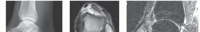
Imaging is mandatory to document the extent of tendinopathy and osseous deformity. Standard weight-bearing lateral radiographs are utilized to evaluate the calcaneal morphology. Objective radiographic measurements include the parallel pitch lines (typically abnormal in Haglund syndrome), the Fowler-Philip angle (pathological if >75 degrees), and the Chauveaux-Liet angle.

Magnetic resonance imaging (MRI) is the gold standard for preoperative planning. It precisely demonstrates the coexistence of retrocalcaneal bursitis, Achilles tendinosis, and calcaneal marrow edema. MRI is crucial for quantifying the percentage of tendon degeneration; if greater than 50% of the tendon insertion is compromised by intrasubstance tearing or calcific tendinosis, an open debridement and reconstruction with possible flexor hallucis longus (FHL) transfer may be indicated over an endoscopic approach.

Ultrasound provides a dynamic assessment and can help rule out non-insertional tendinopathy. A limited bone scan or single-photon emission computed tomography (SPECT) can assist in the differential diagnosis by highlighting focal areas of increased metabolic activity at the posterosuperior tuberosity, confirming the mechanical impingement.

Nonoperative Management Strategies
Nonoperative measures must be trialed for a minimum of 3 to 6 months prior to surgical consideration. Modalities include the use of nonsteroidal anti-inflammatory drugs (NSAIDs), shoe wear modification (utilizing backless shoes or footwear with soft, irregular counters), and heel lifts to induce relative plantarflexion and decrease tendon tension. Physical therapy focuses on eccentric stretching exercises, cryotherapy, and pressure-relieving orthotics. Extracorporeal shockwave therapy (ESWT) has also shown efficacy in chronic cases.
| Parameter | Operative Indications | Non Operative Indications |
|---|---|---|
| Duration of Symptoms | > 6 months of failed conservative care | < 6 months, initial presentation |
| Pain Characteristics | Severe, limits ADLs, night pain present | Mild to moderate, activity-related only |
| Imaging Findings | Prominent Haglund deformity, intact Achilles (<50% tearing), bursitis | Mild bursitis, no significant osseous impingement |
| Functional Status | Altered gait, inability to wear standard footwear | Able to ambulate with minimal compensation |
| Contraindications | Active infection, severe vascular disease, >50% Achilles detachment | None |
Pre Operative Planning and Patient Positioning
Thorough preoperative planning dictates the efficiency and safety of the endoscopic procedure. The surgeon must meticulously review the sagittal MRI slices to understand the exact depth and proximal extent of the retrocalcaneal bursa, as well as the precise location of the Achilles insertion relative to the osseous prominence.
Anesthesia and Operating Room Setup
The procedure is typically performed under general anesthesia or regional anesthesia (popliteal block) combined with monitored anesthesia care. A thigh or proximal calf tourniquet is applied to ensure a bloodless visual field, which is critical for endoscopic orientation. The arthroscopy tower is positioned at the foot of the bed, while the fluoroscopy unit (C-arm) is brought in from the contralateral side, positioned to obtain perfect lateral projections of the operative hindfoot.
Patient Positioning and Landmark Identification
The patient is placed in the prone position. Bilateral sequential compression devices are applied to the upper extremities or contralateral lower extremity. The operative leg is supported with a bump placed under the distal tibia, allowing the ankle to hang freely over the edge of the operating table. This positioning permits unrestricted, dynamic intraoperative dorsiflexion and plantarflexion, which is essential for assessing impingement and facilitating instrument maneuverability.

Precise anatomical landmarks must be outlined with a surgical marker prior to exsanguination. The medial and lateral borders of the Achilles tendon are palpated and marked. The superior aspect of the calcaneal tuberosity is identified. The sural nerve, which courses posterior to the lateral malleolus and lateral to the Achilles tendon, must be carefully considered. It typically crosses the lateral border of the Achilles tendon approximately 9 to 12 cm proximal to the insertion.

Detailed Surgical Approach and Technique
The endoscopic decompression of the retrocalcaneal space utilizes a two-portal paratendinous approach. Normal-appearing and diseased tendons can usually be distinguished endoscopically with high fidelity.
Portal Placement and Safe Zones
The medial and lateral portals are established at the level of the superior aspect of the calcaneal tuberosity, immediately anterior to the Achilles tendon.
- Lateral Portal: The lateral portal is typically created first. A vertical stab incision is made just anterior to the lateral border of the Achilles tendon. Blunt dissection using a mosquito hemostat is performed down to the retrocalcaneal space to avoid injury to the sural nerve or the lesser saphenous vein.
- Medial Portal: A blunt trocar with a 4.0-mm, 30-degree arthroscope is introduced through the lateral portal and directed medially, hugging the anterior surface of the Achilles tendon. Once the medial skin is tented by the trocar, a corresponding vertical incision is made to establish the medial portal.

Endoscopic Bursectomy and Space Preparation
A standard arthroscopic fluid pump is utilized, maintaining pressures between 30 and 40 mm Hg to prevent excessive extravasation into the soft tissues.
The initial endoscopic view is often obscured by hypertrophic, inflamed synovial tissue within the retrocalcaneal bursa. A 4.5-mm full-radius soft tissue shaver is introduced through the medial portal. A systematic bursectomy is performed, clearing the fibrofatty tissue of Kager's fat pad superiorly, the anterior surface of the Achilles tendon posteriorly, and the superior facet of the calcaneus anteriorly.

During this phase, the surgeon must carefully inspect the anterior paratenon and the substance of the Achilles tendon. Fraying, reactive fibrosis, or partial tearing of the anterior Achilles fibers (the "kissing lesion" from the calcaneal impingement) should be gently debrided back to stable, healthy tendon tissue. Radiofrequency wands may be utilized for meticulous hemostasis and to ablate highly vascularized inflammatory tissue.

Calcaneoplasty and Osseous Resection
Once the posterosuperior calcaneal prominence is fully skeletonized and visualized, the calcaneoplasty commences. An arthroscopic burr (typically 4.0 mm or 4.5 mm, hooded) is introduced.
The osseous resection begins at the most superior aspect of the prominence. The burr is swept in a medial-to-lateral and lateral-to-medial direction. The surgeon must maintain spatial awareness to avoid asymmetric resection. The goal is to remove the prominent posterosuperior angle, creating a flat, sloping surface that parallels the anterior border of the Achilles tendon during dorsiflexion.

Care must be taken to avoid violating the actual insertion footprint of the Achilles tendon on the middle facet. The resection is carried anteriorly into the superior facet by approximately 1.5 to 2.0 cm, depending on the preoperative radiographic templating.

Intraoperative Fluoroscopic Verification
Endoscopic visualization alone is insufficient to confirm adequate osseous resection due to the lack of macroscopic perspective. Intraoperative lateral fluoroscopy is mandatory.
The C-arm is utilized to obtain a true lateral view of the calcaneus. The surgeon evaluates the resection by verifying that the posterosuperior prominence has been eliminated and that the newly created osseous contour lies inferior to the parallel pitch lines. The ankle is taken through a full range of motion under live fluoroscopy to ensure that no residual osseous impingement occurs against the Achilles tendon during maximum dorsiflexion.

If residual prominence is noted, the burr is reintroduced, and further resection is performed until a satisfactory radiographic and clinical decompression is achieved. Once complete, the joint space is thoroughly irrigated to remove all bone debris, which could otherwise cause postoperative irritation or heterotopic ossification. The portals are closed with simple non-absorbable sutures, and a compressive sterile dressing is applied.
Complications and Management
While endoscopic retrocalcaneal decompression is associated with lower morbidity than open approaches, complications can still occur. Meticulous surgical technique and adherence to anatomical safe zones are required to minimize these risks.
| Complication | Estimated Incidence | Etiology and Risk Factors | Salvage and Management Strategy |
|---|---|---|---|
| Sural Nerve Injury | 2% - 5% | Improper lateral portal placement, aggressive lateral dissection | Observation for neuropraxia; gabapentinoids; surgical exploration/neurolysis for persistent neuroma. |
| Achilles Tendon Avulsion | < 1% | Over-resection of the calcaneus into the middle facet footprint | Open repair/reconstruction with suture anchors; possible FHL transfer if tissue quality is poor. |
| Under-resection / Persistent Pain | 5% - 10% | Failure to use intraoperative fluoroscopy, inadequate anterior resection | Revision endoscopic or open calcaneoplasty after confirming residual impingement on postoperative imaging. |
| Infection (Superficial/Deep) | 1% - 2% | Standard surgical risk, poor portal healing | Oral or intravenous antibiotics; surgical debridement and lavage for deep space infections. |
| Incisional Neuroma/Scar Pain | 3% - 5% | Portal placement over superficial sensory branches | Desensitization therapy, topical analgesics, corticosteroid injections. |
Sural nerve neuropraxia is the most frequently cited complication, emphasizing the need for blunt dissection during lateral portal establishment. Achilles tendon rupture is a catastrophic but rare complication, entirely preventable by maintaining a clear visual distinction between the superior facet (target of resection) and the middle facet (tendon insertion).
Post Operative Rehabilitation Protocols
The postoperative rehabilitation protocol is designed to protect the healing soft tissues while preventing stiffness and rapidly restoring functional mobility. The endoscopic approach allows for a significantly accelerated recovery timeline compared to open procedures.
Phase One Immediate Postoperative Period
The initial phase (0 to 2 weeks) prioritizes wound healing and edema control. The patient is placed in a short leg splint or a controlled ankle motion (CAM) boot locked in approximately 10 to 15 degrees of plantarflexion to minimize tension on the Achilles tendon and the debrided paratenon. Weight-bearing status is typically restricted to touch-down weight-bearing (TDWB) or non-weight-bearing (NWB) using crutches or a knee scooter. Elevation and strict cryotherapy are mandated. Sutures are removed at the 10 to 14-day mark.
Phase Two Progressive Range of Motion
During the second phase (2 to 6 weeks), the patient remains in the CAM boot but is allowed to progressively transition to a neutral ankle position. Partial weight-bearing is initiated and advanced to full weight-bearing as tolerated by week 4. Active range of motion (ROM) exercises out of the boot are commenced, focusing on gentle plantarflexion and dorsiflexion to the neutral position. Inversion and eversion exercises are also integrated. Aggressive passive dorsiflexion is strictly avoided to prevent mechanical stress on the anterior Achilles fibers.
Phase Three Strengthening and Return to Play
The third phase (6 to 12 weeks) involves weaning from the CAM boot to supportive athletic footwear, often utilizing a temporary 1/4-inch heel lift to reduce initial tendon strain. Physical therapy focuses on progressive resistance exercises. Eccentric loading protocols, central to the rehabilitation of tendinopathy, are cautiously introduced. Proprioceptive training and closed kinetic chain exercises are advanced.
Return to sport or heavy manual labor is highly individualized but is typically achieved between 3 to 6 months postoperatively. Clearance for high-impact activities (running, jumping) requires symmetrical ankle ROM, absence of pain with single-leg heel raises, and complete resolution of retrocalcaneal swelling.
Summary of Key Literature and Guidelines
The transition from open calcaneoplasty to endoscopic retrocalcaneal decompression has been heavily supported by orthopedic literature over the past two decades. Landmark studies by van Dijk et al. established the safety and efficacy of the two-portal endoscopic technique, demonstrating superior early functional outcomes and significantly lower rates of wound complications compared to traditional open approaches.
Systematic reviews comparing open versus endoscopic management of Haglund syndrome consistently reveal that while both techniques provide equivalent long-term pain relief and patient satisfaction, the endoscopic cohort achieves a faster return to work and athletic participation. The complication profile is also markedly different; open procedures carry a higher risk of painful scar formation, skin necrosis, and prolonged recovery, whereas endoscopic complications are largely limited to transient sural nerve neuropraxia and under-resection.
Current clinical consensus guidelines recommend endoscopic decompression as the surgical treatment of choice for patients with Haglund syndrome and retrocalcaneal bursitis who possess structurally intact Achilles tendons (less than 50% anterior substance degeneration). For patients with profound insertional calcific tendinosis or extensive intratendinous tearing, an open approach with tendon detachment, debridement, and formal reconstruction remains the gold standard. The precise execution of the endoscopic technique, coupled with rigorous intraoperative fluoroscopic validation, ensures reproducible, high-quality outcomes for this challenging clinical entity.
Clinical & Radiographic Imaging
You Might Also Like