Mastering Hip Fractures: A Comprehensive Guide to Epidemiology, Classification, and Surgical Anatomy
Key Takeaway
Hip fractures, breaks of the proximal femur, are intracapsular (femoral neck) or extracapsular (intertrochanteric/subtrochanteric). Key classifications like Garden, Pauwels, AO/OTA, and Evans-Jensen guide treatment. Critical surgical anatomy includes the proximal femur's structure and the femoral head's vulnerable retinacular blood supply, significantly impacting avascular necrosis risk.
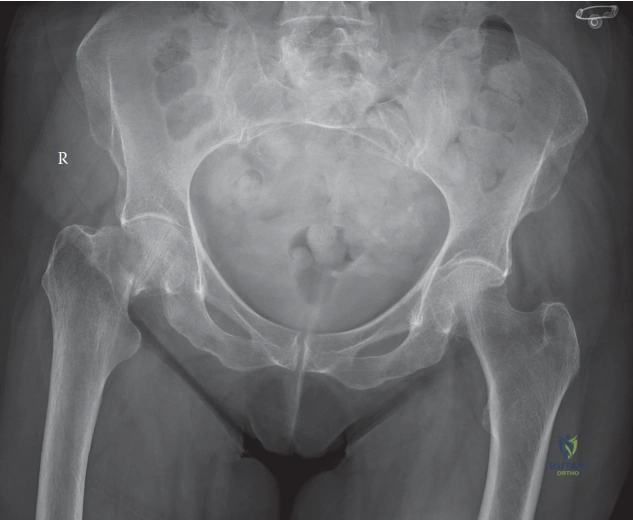
An 82-year-old female presents following a mechanical fall. She is alert, oriented, and complaining of right hip pain. Radiographs confirm a displaced femoral neck fracture (Garden IV). She is medically fit for surgery. Outline your surgical decision-making process for this patient.
Candidate: I would assess her physiological status and pre-injury mobility. Given her age and the displaced fracture, I would discuss the risks and benefits of arthroplasty versus internal fixation. In most elderly patients, hemiarthroplasty is standard, but I might consider a total hip arthroplasty if she is a high-functioning individual without significant cognitive impairment.
Focusing solely on the fracture classification without mentioning the patient's physiological status, cognitive baseline, or pre-injury functional demand. Failing to differentiate why one would choose a hemiarthroplasty versus a total hip arthroplasty.
Structure the answer: 1) Patient factors: Physiological age, comorbidities, cognitive status (using AMTS/MMSE), and pre-injury mobility. 2) Fracture factors: Displaced Garden IV carries high AVN/non-union risk if fixed (ORIF). 3) Treatment strategy: Hemiarthroplasty is the standard for low-demand, elderly patients for rapid mobilization. THA is indicated for physiologically active, high-demand patients, provided they are cognitive intact and independent, to minimize revision rates and improve long-term function.
During the management of an intertrochanteric fracture, you are choosing between a Dynamic Hip Screw (DHS) and a Cephalomedullary Nail (IMN). What are the specific biomechanical considerations?
Candidate: A DHS is suitable for stable fractures with an intact lateral wall. An IMN is superior for unstable patterns, such as reverse obliquity or those with significant posteromedial comminution, because it acts as a load-sharing device and provides a shorter lever arm, reducing bending stress at the implant-bone interface.
Failing to define "stability." Simply stating "IMN is better" without explaining the biomechanical reason (shortening of the lever arm, load-sharing, or the specific risk of lateral wall compromise in DHS).
Classify stability: Stable (intact lateral wall, no posteromedial comminution) -> DHS. Unstable (lateral wall compromise, subtrochanteric extension, reverse obliquity) -> IMN. Emphasize that in unstable fractures, the DHS construct experiences excessive varus-producing bending moments leading to lag screw cutout. The IMN provides central load sharing, is biomechanically superior for unstable patterns, and has a lower reoperation rate in these specific cases.
You are performing a posterior approach for a hemiarthroplasty. What structures are at risk, and how do you ensure the stability of the construct before closing?
Candidate: The sciatic nerve is at risk if retracted too aggressively posteriorly. To ensure stability, I must perform an anatomical repair of the short external rotators (piriformis, obturator internus, gemelli) and the posterior capsule to the greater trochanter, which acts as a posterior buttress.
Ignoring the importance of the soft tissue repair. Simply saying "close the wound" without emphasizing the reattachment of the external rotators, which is the primary deterrent to posterior dislocation in the posterior approach.
Identify the Sciatic nerve as the primary structure at risk. Explain the "Stability Triad" for the posterior approach: 1) Capsulorraphy (repair of the posterior capsule). 2) External rotator repair (reattaching the piriformis/short rotators to the trochanteric bed). 3) Correct component positioning (appropriate version and offset). Mentioning the use of trans-osseous sutures or drill holes for secure reattachment demonstrates surgical maturity.
