Introduction & Epidemiology
The lumbrical plus deformity is a recognized, albeit uncommon, clinical entity in hand surgery, characterized by paradoxical extension of the interphalangeal (IP) joints during attempted active flexion of the involved digit. This unusual kinematic pattern results from an imbalance in the delicate interplay between the flexor digitorum profundus (FDP) tendon and its associated lumbrical muscle. The condition invariably leads to functional impairment, compromising grasp, pinch, and fine motor dexterity.
Epidemiologically, lumbrical plus deformity is most frequently encountered as an iatrogenic complication following flexor tendon surgery. Common scenarios include:
*
Over-tensioning of FDP repairs or grafts
: When the FDP tendon is repaired or reconstructed with excessive tension, its effective excursion is significantly limited. During attempted active flexion, the FDP struggles to pull the distal phalanx directly. Instead, the force is preferentially transmitted to the lumbrical origin, which then acts as a primary extensor of the IP joints while maintaining or increasing MCP flexion.
*
Flexor Digitorum Superficialis (FDS) to FDP transfers
: If the transferred FDS tendon is set with excessive tension, a similar mechanism can arise.
*
Avulsion of the FDP insertion (e.g., "Jersey Finger")
: In cases where the FDP tendon avulses from its insertion and retracts proximally, the lumbrical muscle, which originates from the FDP, becomes functionally unopposed distally. Attempted FDP activation then primarily pulls on the lumbrical, leading to its paradoxical action.
*
Deep lacerations of the FDP distal to the lumbrical origin
: Similar to avulsion injuries, if the FDP tendon is compromised distally, the lumbrical's action can become exaggerated.
*
Congenital anomalies
: Rarely, congenital variations in lumbrical anatomy or tendon attachments can predispose to this deformity.
*
Spasticity or neurological conditions
: In some neurological disorders, altered muscle tone can contribute to lumbrical overactivity.
While precise incidence rates are difficult to ascertain due to its nature as a complication or rare presentation, awareness of its potential occurrence in the context of flexor tendon surgery is paramount for all hand surgeons. Early recognition and appropriate management are crucial for restoring optimal hand function.
Surgical Anatomy & Biomechanics
A thorough understanding of the intricate anatomy and biomechanics of the flexor-extensor system is fundamental to diagnosing and effectively treating lumbrical plus deformity.
Lumbrical Muscles
The four lumbrical muscles are unique in their origin and insertion, positioning them at a critical biomechanical crossroads:
*
Origin
: They arise from the radial side of the FDP tendons within the palm. The first and second lumbricals typically originate from the FDP tendons of the index and middle fingers, respectively, and are unipennate. The third and fourth lumbricals are bipennate, originating from adjacent FDP tendons (e.g., third lumbrical from middle and ring FDP tendons, fourth from ring and small FDP tendons).
*
Insertion
: They pass volar to the deep transverse metacarpal ligament, then extend dorsally along the radial side of the proximal phalanx to insert into the radial lateral band of the extensor apparatus and the base of the proximal phalanx.
*
Innervation
: The first and second lumbricals are innervated by the median nerve. The third and fourth lumbricals are innervated by the ulnar nerve. This dual innervation highlights their independence and specific roles.
*
Action
: The primary actions of the lumbricals are:
*
Flexion of the Metacarpophalangeal (MCP) joints
: By their attachment to the proximal phalanx via the extensor hood, they pull the proximal phalanx into flexion.
*
Extension of the Interphalangeal (IP) joints
: Through their insertion into the lateral bands, they contribute to the extension of both the Proximal Interphalangeal (PIP) and Distal Interphalangeal (DIP) joints.
This combined action, known as the "intrinsic plus" position (MCP flexion, IP extension), is essential for precision grip, maintaining the arches of the hand, and coordinating fine motor movements.
Flexor Digitorum Profundus (FDP)
- Origin : The FDP muscle originates from the anterior and medial surfaces of the ulna and the interosseous membrane.
- Insertion : Its four distinct tendons pass through the carpal tunnel, enter the fibrous flexor sheaths in the digits, and insert onto the palmar base of the distal phalanx.
- Innervation : The FDP to the index and middle fingers is typically innervated by the median nerve (via the anterior interosseous nerve). The FDP to the ring and small fingers is usually innervated by the ulnar nerve.
- Action : Its primary action is flexion of the DIP joint, followed by PIP and MCP joint flexion. It is the sole flexor of the DIP joint.
Extensor Mechanism
The extensor mechanism of the digits is a complex aponeurosis connecting the extensor digitorum communis (EDC) tendon, the lumbricals, and the interossei. It consists of:
*
Sagittal bands
: Stabilize the EDC tendon over the MCP joint and contribute to MCP extension.
*
Central slip
: Inserts into the base of the middle phalanx, extending the PIP joint.
*
Lateral bands
: Formed by contributions from the lumbricals, interossei, and the EDC tendon; they merge distally to form the terminal tendon.
*
Terminal tendon
: Inserts into the base of the distal phalanx, extending the DIP joint.
Biomechanics of Lumbrical Plus Deformity
The pathophysiology of lumbrical plus deformity lies in the disruption of the normal force transmission within the flexor system. When the FDP tendon is abnormally taut (e.g., due to excessive shortening after repair, graft, or severe retraction after avulsion), several critical biomechanical changes occur:
1.
Limited FDP Excursion
: The primary FDP muscle belly contracts, but the excessive tension on the tendon restricts its distal excursion required to effectively flex the DIP joint.
2.
Exaggerated Lumbrical Pull
: Instead of transmitting force primarily to the distal phalanx, the contracting FDP tendon pulls predominantly on the origin of the lumbrical muscle. This effectively shortens the lumbrical and intensifies its pull.
3.
Paradoxical IP Extension
: The now over-active lumbrical, acting with greater mechanical advantage, flexes the MCP joint while simultaneously extending the PIP and DIP joints through its connection to the lateral bands and terminal tendon. This creates the characteristic paradoxical IP extension during attempted active FDP flexion. The FDP, unable to adequately flex the distal phalanx, becomes a proximal motor for the lumbrical.
The deformity is most evident when the patient attempts to flex the involved digit. Instead of smooth, coordinated flexion, the MCP joint flexes, but the PIP and DIP joints paradoxically extend or remain extended, preventing the formation of a functional fist or strong grip.
Indications & Contraindications
Indications for Surgical Intervention
Surgical intervention for lumbrical plus deformity is primarily driven by functional impairment and the failure of conservative management.
*
Functional Impairment
: Patients typically present with an inability to form a full fist, difficulty with grasping objects, diminished pinch strength, or inability to perform specific activities of daily living (ADLs) or occupational tasks. The paradoxical IP extension directly interferes with digit flexion.
*
Pain
: While not a primary symptom, chronic dysfunctional mechanics or compensatory strain on other structures can lead to pain, which may serve as an additional indication if refractory to conservative measures.
*
Clear Etiology
: A definitive diagnosis of lumbrical plus deformity, especially in the context of prior flexor tendon surgery or FDP avulsion, provides a clear target for surgical correction.
*
Persistent Deformity
: The presence of persistent, disabling paradoxical IP extension that has not responded to a trial of non-operative management (e.g., splinting, hand therapy) warrants surgical consideration.
Contraindications for Surgical Intervention
Absolute and relative contraindications must be carefully considered:
*
Asymptomatic Deformity
: Incidental findings or very mild deformities that do not significantly impact function or cause symptoms typically do not require surgical intervention.
*
Poor General Health
: Significant co-morbidities that increase surgical or anesthetic risk (e.g., uncontrolled diabetes, severe cardiovascular disease, active infection) may preclude surgery.
*
Unrealistic Patient Expectations
: Patients must have a realistic understanding of the potential outcomes, rehabilitation commitment, and risks associated with surgery.
*
Active Infection
: Any active infection in the surgical field is an absolute contraindication to elective hand surgery until adequately treated.
*
Severe Irreparable Tendon Damage or Joint Contractures
: If the underlying tendon or joint damage is so extensive that functional improvement is unlikely, or if severe fixed contractures exist that cannot be overcome by lumbrical release alone, alternative strategies or non-operative care may be more appropriate.
*
Early Post-Operative Period
: If the deformity is noted soon after primary flexor tendon surgery, a period of conservative management and observation is often warranted before considering revision surgery, as some dynamic deformities may resolve or improve with therapy as swelling subsides and tissues heal.
Operative vs. Non-Operative Indications
| Indication Type | Operative Management | Non-Operative Management |
|---|---|---|
| Functional Impairment | Significant, persistent loss of grip/pinch, activities of daily living (ADL), occupational/sports demands | Mild, intermittent symptoms; minimal impact on ADL; dynamic deformity improved with therapy |
| Pain | Persistent, interfering with function, unresponsive to conservative analgesia and therapy | Mild to moderate pain managed with NSAIDs, activity modification, splinting |
| Deformity Severity | Fixed, severe paradoxical IP extension during attempted flexion; significantly limits digital cascade | Dynamic, mild extension, correctable with passive range of motion (PROM); improves with active-assistive exercises |
| Etiology | Iatrogenic (over-tensioned FDP repair/graft), traumatic (FDP avulsion with significant retraction), specific congenital variants | Mild post-surgical edema/inflammatory response; early-stage, dynamic cases; mild spasticity amenable to Botox |
| Response to Therapy | Failed appropriate conservative management (splinting, dedicated hand therapy for 6-12 weeks) | Initial management for all new presentations; effective for mild cases or those improving over time |
| Patient Desire | Highly motivated patient seeking functional restoration and understands surgical risks/benefits | Patient preference for non-invasive approach; willing to adapt function; avoids surgical risks |
| Associated Conditions | No severe comorbidities; favorable anesthetic risk; good bone/joint stock for reconstruction if needed | Uncontrolled systemic disease; high anesthetic risk; severe fixed joint contractures making surgical correction futile |
Pre-Operative Planning & Patient Positioning
Meticulous pre-operative planning is essential to ensure a successful outcome and minimize complications.
Pre-Operative Assessment
-
Detailed History
:
- Mechanism of injury : Was there a prior laceration, crush injury, or avulsion?
- Prior surgeries : Document all previous hand surgeries, particularly flexor tendon repairs, grafts, or transfers in the affected digit. Review operative reports to understand the initial management and potential for over-tensioning.
- Onset and progression of symptoms : When was the paradoxical extension first noted? Has it worsened or improved with conservative measures?
- Functional limitations : Quantify the impact on ADLs, work, and recreational activities.
- Pain profile : Characterize the pain, its location, intensity, and aggravating/alleviating factors.
- Hand dominance and occupational demands : Crucial for understanding the patient's functional expectations.
- General medical history : Co-morbidities (diabetes, peripheral vascular disease, smoking) that may affect healing or increase surgical risk.
-
Physical Examination
:
- Visual inspection : Observe the hand at rest and during attempted motion. Look for skin integrity, scarring, and any static deformities.
-
Range of Motion (ROM)
:
- Active ROM (AROM) : Specifically observe attempted active flexion of the affected digit. The hallmark lumbrical plus deformity will manifest as MCP flexion with paradoxical PIP and DIP extension. Document individual joint angles.
- Passive ROM (PROM) : Assess full passive flexion and extension of all joints (MCP, PIP, DIP). This helps differentiate dynamic lumbrical plus from fixed joint contractures. Full PROM of the IP joints suggests the deformity is dynamic and amenable to surgical correction.
- Palpation : Assess for tenderness, swelling, or palpable masses along the flexor tendons.
- Neurovascular Status : Evaluate sensation (two-point discrimination, light touch) and digital perfusion (capillary refill). Assess motor function of intrinsic and extrinsic muscles.
- Specific Tests : Apply gentle resistance to the dorsum of the proximal phalanx while the patient attempts to flex the digit. This can accentuate the paradoxical extension.
-
Imaging Studies
:
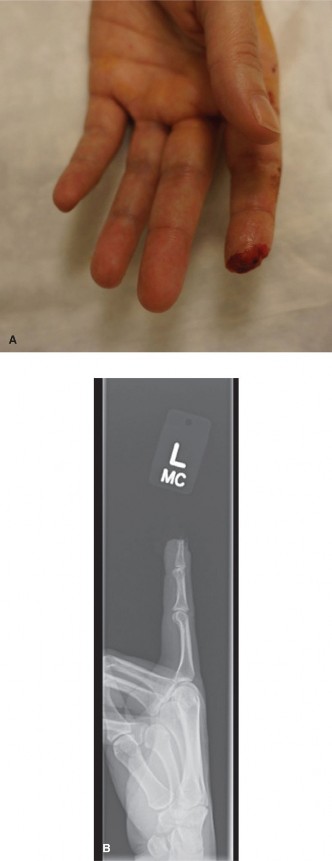
- Plain Radiographs : Standard AP, lateral, and oblique views of the hand and affected digit. These are primarily to rule out bony pathologies, joint degeneration, fractures, or dislocations that might mimic or contribute to functional deficits. They can also reveal signs of previous surgery (e.g., K-wires).
- Ultrasound or MRI : Rarely indicated, but can be useful in ambiguous cases to assess tendon integrity, adhesions, or the precise origin and course of the lumbrical if suspicion of an anomalous structure is high.
- Anesthesia Consultation : Discuss anesthetic options with the patient and anesthesiologist. Regional block (e.g., axillary or supraclavicular block) is often preferred as it provides excellent intraoperative and post-operative analgesia and allows the patient to potentially cooperate with intraoperative assessment of tendon tension if performed with sedation. General anesthesia is an alternative.
- Patient Education : Thoroughly explain the nature of the deformity, the proposed surgical procedure, potential risks (infection, nerve injury, recurrence, stiffness), expected benefits, and the critical role of post-operative hand therapy. Manage expectations regarding the timeline for recovery and functional return.
Patient Positioning
- Operating Table : The patient is positioned supine on the operating table.
- Upper Extremity Position : The affected upper extremity is abducted and externally rotated, placed on a specialized hand table to ensure comfortable access for the surgeon and assistant. The elbow should be slightly flexed.
- Tourniquet Application : A pneumatic tourniquet is applied to the upper arm. This provides a bloodless field, which is essential for meticulous dissection of delicate neurovascular structures and tendons. The tourniquet pressure and inflation time should be monitored carefully.
- Sterile Preparation and Draping : The entire upper extremity, from the shoulder to the fingertips, is prepared with an antiseptic solution and draped to maintain sterility throughout the procedure.
- Magnification : Use of surgical loupes (2.5x to 4.5x magnification) is highly recommended for identifying and protecting critical neurovascular structures and for precise tendon work.
Detailed Surgical Approach / Technique
The primary goal of surgical intervention for lumbrical plus deformity is to eliminate the excessive tension on the lumbrical muscle that causes paradoxical IP extension, thereby restoring effective FDP function and coordinated digital flexion. The most common and generally effective approach is a lumbrical tenotomy, sometimes combined with FDP lengthening in specific iatrogenic cases.
Incision and Exposure
The choice of incision depends on the affected digit and the anticipated extent of exposure required.
1.
Standard Longitudinal Incision
: A straight longitudinal incision over the proximal phalanx or metacarpal, extending distally to the A2 pulley level, offers direct access. However, it carries a higher risk of scar contracture over joints.
2.
Brunner Zig-Zag Incision
: This is generally preferred in the digits as it conforms to the skin creases, allows for excellent exposure without crossing flexion creases perpendicularly, and minimizes the risk of flexion contracture.
* Begin the incision over the proximal phalanx, creating a zig-zag pattern that extends proximally into the palm (if lumbrical origin needs to be addressed more proximally) or distally over the middle phalanx (if FDP lengthening is anticipated).
* Careful skin incision to avoid injury to underlying neurovascular bundles.
* Raise full-thickness skin flaps to expose the underlying subcutaneous tissue and digital neurovascular bundles.
Dissection and Identification of Structures
- Neurovascular Bundle Protection : Meticulously identify and protect the digital neurovascular bundles. These run along the sides of the flexor sheath, volar to the digital arteries and nerves. Retract them dorsally, or radially and ulnarly, depending on the approach.
- Flexor Sheath Exposure : Expose the fibrous flexor sheath. The A1 pulley is located at the MCP joint level, and the A2 pulley lies over the proximal phalanx. The specific level of exposure depends on the intended intervention. If the lumbrical origin needs to be accessed, the sheath may need to be opened proximally. The A2 and A4 pulleys are critical for maintaining the flexor tendon's mechanical advantage and should be preserved whenever possible.
-
Identification of FDP Tendon and Lumbrical
:
- Open the flexor sheath (e.g., release the A1 pulley if it obstructs access, or create a small window between pulleys).
- Retract the flexor digitorum superficialis (FDS) tendons if present (in the index, middle, ring fingers). This will expose the underlying FDP tendon.
- Identify the lumbrical muscle. It typically originates from the radial side of the FDP tendon in the palm, distal to the carpometacarpal joint line, and runs distally along the radial side of the FDP tendon. It appears as a reddish-brown, often fusiform, muscle belly transitioning into a thin tendon that inserts into the extensor mechanism.
Intraoperative Assessment
Before definitive intervention, intraoperative assessment is crucial:
*
Tension Test
: Gently pull the FDP tendon proximally to confirm its tightness and observe if it reproduces or exacerbates the paradoxical IP extension. Conversely, passively extending the wrist and flexing the MCP joint can sometimes reduce the tension on the lumbrical, allowing better IP flexion.
*
Trial Release (if possible)
: In some cases, a temporary clamping or gentle traction on the lumbrical can simulate the effect of release.
Techniques for Lumbrical Release
The choice of technique depends on the underlying etiology and the surgeon's preference.
-
Lumbrical Tenotomy (Transection) :
- This is the most direct and widely accepted method for resolving lumbrical plus deformity.
- Procedure : Identify the lumbrical tendon at its origin from the FDP tendon. Using fine scissors or a scalpel, carefully transect the lumbrical tendon proximally, as close to its origin from the FDP as possible. Ensure no digital nerves or vessels are inadvertently cut.
- Immediate Result : Following transection, reassess the digit. The paradoxical IP extension should immediately resolve, and the digit should be able to achieve full, smooth active flexion of all joints.
- Advantages : Simple, effective, and predictable.
- Disadvantages : Complete loss of lumbrical function to that digit. While this may theoretically impact precision grip, in practice, the loss of function is generally compensated by other intrinsic muscles, and the overall improvement in active flexion far outweighs this potential deficit. The risk of lumbrical insufficiency is low.
-
Lumbrical Advancement/Recession (Partial Tenotomy/Tenodesis) :
- Less commonly performed as the primary solution for established lumbrical plus, these techniques aim to reduce the lumbrical's effective pulling length while attempting to preserve some function.
- Procedure : Involves incising the lumbrical tendon and then re-attaching it more distally (advancement) or proximally (recession) to the FDP tendon or adjacent periosteum, with careful tensioning.
- Advantages : Attempts to retain some lumbrical function.
- Disadvantages : Technically more demanding, higher risk of recurrence if tension is not precisely adjusted, or overtightening leading to new deformities.
-
FDP Tenotomy/Lengthening (for severe iatrogenic cases) :
- If the primary cause of lumbrical plus is definitively an excessively short and tight FDP tendon (e.g., after over-advancement of a flexor tendon repair or an oversized graft), a lumbrical tenotomy alone may not fully resolve the problem or may result in recurrence. In such situations, direct lengthening of the FDP tendon may be indicated.
- Indications : Documented FDP over-tensioning.
-
Procedure
:
- Expose the FDP tendon well, typically in the mid-palmar or forearm region to gain sufficient length for a Z-plasty. A longer incision may be necessary.
- Perform a Z-lengthening of the FDP tendon. The tendon is incised longitudinally in a Z-shape (or step-cut).
- The two ends are then advanced/retracted to achieve the desired length. The ideal tension is achieved when the affected digit can passively extend completely while maintaining a normal resting cascade, and can actively flex without paradoxical extension. A useful clinical test is to ensure the repaired FDP tendon allows the digit to passively extend fully without causing adjacent digits to flex prematurely (quadriga effect).
- Secure the lengthened tendon with strong, non-absorbable sutures (e.g., modified Kessler or Bunnell stitch).
-
Image Integration
:
This image could depict a Z-lengthening of the FDP tendon, showing the step-cut incision and the subsequent lengthening and repair, or illustrate the anatomical relationship of the lumbrical to the FDP tendon in a schematic. - Advantages : Addresses the root cause in specific iatrogenic cases, potentially preventing recurrence.
- Disadvantages : More extensive surgery, longer recovery, higher risk of adhesions, and potential for flexor weakness if over-lengthened. This is generally a secondary procedure after failed primary lumbrical release, or reserved for clear, severe FDP overtensioning.
Intraoperative Reassessment and Closure
- Immediate Post-Intervention Check : After lumbrical tenotomy or FDP lengthening, reassess the digit's active and passive range of motion. Confirm that the paradoxical IP extension is resolved and full active flexion of all digital joints can be achieved. Ensure that the FDP tendon glides freely within its sheath.
- Hemostasis : Achieve meticulous hemostasis using bipolar electrocautery to minimize hematoma formation.
- Irrigation : Irrigate the wound thoroughly with sterile saline.
-
Closure
:
- Close the subcutaneous tissue with fine absorbable sutures.
- Close the skin with interrupted nylon sutures (e.g., 4-0 or 5-0 nylon).
- Apply a sterile dressing.
- Apply a protective splint in a functional position or an intrinsic plus position, depending on the stability and concerns for early contracture.
Complications & Management
Despite meticulous surgical technique, complications can arise following surgery for lumbrical plus deformity. Awareness of these potential issues and their appropriate management is critical for optimizing patient outcomes.
Common Complications, Incidence, and Salvage Strategies
| Complication | Incidence | Clinical Presentation | Salvage Strategy |
|---|---|---|---|
| Recurrence of Deformity | Variable, 5-15% | Return of paradoxical IP extension during attempted active flexion. May be subtle initially or progressively worsen. | Re-exploration and further lumbrical release (if inadequate initial release); consider FDP lengthening if underlying FDP overtension was not fully addressed initially. |
| Infection | Low, <1% | Localized erythema, warmth, swelling, pain, purulent discharge, fever. | Superficial: Oral antibiotics, local wound care. Deep: Surgical debridement, IV antibiotics, wound irrigation, culture-directed therapy. |
| Digital Nerve Injury | Low, <1% | Numbness, paresthesia, or hyperesthesia in the distribution of the affected digital nerve. | Neurapraxia/Axonotmesis : Observation, supportive care, nerve gliding exercises, neuropathic pain management. Neurotmesis (Transection) : Prompt microsurgical repair (primary repair if possible, otherwise nerve graft). |
| Scarring/Adhesions | Moderate, 5-10% | Restricted tendon glide, decreased ROM, pain, cosmetic concerns. | Aggressive hand therapy, scar massage, silicone sheeting, static/dynamic splinting. If severe and refractory to conservative care, surgical tenolysis may be required. |
| Flexion Contracture (PIP/DIP) | Variable, 5-10% | Inability to fully extend the PIP or DIP joint. | Early aggressive hand therapy, serial splinting, dynamic splinting. If fixed and severe, surgical capsulotomy or release of collateral ligaments. |
| Lumbrical Insufficiency/Weakness | Rare | Inability to fully flex the MCP joint while extending the IP joints (loss of intrinsic plus function), subtle grip weakness. | Primarily managed with occupational therapy to maximize function; rarely requires surgical intervention (e.g., intrinsic reconstruction) as overall function usually improves with resolved lumbrical plus. |
| Loss of FDP Function | Rare | Inability to flex the DIP joint, often due to iatrogenic damage to the FDP tendon itself (e.g., during FDP lengthening). | Re-exploration and repair/reconstruction of the FDP tendon if viable. If not, secondary tendon transfer for replacement (e.g., FDS transfer). |
| Complex Regional Pain Syndrome (CRPS) | Low, 1-5% | Persistent, disproportionate pain, swelling, skin color/temperature changes, allodynia, hyperalgesia, sudomotor changes. | Early recognition is key. Multimodal pain management (medications, sympathetic blocks), aggressive hand therapy focusing on desensitization and active motion, psychological support. |
| Hematoma | Low | Localized swelling, tenderness, ecchymosis, may compromise wound healing. | Small hematomas: Observation, compression. Large/expanding: Surgical evacuation, meticulous hemostasis. |
Other Potential Complications
- Stiffness : A general term for reduced ROM in the affected digit or hand, often a result of edema, pain, or insufficient rehabilitation. Addressed by aggressive hand therapy.
- Delayed Wound Healing : Can occur due to infection, poor circulation, tension on the wound, or systemic factors. Managed with wound care, addressing underlying causes.
- Suture Extrusion : Especially if non-absorbable sutures are placed subcutaneously too superficially.
General Management Principles
- Early Recognition : Prompt identification of complications is paramount.
- Multidisciplinary Approach : Close collaboration with hand therapists, pain specialists, and infectious disease specialists is often necessary.
- Patient Education : Clear communication regarding potential complications and the importance of adherence to rehabilitation protocols.
- Preventative Measures : Meticulous surgical technique, proper hemostasis, careful handling of tissues, and appropriate post-operative immobilization and rehabilitation are the best preventative strategies.
Post-Operative Rehabilitation Protocols
Post-operative rehabilitation is a critical determinant of functional outcome following surgery for lumbrical plus deformity. The protocol emphasizes controlled motion, edema and scar management, and progressive strengthening, all under the guidance of a skilled hand therapist. The specific protocol may be tailored based on the stability of the repair (e.g., FDP lengthening versus simple lumbrical tenotomy) and surgeon's preference.
Phase I: Immediate Post-Operative (Day 0-7)
-
Immobilization
:
- A dorsal blocking splint is typically applied. The wrist is positioned in slight flexion (e.g., 20-30 degrees), the MCP joints are in moderate flexion (e.g., 70-90 degrees), and the IP joints are in full extension. This "intrinsic plus" position helps to prevent flexion contractures of the MCPs and facilitates IP flexion once active motion begins.
- For cases involving FDP lengthening, protection against active FDP contraction might be needed, potentially with a more restrictive splint initially or a shorter arc of FDP motion.
-
Edema Management
:
- Strict elevation of the hand above heart level (e.g., resting on pillows) to minimize swelling.
- Gentle active range of motion of uninvolved joints (shoulder, elbow, wrist, and unaffected fingers) to maintain circulation and reduce stiffness.
- Light compression dressings, if appropriate.
-
Pain Management
:
- Prescription pain medication as needed.
- Patient education on signs of infection and other complications.
-
Early Gentle Motion (if stable)
:
- For simple lumbrical tenotomy, gentle active flexion and extension of the affected digit's IP joints may begin within the confines of the splint or a protective arc, as tolerated, to prevent adhesions and restore glide.
Phase II: Early Mobilization (Weeks 1-3)
-
Splinting
:
- Continue dorsal blocking splint or a custom static-progressive splint between exercise sessions and at night.
- May transition to a dynamic splint to assist with IP extension or flexion, if needed.
-
Active Range of Motion (AROM)
:
- Initiate gentle, active flexion and extension exercises for the affected MCP, PIP, and DIP joints. The goal is full, coordinated motion without paradoxical extension.
- Focus on isolated FDP and FDS gliding exercises to prevent adhesions and improve tendon excursion.
- Begin place-and-hold exercises to promote active muscle control.
-
Passive Range of Motion (PROM)
:
- Gentle PROM exercises, ensuring full flexion and extension are achieved without pain or resistance.
-
Edema and Scar Management
:
- Continue elevation.
- Gentle scar massage once the incision is well-healed to minimize adhesions.
- Use of silicone gel sheeting or topical scar creams.
- Nerve Gliding Exercises : Introduce gentle nerve gliding exercises to prevent adhesion of digital nerves.
Phase III: Progressive Strengthening (Weeks 4-6)
-
Splinting
:
- Gradual weaning from splinting, typically worn only at night or during demanding activities.
-
Strengthening
:
- Gradual introduction of light strengthening exercises using therapeutic putty, rubber bands, and light weights.
- Focus on intrinsic muscle strengthening (MCP flexion, IP extension) and extrinsic flexor strengthening (FDP/FDS).
- Progress from light resistance to moderate resistance.
-
Functional Activities
:
- Begin incorporating light functional activities of daily living that promote grip and pinch.
-
Continue Scar Management
:
- Maintain scar massage and other modalities to prevent hypertrophic scarring.
Phase IV: Return to Activity (Weeks 7-12+)
- Full Range of Motion : The goal is to achieve full, pain-free active and passive range of motion in all affected joints.
- Advanced Strengthening : Progress to more demanding strengthening exercises, mimicking occupational or sport-specific movements.
- Functional Integration : Focus on integrating the hand back into all desired activities.
- Sports-Specific Rehabilitation : If applicable, work with the therapist to gradually return to sport-specific training.
- Patient Education : Reinforce home exercise programs and provide guidance on activity modification and long-term self-management to prevent recurrence or secondary issues.
Key Principles of Rehabilitation
- Individualization : Protocols must be tailored to the individual patient's progress, pain level, and the specific surgical procedure performed.
- Communication : Close and frequent communication between the surgeon and the hand therapist is paramount for optimal progression and problem-solving.
- Patient Compliance : The success of rehabilitation heavily relies on the patient's adherence to the prescribed exercises and precautions.
- Early Controlled Motion : Crucial for preventing adhesions, maintaining tendon glide, and restoring joint mobility.
- Protection : Adequate protection of the surgical site is essential, especially in the early phases, to allow for tissue healing.
Summary of Key Literature / Guidelines
The understanding and management of lumbrical plus deformity have evolved through clinical observation and biomechanical studies, emphasizing a consistent approach to diagnosis and treatment. Key literature and guidelines reflect these developments:
Historical Context & Recognition
The paradoxical action of the lumbrical in specific pathological conditions was recognized by early pioneers in hand surgery. Sterling Bunnell described the intrinsic muscles' complex role in hand function. J. William Littler and other prominent hand surgeons further elucidated the specific mechanism of the lumbrical plus deformity, particularly in the context of flexor tendon injuries and repairs. Their work laid the foundation for understanding its biomechanics and guiding surgical intervention.
Diagnostic Criteria
The literature consistently highlights the clinical hallmark: paradoxical extension of the IP joints during attempted active flexion of the digit. This observation, coupled with a thorough history (especially prior flexor tendon surgery or injury), remains the cornerstone of diagnosis. Imaging studies are primarily used to rule out concomitant pathology rather than directly diagnose the lumbrical plus itself.
Surgical Nuances
- Lumbrical Tenotomy as the Standard : The overwhelming consensus in current hand surgery literature is that simple lumbrical tenotomy at its origin from the FDP tendon is the most effective and least morbid surgical intervention for established lumbrical plus deformity. Studies and case series consistently report good to excellent outcomes with resolution of the paradoxical extension and restoration of functional flexion. The loss of a single lumbrical's specific function is generally well-compensated by other intrinsic muscles, and the functional gain from improved FDP excursion is significant.
-
Role of FDP Lengthening
: FDP lengthening (typically via a Z-plasty) is a more involved procedure and is generally reserved for cases where:
- The primary etiology is clearly demonstrated to be iatrogenic overtensioning of the FDP tendon (e.g., after over-advancement or an oversized graft).
- A simple lumbrical tenotomy has failed, suggesting that the underlying FDP tightness was not sufficiently addressed.
-
Intraoperative assessment confirms persistent FDP tightness even after lumbrical release.
Literature supports this more cautious approach, emphasizing that FDP lengthening carries a higher risk of adhesions, flexor weakness if over-lengthened, and a more prolonged rehabilitation.
- Intraoperative Assessment : Guidelines emphasize the critical importance of intraoperative confirmation of the diagnosis and assessment of tendon tension. Visually observing the digital cascade and testing FDP excursion before and after intervention helps ensure the problem is addressed adequately.
- Preventative Measures : In the context of primary flexor tendon surgery, literature stresses the importance of careful and accurate tensioning of FDP repairs and grafts to avoid iatrogenic lumbrical plus deformity. This includes intraoperative assessment of the resting digital cascade.
Outcomes
- Generally Favorable : Surgical correction of lumbrical plus deformity, particularly with lumbrical tenotomy, generally yields good to excellent functional outcomes, with resolution of the paradoxical extension and significant improvement in grip and pinch strength.
- Dependence on Rehabilitation : The success of surgery is highly dependent on meticulous post-operative hand therapy to prevent stiffness, adhesions, and to restore full range of motion and strength.
- Recurrence : While low, recurrence can occur, often pointing to an incomplete release or an unaddressed underlying FDP overtensioning.
Key References & Authors
Classic and contemporary hand surgery texts provide comprehensive discussions:
*
Green's Operative Hand Surgery
: A foundational resource that details the anatomy, biomechanics, and surgical techniques for lumbrical plus, often citing original work by Littler and others.
*
Campbell's Operative Orthopaedics
: Offers another authoritative perspective on hand pathology and surgical management.
*
Journal of Hand Surgery (American and European volumes)
: Publishes numerous case reports, clinical series, and biomechanical studies that further refine our understanding and management strategies.
In summary, the academic literature consistently frames lumbrical plus deformity as a specific biomechanical imbalance amenable to surgical correction, with simple lumbrical tenotomy being the procedure of choice for most cases, reserving FDP lengthening for specific instances of documented overtensioning. The emphasis remains on precise diagnosis, meticulous surgical technique, and dedicated post-operative rehabilitation.
