Pes Cavus Deformity & Ankle Pain: Etiology, Diagnosis, & Management for Orthopedic Professionals
Key Takeaway
Pes Cavus is a foot deformity characterized by an abnormally high arch, often leading to ankle pain and instability. Diagnosis involves clinical examination, gait analysis, and specialized tests like the Coleman Block Test to assess hindfoot flexibility. Management ranges from conservative orthotics and physical therapy to surgical correction in severe cases, aiming to restore proper foot mechanics and alleviate pain.
A 28-year-old patient presents with chronic lateral ankle pain and recurrent sprains. On examination, you notice a high medial arch and a hindfoot in varus. What is your clinical investigation strategy to determine the biomechanical nature of this deformity?
Candidate: I would examine for underlying neurological causes like CMT. I'll perform a Silfverskiöld test to check for equinus and the Coleman block test to assess hindfoot flexibility. I would also order weight-bearing radiographs of the foot and ankle.
The candidate focuses only on the ankle. They fail to mention the neurological "red flags" (e.g., spinal dysraphism, family history), omit the specific radiographic angles (Meary’s, Calc-pitch), or fail to explain the diagnostic logic behind the Coleman block test (differentiating forefoot vs. hindfoot-driven deformity).
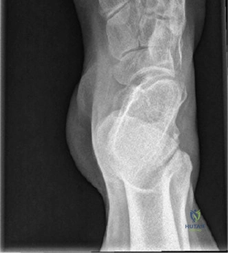
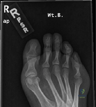
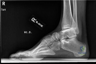
I would approach this systematically: 1) Neurological screening: Examine the spine and perform a full motor/sensory/reflex exam to rule out hereditary/neurological conditions. 2) Physical exam: Silfverskiöld for gastrocnemius contracture, and the Coleman block test—if the hindfoot corrects, the varus is secondary to a plantarflexed 1st ray. 3) Imaging: WB AP/Lateral/Mortise views and Harris heel view. I would specifically quantify Meary’s angle, calcaneal pitch, and Hibbs angle to define the severity and apex of the cavus.
The patient's Coleman block test shows that the hindfoot remains in varus while standing on the block. What does this indicate, and how does it change your surgical planning?
Candidate: This means the hindfoot varus is rigid and fixed, not just compensatory. I would need to perform a calcaneal osteotomy to correct the alignment, rather than just treating the forefoot.
Failing to mention the sequence of surgery. A good answer must emphasize that in a rigid deformity, you must address both the primary driver (1st ray) and the secondary rigid component (calcaneal osteotomy) to achieve a plantigrade foot.
A persistent varus on the Coleman block indicates a rigid, fixed hindfoot deformity, likely due to chronic subtalar and soft tissue contractures. My surgical plan must be 'a la carte': 1) Plantar fascia release (Steindler stripping). 2) First ray dorsiflexion osteotomy to address the primary driver. 3) Lateralizing calcaneal osteotomy (LCO) to shift the mechanical axis and restore the eversion moment arm. 4) Tendon transfer (peroneus longus to brevis) to rebalance the foot, and finally, lateral ligament stabilization.
During your reconstruction, you are performing the lateral calcaneal osteotomy. How do you prevent complications regarding the nerves and soft tissues, and what are the primary risks of this specific maneuver?
Candidate: I would make a lateral incision posterior to the peroneal tendons. The main risk is injury to the sural nerve. I'd perform careful dissection and protect the neurovascular structures.
Ignoring the "tension" issue. Candidates often forget that correcting a varus deformity to valgus significantly increases tension on the lateral wound, making wound dehiscence a high-frequency complication.
Sural nerve protection via meticulous blunt dissection is vital. Regarding complications, the primary risk is wound healing issues; the lateral incision is under significant tension once the varus is corrected. To mitigate this, I perform the soft tissue/medial procedures first, and address the lateral osteotomy and closure with the foot in a corrected, relaxed position. Other risks include sural nerve neuroma, under-correction, and non-union at the osteotomy site, which I minimize with rigid internal fixation using large-fragment headless compression screws.
