Mastering Necrotizing Soft Tissue Diagnosis with LRINEC Criteria
Key Takeaway
Your ultimate guide to Mastering Necrotizing Soft Tissue Diagnosis with LRINEC Criteria starts here. The Laboratory Risk Indicator for Necrotizing Fasciitis (LRINEC) criteria help differentiate necrotizing soft tissue infections from cellulitis. This diagnostic tool evaluates CRP, WBC, hemoglobin, sodium, creatinine, and glucose levels. Importantly, the erythrocyte sedimentation rate (ESR) is not a component of the LRINEC criteria. A LRINEC score of 8 or higher indicates a 75% chance of necrotizing fasciitis.
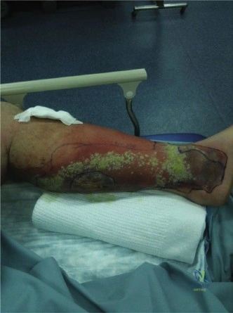
A 55-year-old diabetic patient presents to the Emergency Department with an acutely painful, swollen left lower limb. He is tachycardic, febrile, and hypotensive. On examination, there are violaceous bullae over the calf and the skin appears anesthetic. You suspect a necrotizing soft tissue infection (NSTI).
How do you immediately assess this patient, and what is the role of the LRINEC score in this clinical decision-making process?
Candidate: I would immediately secure the airway, breathing, and circulation. I'd start broad-spectrum antibiotics and fluid resuscitation. I'd calculate the LRINEC score using bloods. If it's 6 or greater, it supports the diagnosis of NSTI, and I would take the patient to theatre for urgent debridement.
Candidates often over-rely on the LRINEC score, suggesting they would "wait for the labs" before acting. They fail to mention that NSTI is a clinical diagnosis and that the LRINEC score is merely an adjunct that should never delay surgical exploration in a patient who is systemically unwell.
A high-scoring answer emphasizes that NSTI is a clinical diagnosis. I would immediately initiate ABCDE resuscitation (fluid, blood cultures, broad-spectrum IV antibiotics including clindamycin for anti-toxin effect). While ordering the LRINEC, I would not wait for results. I would explain that a LRINEC ≥ 6 provides high positive predictive value, but a low score does not rule out the disease. If the clinical index of suspicion is high—as in this patient with bullae, systemic sepsis, and anesthesia—the patient requires an urgent 'finger test' in the OR, regardless of laboratory values.
You are in the operating theatre. You have made a generous incision. You observe grey, friable tissue that separates easily from the underlying fascia with blunt dissection. The muscle underneath appears dull and non-contractile when stimulated with cautery.
Describe your surgical management strategy for this patient.
Candidate: I would perform an aggressive debridement. I'd remove all necrotic fascia, fat, and any muscle that doesn't bleed or contract. I would irrigate thoroughly, leave the wound open, and plan for a second look in 24-48 hours.
Failing to mention the necessity of serial debridements or ignoring the importance of anatomical compartment boundaries. Candidates often forget to mention sending deep tissue samples for microbiology and histopathology.
The goal is source control. I would perform radical excision of all devitalized skin, subcutaneous fat, deep fascia, and necrotic muscle until reaching 'bleeding, healthy tissue.' I would use the 'finger test' to probe fascial planes to ensure no tracking infection is missed. I would ensure samples are sent for aerobic, anaerobic, and fungal cultures, plus histology. I would pack the wound with non-adherent dressings or NPWT and explicitly state the requirement for serial debridements (the 'second-look' principle) every 24-48 hours until the wound is clean, as the infection may continue to progress beyond the initial margins.
