Introduction & Epidemiology
Bunionette deformity, also known as tailor's bunion or lateral bunion, represents a prominent osseous protuberance of the fifth metatarsal head, typically located on its lateral or dorsolateral aspect. This condition is characterized by pain, localized callus formation, bursitis, and difficulty with shoewear, often leading to significant functional impairment. While traditionally attributed to restrictive shoewear, particularly in historical context with tailors sitting cross-legged, the etiology is multifactorial, encompassing genetic predisposition, altered foot biomechanics, and environmental factors.
Epidemiologically, bunionettes are less common than hallux valgus but represent a substantial portion of forefoot pathology. Prevalence estimates vary, with some studies suggesting up to 3-4% of the adult population experience symptoms. The condition affects women more frequently than men, paralleling patterns observed in other forefoot deformities, likely due to a combination of genetic factors and narrower, less accommodating footwear.
Bunionettes are broadly classified into three primary types based on their underlying anatomical abnormality, as described by Coughlin:
*
Type I:
Isolated lateral exostosis of the fifth metatarsal head, often with a normal fourth-fifth intermetatarsal (IM) angle. This variant is primarily an osseous spur causing friction.
*
Type II:
Lateral bowing of the distal fifth metatarsal shaft. The entire distal metatarsal is deviated laterally, increasing the prominence of the metatarsal head. The 4-5 IM angle may be normal or only slightly elevated.
*
Type III:
Increased fourth-fifth intermetatarsal (IM) angle, characterized by medial deviation of the fifth metatarsal base relative to the fourth, pushing the fifth metatarsal head laterally. This is often associated with a splayfoot deformity.
A fourth category, Iselin's disease , refers to an apophysitis of the fifth metatarsal base in adolescents, which, while not a true bunionette, can present with lateral foot pain. Understanding these classifications is crucial for surgical planning, as each type necessitates a distinct operative strategy. The primary goal of intervention, whether conservative or operative, is the alleviation of pain and restoration of functional gait.
Surgical Anatomy & Biomechanics
A thorough understanding of the surgical anatomy and biomechanics of the fifth ray is paramount for effective treatment of bunionette deformities. The fifth metatarsal articulates proximally with the cuboid and medially with the fourth metatarsal, forming the tarsometatarsal (TMT) joint. Distally, it articulates with the proximal phalanx of the fifth digit at the metatarsophalangeal (MTP) joint.
The fifth metatarsal is a unique structure, characterized by its greater mobility compared to the central metatarsals. Its TMT joint is relatively mobile in the sagittal and transverse planes, allowing for pronation and supination of the fifth ray, which is critical for adapting the foot to uneven surfaces. This inherent mobility also contributes to the susceptibility to deformity under abnormal loading.
Key anatomical structures in the vicinity of the fifth metatarsal head include:
*
Musculotendinous Units:
The peroneus brevis tendon inserts onto the lateral aspect of the fifth metatarsal base, while the peroneus longus tendon passes underneath the cuboid to insert on the first metatarsal and medial cuneiform, influencing forefoot pronation/supination. The abductor digiti minimi muscle originates from the calcaneal tuberosity and inserts into the lateral aspect of the fifth proximal phalanx, contributing to fifth toe abduction and stability. Its tendon lies lateral to the joint capsule.
*
Neurovascular Structures:
The lateral dorsal cutaneous nerve (a terminal branch of the sural nerve) courses superficial to the lateral aspect of the fifth metatarsal head and shaft. This nerve is highly susceptible to iatrogenic injury during surgical approaches to the bunionette, leading to painful neuroma or paresthesia. The common digital nerve to the fourth web space also has branches supplying the fifth toe and can be affected. Dorsal metatarsal arteries contribute to local blood supply.
*
Capsuloligamentous Complex:
The fifth MTP joint is stabilized by a joint capsule, medial and lateral collateral ligaments, and the plantar plate. Chronic irritation and deformity can lead to bursitis over the lateral aspect of the metatarsal head.
Biomechanical considerations are central to the pathogenesis and treatment of bunionettes. Normal weight-bearing involves a complex distribution of forces across the forefoot. In a bunionette deformity, the prominent fifth metatarsal head experiences increased pressure during the propulsion phase of gait, particularly when wearing restrictive footwear.
*
Increased 4-5 IM Angle:
A widened intermetatarsal angle between the fourth and fifth metatarsals effectively shifts the fifth metatarsal head laterally, making it more prominent. This often coexists with increased forefoot splay.
*
Lateral Bowing:
A congenital or acquired curvature of the fifth metatarsal shaft laterally exaggerates the prominence, irrespective of the IM angle.
*
Lateral Exostosis:
This isolated bony prominence is often a response to repetitive microtrauma or pressure, leading to an adaptive osteophyte.
*
Plantarflexed Fifth Metatarsal:
While less common, a plantarflexed fifth metatarsal can also contribute to lateral weight-bearing pressure and discomfort.
The interplay of these factors creates a painful pressure point, leading to inflammation of the overlying bursa and soft tissues, skin irritation, and eventual callus formation. Surgical correction aims to realign the fifth metatarsal and/or reduce the bony prominence to restore more physiological weight distribution and eliminate chronic irritation.
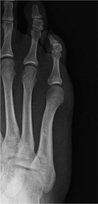
Figure: Anatomical depiction of the fifth metatarsal and its articulations, highlighting the region affected by bunionette deformity and structures at risk.
Indications & Contraindications
The decision-making process for intervention in bunionette deformity involves a careful assessment of symptoms, functional limitations, radiographic findings, and the patient's overall health status and expectations. Non-operative management is invariably the first line of treatment.
Non-Operative Indications (Initial Management)
Non-operative treatment strategies are appropriate for patients with mild to moderate symptoms, those unwilling or unable to undergo surgery, or as an initial attempt to alleviate pain and improve function. The duration of conservative management should be adequate, typically 3-6 months, before considering surgical options.
*
Shoe Modifications:
Wearing wider shoes with a deep toe box to accommodate the forefoot and reduce pressure on the lateral aspect of the fifth metatarsal head.
*
Padding and Orthotics:
Custom or off-the-shelf pads (e.g., gel sleeves, bunionette pads) to cushion the prominent area. Orthotics designed to address underlying biomechanical issues like excessive pronation or forefoot splay.
*
Non-Steroidal Anti-Inflammatory Drugs (NSAIDs):
Oral or topical NSAIDs to manage pain and inflammation associated with bursitis or synovitis.
*
Corticosteroid Injections:
Local injections into an inflamed bursa can provide temporary relief but carry risks of fat atrophy, skin discoloration, and infection, and are not a definitive solution.
*
Physical Therapy:
Modalities like ice, massage, and exercises to improve foot mechanics and muscle balance are rarely effective for significant structural deformities but may assist with soft tissue irritation.
Operative Indications
Surgical intervention is indicated when conservative measures have failed to provide symptomatic relief, when the deformity causes significant functional limitation, or when progressive deformity is evident. The presence of intractable pain, recurrent callus formation, or an inability to wear conventional footwear are key motivators for surgery.
Specific radiographic criteria often guide the decision and choice of procedure:
*
Type I (Exostosis):
Pain from isolated lateral prominence of the fifth metatarsal head, unresponsive to shoe modifications.
*
Type II (Lateral Bowing):
Clinical deformity and pain associated with a laterally bowed fifth metatarsal shaft, often with an increased lateral metatarsal head angle.
*
Type III (Increased 4-5 IM Angle):
Clinical deformity and pain secondary to an elevated intermetatarsal angle between the fourth and fifth metatarsals (e.g., > 8.5 degrees on AP weight-bearing radiograph), with or without an increased lateral metatarsal head angle.
Contraindications
Absolute and relative contraindications must be thoroughly assessed pre-operatively.
*
Absolute Contraindications:
* Active infection (local or systemic).
* Severe peripheral vascular disease or neuropathy that compromises healing.
* Uncontrolled diabetes mellitus.
* Acute Charcot arthropathy.
* Lack of symptoms or unrealistic patient expectations.
*
Relative Contraindications:
* Smoking (significantly impairs bone and soft tissue healing).
* Poor nutritional status.
* Significant obesity.
* Severe systemic illness.
* Poor skin quality over the surgical site.
* Severe psychological conditions impacting adherence to post-operative protocols.
Table: Operative vs. Non-Operative Indications
| Feature | Non-Operative Indication | Operative Indication |
|---|---|---|
| Pain Level | Mild to moderate, intermittent | Severe, chronic, intractable pain |
| Functional Impact | Minor discomfort, manageable with shoe changes | Significant functional limitation, difficulty with ambulation and shoewear |
| Deformity Severity | Mild prominence, no significant structural changes | Moderate to severe prominence, structural deformity (Type I, II, or III) |
| Callus/Bursitis | Incidental, responsive to padding | Recurrent, painful callus/bursitis despite conservative care |
| Duration of Symptoms | Recent onset, short duration | Chronic symptoms (> 6 months) unresponsive to adequate non-operative trials |
| Radiographic Findings | Minor exostosis, normal IM angles | Significant lateral exostosis, lateral bowing, or increased 4-5 IM angle |
| Patient Desire | Preference for non-surgical options | Desire for definitive correction after failed conservative care |
Pre-Operative Planning & Patient Positioning
Meticulous pre-operative planning is essential to ensure optimal surgical outcomes, minimize complications, and select the appropriate procedure for the specific bunionette type.
Pre-Operative Evaluation
-
Clinical Assessment:
- History: Detailed history of pain (onset, character, aggravating/alleviating factors), duration of symptoms, previous treatments, shoe modifications, and functional limitations. Assess patient's activity level and expectations.
-
Physical Examination:
- Inspection: Observe forefoot splay, degree of lateral prominence, presence of erythema, bursitis, or callus over the fifth metatarsal head. Assess alignment of the lesser toes.
- Palpation: Localize tenderness to the fifth metatarsal head, evaluate for an inflamed bursa.
- Range of Motion (ROM): Assess MTP joint ROM, looking for stiffness or pain.
- Neurological Exam: Evaluate light touch and two-point discrimination in the distribution of the lateral dorsal cutaneous nerve to establish a baseline and rule out pre-existing neuropathy.
- Vascular Exam: Palpate pedal pulses (dorsalis pedis, posterior tibial) and assess capillary refill.
- Gait Analysis: Observe the patient's gait for compensatory mechanisms and areas of increased pressure.
-
Radiographic Assessment:
-
Weight-Bearing Anteroposterior (AP), Lateral, and Oblique Views of the Foot:
These are standard and crucial for detailed assessment.
- AP View: Essential for measuring the 4-5 intermetatarsal (IM) angle (normal < 8.5 degrees) and the lateral metatarsal head angle (angle between the articular surface of the fifth metatarsal head and a line perpendicular to the axis of the fifth metatarsal shaft, normal < 5-6 degrees). This view helps classify the bunionette type.
- Lateral View: Assesses sagittal plane alignment of the fifth metatarsal (e.g., plantarflexion deformity).
- Oblique View: Can further delineate the prominence and bone pathology.
- Stress Views: Rarely indicated but can assess stability if concerns exist.
- Advanced Imaging (CT/MRI): Generally not required for routine bunionette management. May be considered in complex cases, suspected tumors, or non-union.
-
Weight-Bearing Anteroposterior (AP), Lateral, and Oblique Views of the Foot:
These are standard and crucial for detailed assessment.
-
Procedure Selection:
Based on the clinical and radiographic findings, the appropriate surgical technique is selected.
- Type I (Exostosis): Simple lateral condylectomy (e.g., prominent bone resection).
- Type II (Lateral Bowing): Corrective osteotomy (e.g., distal chevron, Weil, or oblique diaphyseal osteotomy).
- Type III (Increased 4-5 IM Angle): Proximal osteotomy (e.g., crescentic, opening/closing wedge) or combined distal and proximal osteotomies for severe cases.
- Soft Tissue Procedures: Often performed adjunctively, such as lateral capsuloligamentous release or medial capsular reefing.
Patient Positioning and Preparation
- Anesthesia: General anesthesia or regional anesthesia (e.g., ankle block, popliteal block) is suitable. A combination may be used for post-operative pain control.
- Patient Position: Supine position on the operating table.
- Tourniquet: A thigh-high pneumatic tourniquet is typically applied to achieve a bloodless field, inflated to 100 mmHg above systolic blood pressure or 250-300 mmHg.
- Sterile Preparation and Draping: The foot and ankle are prepped with an antiseptic solution (e.g., chlorhexidine or povidone-iodine) and draped in a sterile fashion, typically isolating the foot.
- Distraction/Manipulation: Traction on the fifth digit may be used to facilitate joint access, though not always necessary.
Detailed Surgical Approach / Technique
The choice of surgical technique is dictated by the bunionette type and severity. The goal is to correct the underlying deformity, reduce pain, and restore proper foot mechanics. Protection of the lateral dorsal cutaneous nerve is paramount in all approaches.
General Principles
- Incision: A dorsolateral longitudinal or curvilinear incision over the fifth metatarsal head and shaft. The length varies based on the chosen osteotomy (distal vs. proximal).
- Soft Tissue Dissection: Careful dissection through skin and subcutaneous tissue. Identify and protect the lateral dorsal cutaneous nerve and its branches, which are typically superficial and dorsolateral. Retract it dorsally or plantarly as needed.
- Capsulotomy: A longitudinal capsulotomy is performed over the MTP joint to expose the metatarsal head.
Specific Surgical Techniques
1. Lateral Condylectomy (for Type I Bunionette)
- Indication: Isolated lateral exostosis of the fifth metatarsal head with a normal 4-5 IM angle and no significant lateral bowing.
-
Technique:
- After exposing the metatarsal head via the dorsolateral incision and capsulotomy, identify the prominent lateral condyle.
- Using an osteotome or sagittal saw, carefully resect the lateral exostosis, ensuring not to compromise the articular surface or the insertion of the abductor digiti minimi tendon if it's not contributing to the prominence.
- The aim is to create a smooth contour, eliminating the lateral prominence.
- Roughened edges are rasped smooth.
- Confirm adequate bone resection by palpation and visual inspection, ensuring no impingement with the fifth phalanx or soft tissues with full range of motion.
- Fixation: No internal fixation is typically required for a simple condylectomy.
2. Distal Metatarsal Osteotomies (for Type II Bunionette and mild Type III)
These osteotomies are performed near the metatarsal head to redirect the articular surface and/or translate the metatarsal head medially.
a. Modified Reverdin-Green-Laird Osteotomy
- Indication: Type II bunionette (lateral bowing) and some Type III bunionettes with moderate increase in 4-5 IM angle. This is an extra-articular osteotomy with a lateral closing wedge.
-
Technique:
- After dorsolateral approach and nerve protection, expose the distal fifth metatarsal.
- Place a Kirschner wire or fine drill bit parallel to the articular surface of the fifth metatarsal head, about 3-5 mm proximal to the articular cartilage.
- A second cut is made with a sagittal saw, extending from the lateral cortex proximally and distally, converging towards a point on the medial cortex just proximal to the physis (to avoid damage). This creates a lateral closing wedge osteotomy. The orientation of the wedge depends on the desired correction. A more medially directed cut allows for greater correction of the IM angle.
- The wedge of bone is resected from the lateral aspect of the metatarsal.
- The distal fragment is then translated medially and/or rotated to correct the deformity. The goal is to reduce the 4-5 IM angle and correct the lateral metatarsal head angle.
- Fixation: Typically stabilized with a single cortical screw (e.g., 2.7 mm or 3.0 mm) placed dorsally or dorsomedially across the osteotomy site. A K-wire can be used temporarily for stabilization during screw placement. Ensure bicortical purchase and flush screw head.
b. Weil Osteotomy (Shortening Osteotomy)
- Indication: When the fifth metatarsal is long relative to the fourth, contributing to MTP joint overload, or when significant medial translation is required without excessive shortening.
-
Technique:
- Expose the distal metatarsal, protecting the nerve.
- The osteotomy is performed using a sagittal saw, typically parallel to the plantar weight-bearing surface (or slightly angled dorsally to prevent dorsal malunion).
- The cut starts dorsally just proximal to the articular cartilage and extends proximally into the metatarsal neck.
- The distal fragment (metatarsal head) is then translated proximally and medially to correct the deformity and shorten the metatarsal. This effectively shifts the weight-bearing axis.
- Fixation: Typically one or two small screws (e.g., 2.0 mm, 2.4 mm, or 2.7 mm) are used, passing from the dorsal aspect of the metatarsal head into the metatarsal shaft.
3. Diaphyseal Osteotomies (for Type II Bunionette with significant bowing)
- Indication: Significant lateral bowing of the fifth metatarsal shaft.
-
Technique:
- Oblique Osteotomy (e.g., Scarf-like): A longer dorsolateral incision is made. The osteotomy is performed in the mid-diaphysis or distal third of the shaft, typically an oblique cut from dorsomedial to plantarolateral. The two fragments are then translated, rotated, and shifted to correct the bowing.
- Fixation: Requires stable fixation, often with two cortical screws (e.g., 2.7 mm or 3.0 mm) for robust stability.
- Considerations: Greater exposure and dissection, increasing risk to the lateral dorsal cutaneous nerve along the shaft.
4. Proximal Metatarsal Osteotomies (for Type III Bunionette)
- Indication: Markedly increased 4-5 IM angle (> 10 degrees) or when a distal osteotomy alone is insufficient. This addresses the deformity at its origin, near the TMT joint.
-
Technique:
- A longer dorsolateral incision is made extending proximally over the fifth metatarsal base.
- Careful dissection to expose the fifth metatarsal base and its articulation with the cuboid. Protection of the sural nerve branches is critical.
- Crescentic Osteotomy: A crescentic saw blade is used to make a curved osteotomy through the metatarsal base, roughly perpendicular to the long axis of the shaft. The distal fragment is then rotated or translated medially to reduce the IM angle.
- Closing Wedge Osteotomy: A triangular wedge of bone is resected from the lateral aspect of the fifth metatarsal base, allowing the distal segment to be translated medially and closed.
- Opening Wedge Osteotomy: A medial wedge is created and opened, often with a bone graft, to medialize the metatarsal.
- Fixation: Proximal osteotomies require robust fixation due to higher biomechanical stresses. Plates (e.g., small fragment locking plate) or multiple screws (e.g., 2.7 mm or 3.0 mm cortical screws) are typically used. Provisional K-wire fixation can aid in achieving optimal position before definitive fixation.
- Considerations: These are more invasive procedures, requiring longer healing times and potentially greater risk of non-union or malunion compared to distal osteotomies.
Adjunctive Procedures
- Lateral Capsulotomy/Release: Often performed as part of the initial exposure, particularly if the MTP joint is tight laterally.
- Medial Capsular Plication/Reefing: Rarely required for bunionettes, but may be considered if there is excessive laxity or an intention to stabilize the joint medially after significant lateral deviation correction.
- Soft Tissue Excision: Resection of inflamed bursa or callus if present.
Closure
- Copious irrigation of the surgical site.
- Layered closure of the joint capsule (if opened), subcutaneous tissue, and skin using absorbable sutures.
- Skin closure with non-absorbable sutures or staples.
- Sterile dressing and appropriate post-operative splint or cast.
Complications & Management
Despite meticulous surgical technique, complications can occur following bunionette correction. A thorough understanding of these potential issues and their management is crucial for all orthopedic surgeons.
Table: Common Complications, Incidence, and Salvage Strategies
| Complication | Incidence (Approximate) | Management / Salvage Strategy |
|---|---|---|
| Recurrence of Deformity | 5-20% | Review original classification and surgical technique. Revision osteotomy (e.g., more aggressive translation, different osteotomy type), consideration of soft tissue balancing. |
| Malunion | 5-15% | Non-symptomatic: Observation. Symptomatic (pain, callus, stiffness): Corrective osteotomy (wedge, rotation, translation), possibly fusion if joint is arthritic. |
| Non-Union/Delayed Union | < 5% | Non-weight bearing, immobilization (boot/cast). Persistent: Revision surgery with debridement, bone grafting (autograft/allograft), rigid internal fixation (plate, screws), pulsed electromagnetic fields (PEMF) or low-intensity pulsed ultrasound (LIPUS). |
| Nerve Injury (Lateral Dorsal Cutaneous Nerve) | 5-15% (transient paresthesia); < 2% (permanent neuroma) | Transient: Reassurance, observation. Persistent paresthesia/neuroma: Conservative (NSAIDs, gabapentin/pregabalin, injections). Surgical: Neurolysis, neurectomy with proximal implantation, nerve grafting. |
| Infection | < 1-2% | Superficial: Oral antibiotics, local wound care. Deep (Osteomyelitis): Surgical debridement, intravenous antibiotics guided by culture, hardware removal if infected. |
| Hardware Irritation/Failure | 5-10% | Symptomatic: Hardware removal after bone healing. Failure with non-union: Revision surgery with new fixation, bone grafting. |
| Stiffness/Limited ROM | 5-10% | Early physical therapy, stretching, mobilization exercises. Persistent: Manipulation under anesthesia, capsulotomy/arthrolysis. |
| Metatarsalgia (Transfer) | 5-15% | Offloading orthotics, shoe modifications. Persistent: Shortening osteotomy of adjacent metatarsal(s) (e.g., Weil osteotomy). |
| Vascular Compromise | Rare | Immediate removal of constrictive dressings, assessment of circulation. Potential for surgical exploration and revascularization. |
| Complex Regional Pain Syndrome (CRPS) | Rare (0.5-2%) | Early diagnosis, pain management (gabapentin/pregabalin), physical therapy, stellate ganglion block, psychological support. |
Management Strategies in Detail
- Recurrence and Malunion: These are often related to inadequate initial correction, failure to address all components of the deformity, or unstable fixation. Re-evaluation with weight-bearing radiographs is crucial. Revision surgery typically involves a more extensive osteotomy, addressing angular and translational components, and ensuring stable fixation. For malunion leading to painful callus, localized contouring or secondary osteotomy may be required.
- Non-Union: Risk factors include smoking, diabetes, poor bone quality, excessive shortening, and unstable fixation. Initial management involves strict non-weight bearing and prolonged immobilization. If non-union persists, surgical intervention with debridement of fibrous tissue, refresh of osteotomy ends, bone grafting (autograft from calcaneus or iliac crest, or allograft), and rigid internal fixation (plate and screws) is indicated. Biologic adjuncts (e.g., PRP) may be considered, though evidence is limited.
- Nerve Injury: The lateral dorsal cutaneous nerve is the most frequently injured structure. Prevention through careful dissection and identification is key. If symptoms are transient, conservative management with reassurance, NSAIDs, and potentially neuropathic pain medications (gabapentin, pregabalin) is appropriate. Persistent or severe neuropathic pain or a palpable neuroma warrants consideration of surgical exploration, neurolysis, or neurectomy. If a neuroma is excised, the nerve stump should be buried in muscle or bone to prevent recurrent neuroma formation.
- Infection: Superficial infections are managed with oral antibiotics and local wound care. Deep infections, especially involving bone (osteomyelitis), require aggressive surgical debridement, often with hardware removal, and prolonged intravenous antibiotic therapy guided by culture and sensitivity results.
- Hardware Irritation: Screws or plates that are prominent or impinge on soft tissues can cause pain. If symptoms persist after bone healing, hardware removal is a simple and effective solution. If hardware failure leads to loss of reduction or non-union, revision surgery is necessary.
- Stiffness: Early, gentle range of motion exercises post-operatively can mitigate stiffness. If significant stiffness or arthrofibrosis develops, aggressive physical therapy, dynamic splinting, or in refractory cases, manipulation under anesthesia or arthroscopic/open capsulotomy may be considered.
- Transfer Metatarsalgia: This occurs when weight-bearing is shifted to an adjacent metatarsal due to excessive shortening or elevation of the corrected metatarsal. Prevention involves careful planning of osteotomy length and inclination. Management includes orthotics with metatarsal pads to offload the painful area. In severe cases, a shortening osteotomy (e.g., Weil) of the adjacent, overloaded metatarsal may be indicated.
Post-Operative Rehabilitation Protocols
A structured and progressive post-operative rehabilitation protocol is critical for maximizing functional recovery, minimizing complications, and achieving optimal long-term outcomes after bunionette surgery. Protocols vary based on the surgical technique, fixation stability, surgeon preference, and patient-specific factors. The following outlines a general framework.
Phase 1: Immediate Post-Operative (Weeks 0-2)
- Goal: Protect surgical site, control pain and swelling, initiate wound healing.
-
Weight Bearing:
- Condylectomy: Usually partial weight-bearing (PWB) in a stiff-soled shoe or surgical boot, or full weight-bearing (FWB) as tolerated if stable.
- Distal Osteotomies (e.g., Reverdin-Green-Laird, Weil): Typically PWB in a post-operative shoe or CAM walker boot. Some surgeons may advocate non-weight bearing (NWB) for the initial 2 weeks for certain osteotomies.
- Proximal/Diaphyseal Osteotomies: Often NWB in a short leg cast or CAM walker boot to protect the more extensive osteotomy and fixation.
- Dressing/Splint: Bulky soft dressing or plaster splint for protection, replaced by a surgical shoe or boot after initial wound check (typically 7-10 days).
-
Pain & Edema Management:
- Elevation above heart level for the majority of the day.
- Ice application for 15-20 minutes, several times daily.
- Oral analgesics (NSAIDs, acetaminophen, opioids as needed).
-
Exercises:
- Gentle, non-weight bearing ankle pumps to reduce swelling and prevent deep vein thrombosis (DVT).
- Toe wiggles (fifth toe only if stable, or all toes if pain allows) to prevent stiffness.
- Wound Care: Keep incision clean and dry. Staples or sutures typically removed at 10-14 days.
Phase 2: Early Mobilization (Weeks 2-6)
- Goal: Gradual increase in weight bearing, restore MTP joint range of motion, reduce swelling.
-
Weight Bearing:
- Condylectomy/Distal Osteotomies: Progress to FWB in a post-operative shoe or CAM walker boot.
- Proximal/Diaphyseal Osteotomies: Progress to PWB in a CAM walker boot, gradually increasing to FWB as tolerated, typically around 4-6 weeks, pending radiographic evidence of early healing.
-
Exercises:
- Active and passive range of motion (ROM) exercises for the fifth MTP joint and other lesser toes, initiated cautiously.
- Gentle stretching of the Achilles tendon and plantar fascia, if indicated.
- Foot intrinsic muscle strengthening exercises (e.g., towel scrunches, marble pick-ups) once incision is well-healed.
- Modalities: Continued use of ice and elevation. Compression stockings may be introduced to manage edema.
- Therapy: Formal physical therapy often begins in this phase, focusing on ROM, gentle strengthening, and gait training.
Phase 3: Progressive Strengthening & Functional Return (Weeks 6-12)
- Goal: Improve strength, balance, proprioception, and prepare for return to activity.
- Weight Bearing: Transition out of surgical boot into comfortable, supportive athletic shoes. Gradual return to FWB and normal gait mechanics.
-
Exercises:
- Progressive strengthening exercises for intrinsic and extrinsic foot muscles (e.g., calf raises, resistive band exercises).
- Balance and proprioception training (e.g., single-leg stance, wobble board).
- Gait retraining to normalize foot-strike and push-off.
- Low-impact cardiovascular activities (e.g., swimming, cycling).
- Modalities: Continue with self-management for pain and swelling.
- Therapy: Continued formal physical therapy, focusing on restoring pre-injury strength and function.
Phase 4: Return to Activity/Sports (Weeks 12+)
- Goal: Gradual and safe return to full activity, including sports, if desired.
- Progression: Progressive increase in activity intensity and duration. Gradual reintroduction of higher-impact activities (e.g., jogging, jumping, cutting sports) under the guidance of the surgeon and therapist.
- Monitoring: Continued monitoring for pain, swelling, or signs of recurrence. Emphasis on wearing appropriate, supportive footwear.
- Long-Term: Maintenance of foot strength and flexibility. Awareness of shoe choices to prevent recurrence.
Key Considerations:
*
Smoking Cessation:
Crucial for bone healing. Patients should be counseled pre- and post-operatively.
*
Diabetes Control:
Essential for wound healing and infection prevention.
*
Patient Compliance:
Adherence to rehabilitation protocols significantly impacts outcomes.
*
Radiographic Healing:
For osteotomies, radiographic evidence of bone healing is typically required before discontinuing protective weight bearing and advancing to higher impact activities. This is often assessed at 6-8 weeks and again at 12 weeks.
Summary of Key Literature / Guidelines
The body of literature on bunionette deformities and their surgical management, while not as extensive as for hallux valgus, provides a framework for evidence-based practice. Historically, simple exostectomy was common, but with a deeper understanding of biomechanics, osteotomies have become the mainstay for correcting structural deformities.
- Classification and Diagnosis: Coughlin's classification (Type I, II, III) remains widely accepted and guides surgical decision-making. Radiographic parameters, particularly the 4-5 IM angle and lateral metatarsal head angle, are critical for accurate diagnosis and pre-operative planning.
-
Comparative Studies of Osteotomies:
- Distal Osteotomies: Studies have shown good to excellent outcomes for distal metatarsal osteotomies (e.g., chevron, Reverdin-Green-Laird, Weil) for Type I and II deformities, and mild Type III deformities. These procedures generally offer quicker recovery, lower complication rates, and predictable correction for localized problems. The Weil osteotomy is particularly useful when metatarsal shortening is also desired or when significant plantarflexion of the metatarsal exists.
- Proximal Osteotomies: For severe Type III deformities with a significantly increased 4-5 IM angle, proximal osteotomies (e.g., crescentic, closing/opening wedge) are more effective at correcting the underlying intermetatarsal divergence. These are technically more demanding and are associated with a higher risk of non-union, delayed union, and potentially longer recovery periods compared to distal osteotomies.
- Diaphyseal Osteotomies: Oblique diaphyseal osteotomies (e.g., "Scarf-like" osteotomies) are effective for Type II lateral bowing but require stable fixation and careful soft tissue handling due to the proximity of the lateral dorsal cutaneous nerve along the shaft.
- Combined Procedures: In complex cases with multiple deformity components (e.g., severe lateral bowing and increased IM angle), a combination of distal and proximal osteotomies may be necessary, although this increases the complexity and potential for complications.
-
Outcomes and Complications:
- Overall satisfaction rates after bunionette surgery are generally high, ranging from 80-95%.
- Recurrence rates vary by technique but are a notable concern, especially with inadequate initial correction. Malunion and non-union are more common with proximal osteotomies.
- Nerve injury, particularly to the lateral dorsal cutaneous nerve, is the most frequently reported soft tissue complication across all techniques.
- Transfer metatarsalgia is a recognized complication, especially with excessive shortening or elevation of the fifth metatarsal.
- Evidence Levels: Much of the literature consists of Level IV (case series) and Level III (retrospective comparative studies) evidence. High-level (Level I or II) prospective, randomized controlled trials comparing different bunionette surgical techniques are limited, making definitive recommendations challenging. However, consensus guidelines generally support tailored surgical approaches based on the specific classification of the bunionette.
- Fixation Methods: Screw fixation (cannulated, headless, cortical) is generally preferred for its stability, allowing for earlier mobilization. Kirschner wires are sometimes used for temporary fixation or in cases of very small osteotomies, but offer less rigid stability. Plate fixation is increasingly used for more complex proximal and diaphyseal osteotomies to ensure robust stabilization.
- Rehabilitation: While specific protocols vary, the emphasis across the literature is on early controlled mobilization to prevent stiffness, followed by progressive weight-bearing and strengthening. Protection during the initial healing phase is critical, especially for osteotomies.
In summary, surgical correction of bunionette deformity is a well-established procedure with predictable outcomes when appropriately indicated and meticulously performed. The key to success lies in accurate pre-operative classification of the deformity, selection of the optimal surgical technique (condylectomy, distal, diaphyseal, or proximal osteotomy, or a combination), stable internal fixation, careful protection of neurovascular structures, and a structured post-operative rehabilitation program. Future research should focus on high-level comparative studies to further refine surgical indications and optimize long-term results.
