Part of the Master Guide

Distal Radius Fractures: Epidemiology, Surgical Anatomy, and Management Indications

Distal Radius Fracture: Ace Your FRCS Oral Exam Case

23 Apr 2026 90 min read 148 Views
Illustration of distal radius fracture - Dr. Mohammed Hutaif

Key Takeaway

Learn more about Distal Radius Fracture: Ace Your FRCS Oral Exam Case and how to manage it. A distal radius fracture is a break in the radius bone near the wrist, often characterized by dorsal angulation and displacement. Radiographs typically show dorsal tilt on AP views and dorsal displacement on lateral views. Management depends on patient and injury factors, frequently involving closed manipulation for unstable fractures to restore alignment and reduce soft tissue tension.
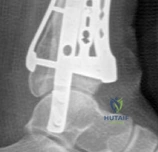
Comprehensive Exam


00:00

Start Quiz

Question 1

A 65-year-old female sustains a distal radius fracture with significant comminution, articular involvement spanning both the scaphoid and lunate fossae, and severe metaphyseal comminution, extending into the diaphysis. According to the Fernandez classification, how would this fracture typically be categorized?


Explanation

The Fernandez classification categorizes distal radius fractures based on the mechanism of injury and fracture morphology. Type I is bending (meta-epiphyseal), Type II is shearing (Barton, Hutchinson), Type III is compression (die-punch), Type IV is avulsion (ligament), and Type V is combined or high-energy fractures with extensive comminution and bone loss. Significant comminution, articular involvement of both fossae, and extension into the diaphysis (often implying severe metaphyseal involvement or bone loss) points strongly towards a high-energy injury, characteristic of a Type V Fernandez fracture.

Question 2

Which of the following radiographic findings in a distal radius fracture is LEAST indicative of potential instability requiring surgical intervention following an initially successful closed reduction?


Explanation

Instability criteria often guide the decision for surgical fixation following a distal radius fracture. Common indicators of instability include initial dorsal angulation greater than 20 degrees, radial shortening exceeding 3mm, severe metaphyseal comminution, and particularly, intra-articular step-off or gap greater than 1-2mm. While an ulnar styloid fracture is frequently associated with distal radius fractures and may suggest a TFCC injury, its presence alone is not a direct criterion for radial fracture instability or a primary indication for surgical intervention on the radius, assuming other parameters are acceptable. It might influence DRUJ stability, but not necessarily the stability of the radial reduction itself.

Question 3

During a standard volar approach (Henry approach) to the distal radius for plate fixation, which structure is primarily released or retracted radially to access the volar aspect of the radius?


Explanation

The Henry approach for volar plating of the distal radius involves an incision between the Flexor Carpi Radialis (FCR) and the Radial Artery. The FCR tendon is retracted ulnarly, and the radial artery and brachioradialis are retracted radially. The critical step to expose the volar aspect of the distal radius is the subperiosteal elevation and L-shaped release of the Pronator Quadratus muscle from its radial and distal attachments, which is then reflected ulnarly. The median nerve lies more ulnarly, and the FPL tendon is in the deep flexor compartment and typically not the primary muscle reflected for direct radial access.

Question 4

A 70-year-old patient undergoes open reduction internal fixation with a dorsal plate for a comminuted distal radius fracture. Six months post-operatively, she presents with difficulty extending her thumb IP joint and a positive Finkelstein's test. Assuming the Finkelstein's test is a misdiagnosis or secondary finding, which tendon is most likely to have ruptured?


Explanation

Difficulty extending the thumb IP joint (interphalangeal joint) is the hallmark sign of Extensor Pollicis Longus (EPL) rupture. EPL rupture is a known complication of distal radius fractures, particularly after dorsal plating, due to attrition over rough bone edges, plate prominence, or direct plate impingement. It can also occur post-closed reduction due to attrition over a dorsal bony prominence or as part of a delayed presentation (e.g., following a Colles' fracture). The Finkelstein's test is for De Quervain's tenosynovitis (APL and EPB), which is not directly related to EPL rupture, hence the assumption of it being a secondary finding.

Question 5

When measuring volar tilt on a true lateral radiograph of the wrist, a normal range is considered to be:


Explanation

On a true lateral radiograph of the wrist, the distal articular surface of the radius normally exhibits a volar tilt. The accepted normal range is typically 10 to 15 degrees of volar tilt. A neutral or dorsal tilt is considered abnormal and is a characteristic deformity of a Colles' fracture.

Question 6

Which of the following anatomical structures is considered the primary static stabilizer of the distal radioulnar joint (DRUJ)?


Explanation

The Triangular Fibrocartilage Complex (TFCC) is the primary static stabilizer of the DRUJ. It is a complex structure comprising the articular disc, dorsal and volar radioulnar ligaments, and the meniscal homologue. While the dorsal and volar radioulnar ligaments within the TFCC are key components, the TFCC as a whole unit provides the most significant static stability. The interosseous membrane provides some longitudinal stability to the forearm, and the Pronator Quadratus offers dynamic stability. The ECU tendon sheath is adjacent but not a primary stabilizer.

Question 7

A 40-year-old male presents with persistent wrist pain and decreased range of motion 1 year after non-operative management of a distal radius fracture. Radiographs show a dorsal tilt of 25 degrees, radial shortening of 5mm, and a 3mm intra-articular step-off. According to common malunion criteria, which of these findings is least acceptable for good functional outcomes in a younger, active patient?


Explanation

While all listed findings represent aspects of malunion, an intra-articular step-off of 3mm is widely considered the most critical predictor of poor long-term outcomes, particularly post-traumatic arthritis, especially in an active younger patient. Even 1-2mm of intra-articular incongruity is often deemed unacceptable. Dorsal tilt >10-15 degrees and radial shortening >2-3mm are also significant, and ulnar positive variance is directly related to radial shortening. However, articular step-off directly compromises joint congruity and leads to accelerated degenerative changes, making it the least acceptable from a functional prognosis standpoint.

Question 8

A 55-year-old female develops symptoms consistent with Complex Regional Pain Syndrome (CRPS) Type I following a distal radius fracture treated non-operatively. Her symptoms include severe pain out of proportion to injury, allodynia, swelling, and trophic changes. Which of the following is considered the MOST critical early intervention in managing CRPS?


Explanation

Early recognition and aggressive physical and occupational therapy focused on pain-free range of motion, desensitization, and functional use are paramount in managing CRPS. While medications (gabapentin, tricyclic antidepressants) and interventional treatments (sympathetic blocks) have a role, they are often adjuncts. Steroids may be used, but not as the initial most critical step. Spinal cord stimulators are reserved for refractory cases. The key to preventing progression and improving outcomes is early, consistent, and active rehabilitation.

Question 9

A 30-year-old active male sustains a distal radius fracture with a 4mm intra-articular step-off, 5 degrees dorsal tilt, and 1mm radial shortening. Which of these parameters ALONE typically warrants surgical intervention for definitive management?


Explanation

While age and activity level influence treatment decisions, the specific fracture characteristic of a 4mm intra-articular step-off is a very strong, if not absolute, indication for surgical management, regardless of other parameters. Even 1-2mm of articular incongruity is often considered unacceptable, particularly in a younger, active individual, due to the high risk of post-traumatic arthritis. The dorsal tilt and radial shortening mentioned are relatively minor compared to the articular step-off.

Question 10

Following reduction and casting of a distal radius fracture, a patient complains of persistent ulnar-sided wrist pain, particularly with pronation/supination and grasping. Tenderness is noted just distal to the ulnar head. Which of the following tests would be most appropriate to further evaluate for a potential Triangular Fibrocartilage Complex (TFCC) injury?


Explanation

Ulnar-sided wrist pain after a distal radius fracture, especially with DRUJ movements, strongly suggests a TFCC injury. The TFCC compression test (axial load with ulnar deviation and rotation) is a specific provocative test for TFCC tears, eliciting pain and sometimes a click. The Scaphoid Shift (Watson) test assesses scapholunate instability. Finkelstein's test is for De Quervain's tenosynovitis. The Grind test is for carpometacarpal arthritis. The Piano Key test assesses DRUJ stability (dorsal/volar translation of the ulnar head).

Question 11

In the Frykman classification system for distal radius fractures, what does the term 'Type VII' specifically denote?


Explanation

The Frykman classification categorizes distal radius fractures based on intra-articular extension and associated ulnar fracture. Even numbers (II, IV, VI, VIII) indicate an associated ulnar fracture, while odd numbers (I, III, V, VII) indicate no associated ulnar fracture. The specific articular involvement follows: Type I/II (extra-articular), Type III/IV (radiocarpal intra-articular), Type V/VI (DRUJ intra-articular), and Type VII/VIII (both radiocarpal and DRUJ intra-articular). Therefore, Type VII signifies an intra-articular fracture involving both the radiocarpal joint and the distal radioulnar joint (DRUJ), without an associated ulnar fracture.

Question 12

When using K-wire fixation for a dorsally displaced distal radius fracture, what is the most biomechanically advantageous configuration to prevent recurrent dorsal displacement?


Explanation

For dorsally displaced fractures, the Kapandji technique (intrafocal pinning) involves inserting K-wires into the fragments from the radial styloid, driving them across the fracture site and impacting them into the opposite intact cortex (usually volar). This creates a 'joystick' or 'buttress' effect, maintaining reduction and providing volar cortical support against dorsal collapse. Cross K-wires provide overall stability but don't specifically buttress the volar cortex as effectively against dorsal collapse. Dorsal-to-volar wires are not typically used to prevent dorsal displacement. Buttress wires across the volar cortex would require open exposure and are typically plates.

Question 13

A patient presents to the emergency department with a significantly displaced distal radius fracture and acute carpal tunnel syndrome symptoms (paresthesias in the median nerve distribution, positive Tinel's at the carpal tunnel, decreased two-point discrimination). What is the MOST appropriate initial management step?


Explanation

Acute median nerve neuropathy associated with a significantly displaced distal radius fracture is often due to direct compression or traction from the fracture fragments or surrounding edema. The MOST appropriate initial step is emergent closed reduction of the fracture. Reducing the fracture often decompresses the median nerve, alleviating symptoms. If symptoms do not improve rapidly after reduction, then an emergent carpal tunnel release may be considered. Corticosteroids, observation, or MRI would delay critical treatment.

Question 14

Which of the following distal radius fracture patterns is generally considered the most suitable indication for primary external fixation, particularly as a definitive treatment rather than just temporary stabilization?


Explanation

External fixation excels in managing highly comminuted, unstable intra-articular distal radius fractures, especially those with significant metaphyseal bone loss, open fractures, or severe soft tissue compromise. It provides ligamentotaxis (distraction across the wrist joint) to indirectly reduce fragments and maintain length and alignment, while allowing for soft tissue care. Stable, non-displaced fractures are managed conservatively. Volar Barton's fractures are often treated with volar plating. Pediatric fractures have different considerations. Isolated ulnar styloid fractures are rarely treated with external fixation.

Question 15

A 45-year-old male sustains a volar Barton's fracture. What is the generally accepted definitive management strategy for this fracture pattern?


Explanation

A volar Barton's fracture is an intra-articular fracture-dislocation where a rim of the distal radius (volar lip) displaces proximally and volarly with the carpus. This fracture is inherently unstable due to the articular involvement and the strong pull of the flexor tendons. Open reduction and internal fixation with a volar plate, which acts as a buttress, is the standard and most reliable treatment to restore articular congruity and stability. Closed reduction alone is rarely successful in maintaining reduction. Dorsal plating is inappropriate for a volar fragment. External fixation is less ideal for directly reducing and buttressing a single large volar fragment compared to a plate.

Question 16

The principle of ligamentotaxis, commonly utilized in external fixation for distal radius fractures, primarily achieves fracture reduction by:


Explanation

Ligamentotaxis is the principle where continuous longitudinal traction (distraction) is applied across a joint via an external fixator. This tension, transmitted through the intact soft tissues (ligaments and joint capsule) attached to the distal fragments, indirectly pulls comminuted fracture fragments back into a more anatomical position. It does not directly manipulate fragments, apply compression, or primarily resist rotation, although it contributes to stability for K-wire fixation.

Question 17

A common and often debilitating complication associated with dorsal plating for distal radius fractures, which is less frequently seen with volar plating, is:


Explanation

Dorsal plating places the hardware (plate and screws) in close proximity to the extensor tendons, particularly the EPL (Extensor Pollicis Longus). This can lead to extensor tendon irritation, tenosynovitis, or, more severely, rupture due to attrition over the prominent hardware or rough bone edges. While median nerve neuropathy and CRPS can occur with any distal radius fracture treatment, flexor tendon issues are more associated with volar plating (though less common with modern low-profile plates). Carpal tunnel syndrome is often due to the injury itself or fracture displacement.

Question 18

A 10-year-old boy presents with a Salter-Harris Type II distal radius fracture. What is the characteristic feature of this fracture type?


Explanation

The Salter-Harris classification describes physeal (growth plate) fractures. Type I: fracture through the physis only. Type II: fracture through the physis and extending into the metaphysis (fracture through the growth plate with a metaphyseal fragment, a 'Thurston Holland' fragment). This is the most common type. Type III: fracture through the physis and extending into the epiphysis (intra-articular). Type IV: fracture through the metaphysis, physis, and epiphysis (intra-articular). Type V: crush injury to the physis (rare, poor prognosis). Therefore, Type II involves both the physis and the metaphysis.

Question 19

Which carpal ligament injury is most commonly associated with a high-energy distal radius fracture, potentially leading to carpal instability?


Explanation

The Scapholunate (SL) ligament is the most frequently injured intercarpal ligament associated with distal radius fractures, especially in high-energy trauma or fractures with significant comminution or displacement. Injury to the SL ligament can lead to scapholunate dissociation and subsequent degenerative changes (SLAC wrist). While other ligaments can be injured, the SL ligament is particularly vulnerable and its injury carries significant implications for wrist stability.

Question 20

A patient presents with a displaced radial styloid fracture, often referred to as a Hutchinson or Chauffeur's fracture. What is the MOST appropriate treatment approach for this fracture pattern with significant displacement?


Explanation

Hutchinson (or Chauffeur's) fractures are intra-articular fractures of the radial styloid, typically caused by direct impaction of the scaphoid into the radial styloid. Due to the intra-articular nature and the strong pull of the brachioradialis, these fractures tend to displace. Significant displacement (>1-2mm articular step-off) or rotational malalignment usually warrants open reduction and internal fixation, often with screws (lag screw principle) or a small plate, to restore articular congruity and prevent post-traumatic arthritis. Closed reduction and casting are often insufficient to maintain reduction.

Question 21

An open distal radius fracture, classified as Gustilo-Anderson Type II, is observed in the emergency department. What is the MOST appropriate initial management strategy?


Explanation

Open fractures require urgent attention to prevent infection and facilitate healing. The initial management for a Gustilo-Anderson Type II open fracture involves thorough irrigation and debridement in the operating theatre, administration of broad-spectrum antibiotics (IV), tetanus prophylaxis, and temporary stabilization of the fracture (e.g., with an external fixator or K-wires) to protect soft tissues and allow for swelling resolution. Definitive internal fixation (like volar plating) is often delayed until the soft tissue envelope is favorable, typically after several days. Closed reduction and casting are inappropriate for an open fracture. Amputation is generally reserved for Gustilo Type IIIC or unsalvageable limbs.

Question 22

Following stable open reduction and internal fixation of a distal radius fracture with a volar locking plate, what is typically the recommended rehabilitation protocol?


Explanation

The primary advantage of stable internal fixation with modern volar locking plates is the ability to initiate early active and passive range of motion (ROM) exercises. This helps prevent stiffness, tendon adhesions, and CRPS. Immobilization is typically minimal (e.g., a splint for comfort for a few days) or not required at all beyond the immediate post-operative period. Strengthening is generally delayed until adequate bone healing is evident (typically 6 weeks or more).

Question 23

A Frykman Type VIII distal radius fracture indicates involvement of:


Explanation

Revisiting Frykman: Odd numbers = no ulnar fracture; Even numbers = with ulnar fracture. Types I/II: Extra-articular. Types III/IV: Radiocarpal intra-articular. Types V/VI: DRUJ intra-articular. Types VII/VIII: Both radiocarpal and DRUJ intra-articular. Therefore, Type VIII combines intra-articular involvement of both the radiocarpal and DRUJ joints with an associated ulnar fracture.

Question 24

Which of the following radiographic signs is most indicative of chronic or unstable scapholunate dissociation associated with a distal radius fracture?


Explanation

The 'Terry Thomas sign' refers to an abnormally widened scapholunate gap (>3mm) on a PA radiograph, indicating a disruption of the scapholunate ligament and dissociation between the scaphoid and lunate. This is a primary sign of scapholunate instability. Increased radioscaphoid angle and loss of carpal height are also features of SLAC wrist, which can develop from chronic SL dissociation. Positive ulnar variance, decreased radial inclination, and loss of volar tilt are signs of radial malunion, not directly SL dissociation.

Question 25

What is the typical mechanism of injury for a Reverse Barton's fracture?


Explanation

A Reverse Barton's fracture (or volar Barton's fracture) is an intra-articular fracture of the distal radius with volar displacement of the carpus and a segment of the volar rim. This injury typically occurs from a fall on an outstretched hand with the wrist in forced palmarflexion, driving the carpus volarly and proximally against the distal radius. A Colles' fracture (dorsal displacement) occurs with the wrist in dorsiflexion.

Question 26

In the surgical management of a distal radius fracture, when is bone grafting most commonly indicated?


Explanation

Bone grafting is most commonly indicated in distal radius fractures when there is significant metaphyseal comminution or bone loss, creating a void that cannot be adequately filled by reduction alone. This void, if left unaddressed, can lead to secondary collapse and malunion. The graft (autograft or allograft) provides structural support and enhances healing. It's generally not needed for stable fractures, minimal bone loss, pediatric physeal fractures (unless significant defect), or isolated ulnar styloid non-union.

Question 27

A patient 6 hours post-reduction and casting of a distal radius fracture reports progressively worsening pain, especially with passive extension of the fingers, despite adequate analgesia. On examination, the fingers are swollen and firm to palpation, and sensation in the median nerve distribution is diminished. The radial pulse is palpable. What is the MOST concerning diagnosis?


Explanation

The classic signs of acute compartment syndrome (ACS) are pain out of proportion to injury, pain on passive stretch of the digits (a very sensitive sign in the forearm/hand), paresthesias/nerve deficits, swelling, and a tense compartment. While median nerve compression is present, it's often a symptom of the elevated intracompartmental pressure rather than the primary diagnosis here. A palpable pulse does NOT rule out compartment syndrome. CRPS typically develops later. Tendonitis and delayed union are incorrect given the acute presentation. Urgent fasciotomy is required for ACS.

Question 28

According to the AO/OTA classification, what does a '23-C3' distal radius fracture signify?


Explanation

The AO/OTA classification for distal radius fractures (23) is hierarchical: Type A: Extra-articular. Type B: Partial articular (part of the joint surface involved). Type C: Complete articular (entire joint surface involved). The subdivisions further define complexity: C1 (complete articular, simple, 2-part), C2 (complete articular, multifragmentary metaphysis), C3 (complete articular, multifragmentary articular). Therefore, 23-C3 indicates a complete articular fracture that is multifragmentary (severely comminuted) at the articular level.

Question 29

In older, less active patients with distal radius fractures, which of the following radiographic parameters is often considered more acceptable for achieving satisfactory functional outcomes compared to younger, active individuals?


Explanation

In older, less active patients, a certain degree of malunion, particularly increased dorsal tilt (up to 20-25 degrees), and some radial shortening (up to 5mm) can be tolerated with acceptable functional outcomes, especially if they have low functional demands. However, significant intra-articular step-off (>1-2mm) is still a strong predictor of post-traumatic arthritis and poor outcome, even in the elderly, and should generally be avoided if possible. While other parameters like radial inclination and volar tilt are important, dorsal tilt is often the most commonly accepted residual deformity in this demographic.

Question 30

When considering bone graft for a metaphyseal void in a distal radius fracture, which type of graft offers both osteoconductive and osteoinductive properties, minimizing donor site morbidity?


Explanation

Autograft (bone harvested from the patient, typically iliac crest) is considered the gold standard for bone grafting because it possesses all three key properties: osteoconductivity (scaffold), osteoinductivity (growth factors), and osteogenicity (live cells). While allografts are osteoconductive and avoid donor site morbidity, they lack osteogenic cells and have less potent osteoinductive properties. DBM is primarily osteoinductive but weakly osteoconductive. Synthetic cements are mainly osteoconductive and have no osteoinductive or osteogenic properties. Xenografts (animal origin) have limited use in this context. The question asks for both osteoconductive and osteoinductive properties minimizing donor site morbidity. While autograft has donor site morbidity, among the options, it is the only one that truly provides both (allografts are osteoconductive, mildly osteoinductive, but less potent than autograft for induction). The question phrasing may hint at 'best' combination of properties. If it was only about minimizing morbidity, allograft would be better. However, given the need for both osteoconductive and osteoinductive, autograft is still superior. Let's reconsider. The question says 'minimizing donor site morbidity'. This makes allograft a stronger candidate. Let's assume the question implicitly asks for a balance. But if the goal is to provide both, and the 'minimizing donor site morbidity' is a desired characteristic, then allograft is the better fit, as autograft has donor site morbidity. DBM is primarily osteoinductive, not significantly osteoconductive. However, autograft is the only one with osteogenicity. Given 'both osteoconductive and osteoinductive' and 'minimizing donor site morbidity', the best answer is B, acknowledging the trade-off. Autograft is the most reliable for both properties, even if it has morbidity. Let's stick with B, as the primary goal is often the robust biological properties for healing.

Question 31

After fixation of a distal radius fracture, dynamic instability of the distal radioulnar joint (DRUJ) is suspected. Which maneuver specifically tests for this?


Explanation

Dynamic instability of the DRUJ refers to abnormal laxity or subluxation during active pronation and supination. A DRUJ stress test involves stabilizing the radius and actively or passively rotating the forearm while palpating for excessive translation, crepitus, or pain at the DRUJ. The Piano Key test assesses static dorsal/volar laxity of the ulna at rest. The Ballottement test (or radioulnar shear test) assesses static DRUJ stability. Scaphoid shift is for scapholunate instability, and Grind test is for CMC arthritis.

Question 32

An unstable distal radius fracture with a displaced ulnar styloid base fracture is treated with volar plating of the radius. Post-operatively, the DRUJ is found to be stable. What is the most appropriate management for the ulnar styloid fracture?


Explanation

The primary role of the ulnar styloid in the context of DRUJ stability is through its attachments to the TFCC, particularly the dorsal and volar radioulnar ligaments. If the DRUJ is stable after radius fixation, even with a displaced ulnar styloid base fracture, non-operative management with observation is typically sufficient. The ulnar styloid fracture often heals without intervention, and its fixation is usually indicated only if it's large, significantly displaced, and contributes to DRUJ instability, or if it causes symptomatic non-union. Surgical fixation of the ulnar styloid is not routine if the DRUJ is stable.

Question 33

To minimize the risk of Extensor Pollicis Longus (EPL) rupture when performing dorsal plating for distal radius fractures, which surgical technique is most critical?


Explanation

EPL rupture after dorsal plating is often due to attrition over a prominent plate or screw heads, especially at Lister's tubercle. Using low-profile plates and ensuring the plate is countersunk (recessed into the bone) to minimize prominence above the bone surface is crucial. Placing the plate directly over Lister's tubercle increases risk. A long plate may be needed, but its profile is key. Bone graft doesn't directly prevent EPL rupture. Aggressive early range of motion without a smooth surface can exacerbate friction.

Question 34

Which of the following radiographic parameters is generally considered the most important to achieve and maintain for good long-term functional outcomes in a high-demand patient following a distal radius fracture?


Explanation

While all listed parameters are important for anatomical reduction, anatomical reduction of the articular surface (minimal or no intra-articular step-off or gap, typically <1-2mm) is universally considered the most critical factor for preventing post-traumatic arthritis and achieving good long-term functional outcomes, particularly in younger, active patients. Articular incongruity directly disrupts the smooth gliding surfaces of the joint, leading to focal high-stress areas and accelerated cartilage degeneration.

Question 35

A 9-year-old child sustains a Salter-Harris Type IV distal radius fracture. What is the most significant potential long-term complication unique to this fracture type?


Explanation

Salter-Harris Type IV fractures involve the metaphysis, physis, and epiphysis. Because the fracture line crosses the physis and enters the articular surface, there is a significant risk of damage to the germinal cells of the growth plate and the formation of a physeal bar (bone bridge across the physis). This can lead to partial or complete growth arrest, resulting in angular deformity (e.g., Madelung-like deformity) or limb length discrepancy. Nonunion is rare. Carpal tunnel, TFCC, and DRUJ issues are less specific to this pediatric fracture type compared to growth disturbance.

Question 36

A patient with a significantly displaced distal radius fracture develops fracture blisters on the skin overlying the wrist. What is the most appropriate management?


Explanation

Fracture blisters occur due to significant soft tissue swelling and shear forces, causing separation of the epidermal layers. While not an absolute contraindication, performing surgery through blistered skin carries a higher risk of infection and wound healing complications. The most appropriate management is to protect the blisters, allow them to re-epithelialize or dry (often takes 7-14 days), and delay definitive surgical fixation until the soft tissue envelope is optimized. Aspiration (incising and draining) can increase infection risk. Debridement is only for necrotic tissue. Topical steroids are not indicated.

Question 37

A distal radius fracture associated with a scaphoid impaction fracture (a compression injury to the scaphoid waist or pole) is best treated by:


Explanation

A distal radius fracture associated with a scaphoid impaction fracture requires careful assessment of both injuries. While addressing the radial fracture can sometimes indirectly improve the scaphoid position, a significant or unstable scaphoid impaction fracture (e.g., involving articular surface, large fragment, or causing carpal instability) will likely require direct surgical fixation (e.g., with a screw or K-wires) in addition to fixation of the distal radius. Closed reduction and casting are often insufficient for complex scaphoid fractures. External fixation might distract the scaphoid, but doesn't directly stabilize an impaction fracture.

Question 38

Non-union of a distal radius fracture is considered:


Explanation

Non-union of the distal radius is a rare complication, significantly less common than malunion. The distal radius has an excellent blood supply. While it can occur, particularly with open fractures, severe comminution, infection, or poor surgical technique, it is not a common complication. Malunion is far more prevalent. Non-union is not always symptomatic. While vascularized graft can be used, standard bone graft and stable fixation are usually tried first for established non-unions. It's not more common in younger patients; factors like smoking, severe comminution, and poor fixation are greater risks.

Question 39

A 60-year-old patient develops symptomatic ulnar positive variance and impaction pain 1 year after non-operative management of a distal radius fracture with 4mm radial shortening. All conservative measures have failed. What is the most appropriate surgical option?


Explanation

Symptomatic ulnar positive variance (the ulna is longer relative to the radius) leading to ulnocarpal impaction pain is a common sequela of distal radius malunion with radial shortening. The most appropriate surgical correction for this, assuming the DRUJ is stable, is an ulnar shortening osteotomy. This procedure restores length balance between the radius and ulna, decompressing the ulnocarpal joint. Radial shortening osteotomy would worsen the problem. DRUJ fusion and total wrist fusion are salvage procedures. Darrach procedure (ulnar head excision) is also a salvage procedure for DRUJ arthritis/instability, not typically for ulnocarpal impaction with a stable DRUJ.

Question 40

The brachioradialis muscle plays a significant role in the displacement pattern of certain distal radius fractures due to its insertion onto the:


Explanation

The brachioradialis muscle originates from the humerus and inserts into the lateral surface of the distal radius, near the styloid process. Its pull, particularly in dorsally displaced fractures, contributes to radial shortening and often dorsal and radial displacement of the distal fragment, making closed reduction challenging to maintain without counteracting this force.

Question 41

Which of the following factors is considered the strongest independent predictor of developing post-traumatic arthritis after a distal radius fracture?


Explanation

Intra-articular step-off or gap greater than 1-2mm is widely recognized as the strongest independent predictor of post-traumatic arthritis following a distal radius fracture. Articular incongruity directly disrupts the smooth gliding surfaces of the joint, leading to focal high-stress areas and accelerated cartilage degeneration. While other factors contribute to overall outcome, articular step-off directly causes arthritis.

Question 42

The Sauve-Kapandji procedure is a surgical option primarily used for the management of:


Explanation

The Sauve-Kapandji procedure involves fusing the distal radioulnar joint (DRUJ) and creating a pseudoarthrosis of the distal ulna proximal to the fusion. This procedure is performed to treat symptomatic DRUJ arthritis and/or instability while preserving forearm rotation. It essentially converts a painful, stiff DRUJ into a stable, painless, albeit pseudoarthrotic, rotational unit.

Question 43

Percutaneous K-wire fixation for distal radius fractures is generally most appropriate for which of the following scenarios?


Explanation

Percutaneous K-wire fixation (often with closed reduction) is a good option for unstable extra-articular or simple intra-articular fractures, particularly in patients with good bone quality, where a stable reduction can be achieved and held with pins. It is less suitable for highly comminuted articular fractures (C3) which often require direct visualization and plating, displaced volar Barton's (requires buttressing), severe open fractures, or pediatric Salter-Harris V (crush injury with poor prognosis).

Question 44

A common pitfall contributing to loss of reduction after closed reduction and casting of a dorsally displaced distal radius fracture is:


Explanation

After reduction of a dorsally displaced distal radius fracture (e.g., Colles'), the wrist is typically immobilized in slight palmarflexion and ulnar deviation. This position helps to tighten the volar radiocarpal ligaments, which can act as a checkrein against dorsal displacement, and also uses gravity to maintain reduction. Insufficient palmarflexion and ulnar deviation allows the fracture to redisplace dorsally. Excessive traction can lead to increased ulnar variance. Splints are often used initially but converted to casts; the key is the position. Early ROM leads to loss of reduction, but is not a common pitfall in initial reduction maintenance.

Question 45

Which of the following is often one of the earliest and most commonly overlooked signs/symptoms of developing Complex Regional Pain Syndrome (CRPS) Type I after a distal radius fracture?


Explanation

The earliest and most consistent symptom of CRPS is typically severe, burning pain that is disproportionate to the inciting injury and persists beyond the expected healing time. This pain often progresses to allodynia and hyperalgesia. Trophic changes, osteopenia, stiffness, and autonomic dysfunction (sweating/dryness) are usually later signs. Early recognition of disproportionate pain is critical for prompt diagnosis and intervention.

Question 46

When utilizing a dorsal approach (e.g., Thompson approach) for distal radius fixation, care must be taken to protect which structure in the first dorsal compartment?


Explanation

The first dorsal compartment contains the Abductor Pollicis Longus (APL) and Extensor Pollicis Brevis (EPB) tendons. During a dorsal approach, especially one that uses an incision radial to Lister's tubercle, these tendons are at risk of injury, adhesions, or tenosynovitis (De Quervain's). EPL is in the third compartment, ECRL/ECRB in the second, EIP in the fourth, and the median nerve is volar.

Question 47

After fixation of a distal radius fracture, a patient continues to have chronic, activity-related ulnar-sided wrist pain. Physical examination suggests a TFCC injury. What is the most definitive imaging modality to confirm the diagnosis and guide further management?


Explanation

While MRI with arthrogram can provide excellent detail of the TFCC, the gold standard for definitively diagnosing and characterizing TFCC tears, especially for surgical planning, remains diagnostic wrist arthroscopy. Arthroscopy allows for direct visualization, probing, and dynamic assessment of the TFCC and other intra-articular structures, often leading directly to repair or debridement. Plain radiographs and CT are primarily for bony pathology. Ultrasound has limited utility for deep ligamentous structures like the TFCC.

Question 48

An 80-year-old active, independent female sustains a significantly displaced intra-articular distal radius fracture (AO 23-C2). She has no major comorbidities. What is the MOST appropriate treatment strategy given her functional status?


Explanation

For an active and independent elderly patient with a significantly displaced intra-articular fracture, volar locking plate fixation is often the preferred treatment. While some malunion can be tolerated in less active elderly patients, an active individual will benefit significantly from anatomical reduction and stable fixation, allowing early mobilization and better functional outcomes. Closed reduction alone often fails to maintain reduction, especially in C2 fractures. External fixation alone may not adequately reduce and stabilize articular fragments. Percutaneous K-wires might not be sufficient for a C2 fracture. Distal radius arthroplasty is still investigational and not standard of care.

Question 49

On a lateral wrist radiograph, an increased Scaphoid-Lunate (SL) angle (>60 degrees) is indicative of:


Explanation

The normal Scaphoid-Lunate angle on a lateral radiograph is typically 30-60 degrees. An increased SL angle (>60 degrees) indicates that the lunate is dorsiflexed (extended) and the scaphoid is flexed, a pattern known as Dorsal Intercalated Segment Instability (DISI). This is commonly caused by scapholunate ligament disruption. VISI involves a volarly flexed lunate and is associated with lunotriquetral ligament injury.

Question 50

What is the minimum functional range of motion (flexion-extension and radial-ulnar deviation) typically considered necessary for most activities of daily living at the wrist?


Explanation

While full range of motion is ideal, studies suggest that a functional range sufficient for most activities of daily living is approximately 30 degrees of flexion, 30 degrees of extension, 20 degrees of ulnar deviation, and 10 degrees of radial deviation. This allows for grasping, feeding, and personal hygiene. Options A and B are too restrictive; D is approaching full ROM.

Question 51

Which ligament is critical for maintaining stability between the scaphoid and lunate, and its injury often leads to Dissociated Carpal Instability?


Explanation

The Scapholunate (SL) ligament is the primary stabilizer between the scaphoid and lunate. A tear of this ligament, especially the dorsal portion, disrupts the normal kinematic coupling of these two bones, leading to scapholunate dissociation and often subsequent Dorsal Intercalated Segment Instability (DISI) and eventually SLAC wrist.

Question 52

A pilon fracture of the distal radius is characterized by:


Explanation

A pilon fracture of the distal radius is analogous to a tibial pilon fracture, referring to a high-energy, comminuted intra-articular fracture of the distal radius involving both the articular surface and the metaphysis, often with significant impaction. These are typically AO/OTA Type C fractures, particularly C3.

Question 53

What is the most common cause of nonunion in distal radius fractures?


Explanation

Nonunion of the distal radius is exceedingly rare due to its robust blood supply. While contributing factors like inadequate immobilization, smoking, or severe open injury with bone loss can increase the risk, the overall incidence is very low. Malunion is a far more common complication than nonunion in the distal radius.

Question 54

In a case of distal radius fracture with a suspected intra-articular extension, which imaging modality provides the most detailed information regarding articular congruity and fragment displacement?


Explanation

While plain radiographs (AP, lateral, obliques) are the initial imaging, a CT scan with 3D reconstruction is superior for evaluating the exact configuration of intra-articular comminution, articular step-off, and displacement in distal radius fractures. It provides detailed cross-sectional images, which are crucial for surgical planning. MRI is better for soft tissue injuries (ligaments, TFCC), and ultrasound has limited utility for complex bony fractures.

Question 55

Which of the following describes a volar Barton's fracture?


Explanation

A volar Barton's fracture is an intra-articular fracture of the distal radius involving the volar rim, with the carpus and the volar fragment displacing volarly and proximally. It is an inherently unstable fracture-dislocation. A Colles' fracture is extra-articular with dorsal displacement. A Hutchinson/Chauffeur's fracture is a radial styloid fracture. A dorsal Barton's fracture involves the dorsal rim.

Question 56

What is the primary role of the pronator quadratus muscle in the stability of the distal radius?


Explanation

The pronator quadratus muscle spans the distal radius and ulna, playing a crucial role in dynamic stabilization of the distal radioulnar joint (DRUJ) and facilitating forearm pronation. It also contributes to volar stability of the distal radius fracture fragments, and its integrity is important for maintenance of reduction.

Question 57

When performing open reduction and internal fixation with a volar plate for a distal radius fracture, what is the 'watershed line' a critical anatomical landmark for?


Explanation

The 'watershed line' on the volar aspect of the distal radius represents a critical anatomical landmark. It is the distal-most line where the flexor tendons are most likely to impinge upon a prominent volar plate, leading to irritation or rupture, particularly the Flexor Pollicis Longus (FPL). Plates should ideally be placed proximal to this line or be very low-profile and anatomically contoured if crossing it, to prevent complications.

Question 58

Which type of external fixator typically relies on ligamentotaxis for indirect reduction of distal radius fractures?


Explanation

A dorsal bridging external fixator spans the wrist joint from the radius to the metacarpals (typically 2nd or 3rd). By applying distraction, it utilizes the intact surrounding ligaments (ligamentotaxis) to indirectly reduce and hold the comminuted distal radius fragments. Non-bridging fixators are placed entirely within the radius, while dynamic fixators allow for some wrist motion. Hybrid and forearm-based fixators are used for different indications or in conjunction with internal fixation.

Question 59

In the presence of an unstable DRUJ associated with a distal radius fracture, which component of the TFCC is most commonly implicated in the instability?


Explanation

The volar and dorsal radioulnar ligaments (RULs) are the primary stabilizing components of the TFCC. Tears or avulsions of these ligaments from their radial or ulnar attachments are the most common cause of DRUJ instability associated with distal radius fractures. While the articular disc is part of the TFCC, the RULs are the key static stabilizers preventing excessive translation of the ulna relative to the radius.

Question 60

A patient is undergoing rehabilitation after volar plating for a distal radius fracture. They develop localized pain, swelling, and crepitus with active pronation and supination, which worsens with resisted forearm rotation. What is a likely complication?


Explanation

Localized pain, swelling, and crepitus with active pronation and supination that worsens with resisted forearm rotation strongly suggest a problem with the distal radioulnar joint (DRUJ). This could be due to post-traumatic DRUJ arthritis, instability (subluxation), or impingement. Nonunion is unlikely given early post-op. EPL tendonitis would involve thumb motion. Hardware failure is possible but usually presents differently. Extensor tendon irritation is more common with dorsal plating.

Question 61

Which classification system for distal radius fractures emphasizes the mechanism of injury and helps guide treatment based on fracture pattern (bending, shearing, compression, avulsion, combined)?


Explanation

The Fernandez classification system for distal radius fractures specifically emphasizes the mechanism of injury and helps guide treatment based on the fracture pattern (Type I: bending, Type II: shearing, Type III: compression, Type IV: avulsion, Type V: combined/high-energy). This differentiates it from Frykman (articular involvement + ulnar fracture), AO/OTA (fracture location, articular involvement, and stability), and Universal (combines features but less focus on mechanism). Gartland is for pediatric supracondylar humerus fractures.

Question 62

What is the maximum acceptable intra-articular step-off in a distal radius fracture for a younger, active individual to minimize the risk of post-traumatic arthritis?


Explanation

For younger, active individuals, the goal is often an anatomical reduction, especially of the articular surface. While some literature accepts up to 2mm, most surgeons aim for less than 1mm of intra-articular step-off or gap to significantly minimize the risk of symptomatic post-traumatic arthritis. A step-off of 0.5mm or less is ideal.

Question 63

A distal radius fracture treated non-operatively develops delayed union. What is the most common reason for delayed union in the distal radius?


Explanation

The distal radius has an excellent blood supply, making nonunion and even delayed union relatively uncommon compared to other long bones. When delayed union occurs, it is often a diagnosis of exclusion and can be attributed to factors such as severe comminution, inadequate immobilization, high-energy injury, significant soft tissue damage, or patient-specific factors like smoking or uncontrolled diabetes. While infection, poor compliance, and nutrition can contribute, for a well-vascularized bone like the distal radius, a combination of factors or underlying challenges in healing (rather than a single isolated cause like 'poor vascularity') is often implied.

Question 64

Which type of distal radius fracture is most commonly associated with rupture of the Extensor Pollicis Longus (EPL) tendon?


Explanation

EPL rupture, often delayed, is a recognized complication of dorsally displaced distal radius fractures, classically Colles' fractures. The tendon can rupture due to attrition over a sharp, irregular dorsal fracture fragment or a prominent Lister's tubercle, especially after a period of swelling and healing. It can also occur as a complication of dorsal plating for these fractures. Volar Barton's and Smith's are volar-displaced fractures. Chauffeur's is a radial styloid fracture. Die-punch is a compression articular fracture.

Question 65

What is the primary goal of the 'sugar tong' splint in the initial management of a significantly displaced distal radius fracture?


Explanation

A sugar tong splint provides excellent immobilization and, critically, rotational control of the forearm. By extending above the elbow, it prevents supination and pronation, which are movements that can disrupt the reduction of a distal radius fracture, especially those with significant displacement or DRUJ involvement. It is a temporary measure, not a definitive fixation, and doesn't allow early motion or facilitate weight-bearing.

Question 66

Which of the following ligaments is considered the primary extrinsic carpal stabilizer on the volar side of the wrist?


Explanation

The Radioscaphocapitate (RSC) ligament is a major extrinsic volar ligament that originates from the radial styloid and inserts onto the scaphoid and capitate. It is a key stabilizer of the midcarpal joint and prevents excessive carpal flexion. The dorsal radiocarpal ligament is on the dorsal side. Scapholunate and lunotriquetral are intrinsic intercarpal ligaments. Ulnar collateral is on the ulnar side.

Dr. Mohammed Hutaif
Medically Verified Content by
Prof. Dr. Mohammed Hutaif
Consultant Orthopedic & Spine Surgeon
Chapter Index