Comprehensive Introduction and Patho-Epidemiology
The management of congenital anomalies of the foot and lower extremity requires a profound, nuanced understanding of pediatric biomechanics, embryological development, and longitudinal growth patterns. While congenital anomalies of the hip, pelvis, trunk, and upper extremities present their own unique reconstructive challenges, pedal anomalies directly impact the developing child's gait mechanics, weight-bearing distribution, and future shoe wear. The pediatric foot is not merely a miniaturized adult foot; it is a highly dynamic, cartilaginous structure that undergoes rapid ossification and morphological adaptation in response to mechanical loading. Consequently, any congenital deviation from standard pedal architecture has the potential to cascade into severe, debilitating biomechanical derangements as the child transitions to bipedal ambulation.
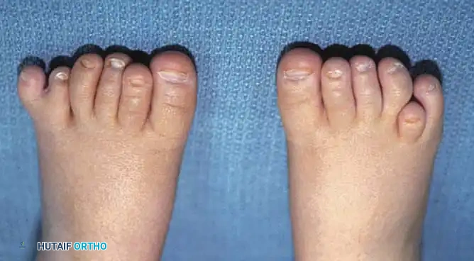
The most frequently encountered congenital anomaly of the forefoot is polydactyly—the presence of supernumerary digits. Polydactyly of the toes is a relatively common congenital deformity, with an overall incidence of approximately two cases per 1,000 live births, though this incidence varies significantly across different ethnic populations. While it may manifest as a component of established genetic syndromes (such as Ellis-van Creveld syndrome, Trisomy 13, Bardet-Biedl syndrome, or McKusick-Kaufman syndrome), it most frequently occurs as an isolated, non-syndromic trait. In its isolated form, pedal polydactyly typically demonstrates an autosomal dominant inheritance pattern with variable phenotypic expression and incomplete penetrance, meaning the severity and exact anatomical configuration can differ markedly even among first-degree relatives.
The embryological insult resulting in polydactyly occurs precisely between the fourth and eighth weeks of gestation during the critical formation of the apical ectodermal ridge (AER) and the zone of polarizing activity (ZPA). The ZPA, located at the posterior margin of the developing limb bud, secretes the Sonic Hedgehog (SHH) morphogen, which establishes a concentration gradient dictating the anteroposterior axis of the limb. Abnormalities in the SHH signaling pathway, or mutations in downstream transcription factors such as GLI3, lead to an ectopic expression of these morphogens, culminating in the aberrant duplication of digital rays. Understanding this precise embryological timeline is critical for the orthopaedic surgeon, as it explains the frequent concomitant presence of other anomalies originating during the same gestational window, such as syndactyly or cleft foot.
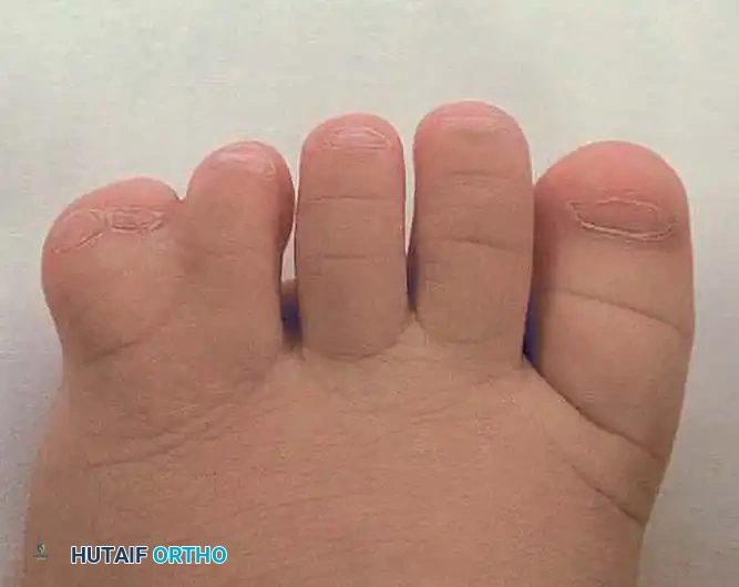
Surgical intervention for these conditions is rarely purely cosmetic; it is fundamentally reconstructive and prophylactic against future morbidity. When evaluating a pediatric patient for forefoot reconstruction, the orthopaedic surgeon must balance three primary objectives: the alleviation of pain, the facilitation of standard shoe wear, and the optimization of cosmesis. A satisfactory clinical result must comprehensively address all three parameters without compromising the biomechanical integrity of the foot. Failure to achieve this balance can result in chronic impingement, adventitial bursitis, and progressive angular deformities that are exponentially more difficult to correct in the older child or adolescent.
Detailed Surgical Anatomy and Biomechanics
The osteology of the forefoot is an intricate arrangement of metatarsals and phalanges designed to distribute weight-bearing forces efficiently during the stance phase of gait. In the normal foot, the metatarsal heads form a transverse arch that flattens dynamically under load, supported by the intrinsic musculature and the plantar aponeurosis. In the setting of polydactyly, the presence of a supernumerary digit disrupts this transverse metatarsal arch and significantly widens the forefoot splay. The accessory digit may present with a variety of skeletal configurations, ranging from a fully formed accessory metatarsal and phalanges to a simple distal phalangeal duplication, or even a "floating" soft tissue appendage devoid of skeletal attachments. These variations fundamentally alter the distribution of forces across the forefoot.
The ligamentous and capsular anatomy of the forefoot is equally complex and critically important during surgical reconstruction. The deep transverse metatarsal ligament (DTML) connects the plantar plates of adjacent metatarsophalangeal (MTP) joints, acting as a primary restraint against excessive forefoot splaying. When an accessory digit is interposed between normal rays, or when an accessory metatarsal is present, the DTML is either stretched, attenuated, or entirely bifurcated. During the surgical excision of a central or postaxial digit, meticulous preservation or reconstruction of the DTML is paramount to restore the normal intermetatarsal distance and prevent postoperative splaying, which would otherwise defeat the purpose of the narrowing procedure.
Musculotendinous anomalies are ubiquitous in pedal polydactyly and pose a significant challenge during reconstruction. The flexor and extensor tendons may bifurcate to insert on both the normal and supernumerary digits, or they may insert aberrantly, creating an imbalance of dynamic forces. This is particularly critical in preaxial polydactyly, where the abductor hallucis tendon—a vital dynamic stabilizer of the medial longitudinal arch and the first MTP joint—may insert onto the duplicated medial digit. If the medial digit is excised without reattaching the abductor hallucis to the retained lateral hallux, the unopposed adductor hallucis will inevitably drive the great toe into a severe, progressive hallux varus deformity.
Biomechanically, the normal foot relies on the windlass mechanism, described by Hicks, wherein dorsiflexion of the toes during terminal stance tightens the plantar aponeurosis, elevating the longitudinal arch and converting the foot into a rigid lever for push-off. Shared tendinous insertions, bifurcated metatarsal heads, or incongruent MTP joints associated with polydactyly can severely alter this mechanism. The altered kinematics lead to a dysfunctional push-off, abnormal pressure distribution beneath the metatarsal heads, and a compensatory gait pattern. Therefore, the surgical goal is not merely the amputation of an extra appendage, but the meticulous restoration of these intricate anatomical and biomechanical relationships to ensure a functional, pain-free gait cycle.
Exhaustive Indications and Contraindications
The decision to proceed with surgical intervention in pediatric pedal polydactyly is guided by a combination of functional, biomechanical, and psychosocial factors. The primary, absolute indication for surgery is the inability to fit the child into standard, commercially available footwear. The widening of the forefoot caused by the supernumerary digit inevitably leads to impingement against the toe box of standard shoes, resulting in painful blistering, adventitial bursa formation, and chronic ulceration. Furthermore, pain associated with impingement or the rubbing of the extra digit against adjacent toes is a compelling indication for surgical excision, as it directly impairs the child's willingness and ability to ambulate normally.
Psychosocial and cosmetic concerns, while often secondary to functional deficits, are highly valid indications for surgery. As children reach school age, peer awareness and body image become critical components of psychological development. Visible congenital deformities can lead to significant emotional distress and social isolation. Therefore, correcting the deformity before the child develops a profound awareness of the anomaly is highly desirable. The optimal timing for this prophylactic reconstruction is typically between 9 and 12 months of age. At this stage, the child is large enough to safely undergo general anesthesia, the anatomical structures are sufficiently developed to allow for meticulous surgical dissection, and the intervention occurs prior to the onset of independent ambulation, thereby preventing the development of compensatory, abnormal gait patterns.
Contraindications to surgical intervention, while rare, must be carefully evaluated. Absolute contraindications include severe, life-limiting medical comorbidities that render general anesthesia an unacceptable risk. In patients with profound syndromic presentations (e.g., severe forms of Trisomy 13) where life expectancy is severely truncated, the risks of elective orthopaedic surgery generally outweigh the benefits. Active local infection or severe vascular compromise of the limb are also absolute contraindications that must be resolved prior to any elective reconstructive procedure.
Relative contraindications require nuanced clinical judgment. In cases of mild postaxial soft-tissue duplication (a "floating" digit) that does not widen the forefoot or impede shoe wear, observation may be a reasonable alternative, though most parents still opt for excision for cosmetic reasons. Additionally, in syndromic patients with severe global developmental delay who are not expected to achieve independent ambulation, the functional indications for surgery are diminished. In such scenarios, the orthopaedic surgeon must engage in extensive, shared decision-making with the multidisciplinary pediatric team and the family to determine if the purely cosmetic and shoe-wear benefits justify the surgical and anesthetic risks.
| Category | Indications for Surgical Intervention | Contraindications to Surgical Intervention |
|---|---|---|
| Functional / Biomechanical | Inability to accommodate standard footwear due to forefoot splay. | Non-ambulatory patient with no shoe-wear difficulties (Relative). |
| Clinical Symptoms | Pain, impingement, blistering, or adventitial bursitis. | Active local or systemic infection (Absolute). |
| Psychosocial / Cosmetic | Prevention of psychosocial distress prior to school age. | Severe, life-limiting syndromic comorbidities (Absolute). |
| Anatomical | Progressive angular deformity of adjacent normal digits. | Severe peripheral vascular compromise (Absolute). |
Pre-Operative Planning, Templating, and Patient Positioning
Clinical assessment must begin with a highly detailed family history and a comprehensive systemic examination to rule out syndromic associations. The orthopaedic surgeon must act as a diagnostician, recognizing that pedal polydactyly may be the sentinel presentation of a broader genetic disorder. The physical examination of the foot must be exhaustive. The surgeon must document the exact number of digits, the presence and extent of any concomitant syndactyly (webbing), the active and passive range of motion of the involved MTP and interphalangeal joints, and the vascular integrity of all toes. Capillary refill and distal pulses must be meticulously assessed, as the vascular supply to the retained digit may be anomalous or shared with the duplicated digit.
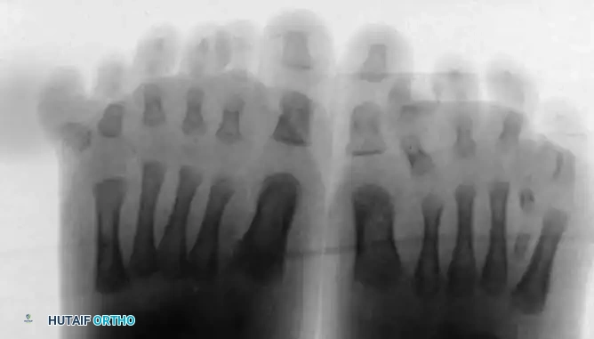
Standard weight-bearing (or simulated weight-bearing in infants) anteroposterior, lateral, and oblique radiographs of the foot are absolutely mandatory. Never proceed with the amputation of a supernumerary digit based on clinical appearance alone. Preoperative radiographs are critical to detect any extra metatarsals, bifurcated metatarsal heads, or shared articular surfaces. The Venn-Watson Classification is utilized here to categorize the pathoanatomy, directing the surgeon's attention to whether the duplication involves a T-shaped metatarsal, a Y-shaped metatarsal, a complete duplication of the ray, or merely soft tissue. Failure to identify and resect an accessory metatarsal will result in a persistent bony prominence, entirely defeating the purpose of the surgery and necessitating complex revision procedures.
Surgical templating involves a strategic decision regarding which digit to resect and which to retain. In postaxial polydactyly, the most lateral digit is typically excised to narrow the foot, provided the retained fifth toe has a congruent MTP articulation and adequate longitudinal alignment. In preaxial polydactyly, the decision is more complex. Usually, the most medial great toe is excised, and the lateral duplicated toe is preserved because it generally maintains a more congruent articulation with the first metatarsal and preserves the critical first web space. However, if the lateral digit is severely dysplastic, the medial digit may be retained, requiring extensive realignment osteotomies. In central duplications with shared articular surfaces, a Bilhaut-Cloquet procedure may be templated, requiring precise geometric calculations to ensure the amalgamated digit matches the contralateral side in girth and length.
Patient positioning and operating room setup are critical for the execution of these delicate pediatric procedures. The patient is placed in the supine position on a standard operating table. A well-padded pediatric calf tourniquet is applied to provide a completely bloodless surgical field, which is absolutely critical for identifying delicate pediatric neurovascular structures, which can be less than a millimeter in diameter. General anesthesia is preferred, supplemented by a regional ankle block or popliteal block administered by the anesthesia team for robust, opioid-sparing postoperative analgesia. The surgeon must utilize loupe magnification (minimum 2.5x to 3.5x) and a dedicated pediatric or hand surgery instrument tray containing fine tenotomy scissors, micro-adson forceps, and miniature osteotomes to ensure tissue handling is atraumatic.
Step-by-Step Surgical Approach and Fixation Technique
Postaxial and Central Polydactyly Excision
The surgical approach for postaxial or central polydactyly begins with meticulous incision design. At the base of the toe selected for amputation, an oval or racquet-shaped incision is designed through the skin and superficial fascia. The handle of the racquet is oriented proximally over the dorsal aspect of the metatarsal to allow for proximal extension if an accessory metatarsal requires resection. A critical clinical pearl is to ensure that the plantar skin flap is designed slightly longer than the dorsal flap. This strategic geometry ensures that the final surgical closure line is positioned dorsally, entirely away from the weight-bearing plantar surface, thereby preventing painful postoperative cicatrix formation.
Once the skin flaps are elevated, deep dissection commences to manage the tendinous structures. The extensor and flexor tendons of the supernumerary digit are identified. Traction is applied to these tendons, drawing them distally as far as possible before they are divided sharply under tension. This maneuver allows the transected tendon ends to retract proximally deep into the soft tissues, effectively preventing postoperative tethering, bowstringing, or the formation of a painful tendinous nodule. Following tendon management, the capsule of the MTP joint is identified and incised transversely. The joint is disarticulated, and the supernumerary phalanges are removed in their entirety, with extreme care taken to protect the neurovascular bundles supplying the adjacent, retained digit.
Bony contouring is the next critical phase. The remaining metatarsal head is inspected; frequently, it will possess a widened, bifurcated, or protruding articular facet that previously supported the amputated phalanx. Using a sharp pediatric osteotome or bone-cutting forceps, this protruding bone is sharply resected. The objective is to create a smooth, anatomically contoured metatarsal head that prevents the formation of a painful adventitial bursa postoperatively. If preoperative radiographs revealed a complete accessory metatarsal, the incision is extended proximally, and a subperiosteal dissection is performed to disarticulate the accessory metatarsal at its tarsometatarsal joint. Leaving a proximal metatarsal stump is a severe technical error, as it will inevitably lead to a painful plantar prominence as the child grows.
Preaxial Polydactyly Reconstruction
Preaxial duplication presents a substantially more complex reconstructive challenge due to the critical biomechanical role of the hallux in weight-bearing and terminal propulsion. In the majority of preaxial polydactyly cases, the most medial great toe is excised. A medial longitudinal or racquet-shaped incision is utilized. During the soft tissue dissection, the abductor hallucis tendon must be meticulously identified and tagged with a non-absorbable suture before the medial digit is disarticulated. Excision of the medial digit inevitably detaches this tendon and compromises the medial collateral ligament of the MTP joint.
Following the excision of the medial digit and the resection of any prominent medial metatarsal condyle, the medial capsule must be robustly imbricated. The critical reconstructive step is the tendon transfer: the tagged abductor hallucis is advanced and securely sutured to the base of the proximal phalanx of the retained lateral hallux. This transfer provides essential dynamic medial stability. Failure to perform this step will result in a progressive, iatrogenic hallux varus deformity driven by the unopposed adductor hallucis. To protect this delicate soft tissue repair, the MTP joint is stabilized with a transarticular Kirschner wire (K-wire), driven antegrade through the phalanx and retrograde into the metatarsal, maintaining the joint in neutral alignment.
Management of Complex Polydactyly and Syndactyly
Occasionally, polydactyly is complicated by concurrent syndactyly (webbing) of the adjacent digits. These combined deformities require highly individualized, complex surgical planning that often blends orthopaedic and plastic surgery principles. In cases of complex polydactyly-syndactyly, simple amputation is vastly insufficient and will result in severe skin deficits. The surgeon must meticulously resect the more peripheral or dysplastic digit while carefully harvesting its skin as a vascularized, full-thickness flap to reconstruct the commissure and provide durable, sensate coverage for the retained digit.
In rare instances of central duplication where the digits are nearly identical in size and share a common, broadened articular surface, a Bilhaut-Cloquet procedure may be indicated. This technically demanding procedure involves the central wedge resection of the duplicated bone and soft tissues from both digits, followed by the precise amalgamation of the two remaining lateral halves to create a single, appropriately sized digit. The primary advantage of this technique is the preservation of the collateral ligaments and neurovascular bundles on the outer margins of the newly constructed digit. However, it carries a high risk of physeal arrest, nail bed deformities, and joint stiffness, requiring exact execution and rigid internal fixation, typically with miniature K-wires or absorbable pins.
Complications, Incidence Rates, and Salvage Management
While the surgical management of pedal polydactyly is generally highly successful when executed with meticulous technique, it carries specific, potentially devastating risks. The most common complication is a residual bony prominence, which occurs in approximately 10% to 15% of cases. This typically results from inadequate resection of a bifurcated metatarsal head, the failure to recognize and excise a complete accessory metatarsal, or the failure to resect the periosteal sleeve, leading to heterotopic ossification. Clinically, this presents as a painful, hard mass that causes recurrent shoe-wear impingement. Salvage management requires a revision surgery to perform a definitive exostectomy or complete ray resection, ensuring the entire anomalous bony and periosteal tissue is eradicated.
Hallux varus is a catastrophic complication following preaxial polydactyly excision, with an incidence rate approaching 20% if the medial soft tissue structures are not adequately reconstructed. The pathomechanics involve the failure to reattach the abductor hallucis tendon and the inadequate imbrication of the medial MTP capsule, allowing the adductor hallucis to pull the great toe into a severe varus alignment. This deformity is not merely cosmetic; it disrupts the windlass mechanism and causes severe impingement. Salvage management is complex and depends on the flexibility of the deformity. Flexible deformities may be salvaged with a medial soft tissue release and a split extensor hallucis brevis tendon transfer. Rigid deformities or those with joint subluxation often require a corrective metatarsal osteotomy or, in severe cases approaching skeletal maturity, an MTP joint arthrodesis.
Neuroma formation and wound complications are less frequent but significant risks. Inadequate proximal resection of the digital nerves during amputation can lead to painful stump neuromas, which present as exquisitely tender nodules at the surgical site. Prevention dictates identifying the digital nerves, applying traction, and sharp transection to allow proximal retraction deep into the intrinsic musculature. Wound dehiscence and infection occur in roughly 2% to 5% of cases, primarily driven by inadequate hemostasis leading to hematoma formation. Meticulous bipolar electrocautery before tourniquet release is essential. Salvage of a neuroma requires surgical excision and burying of the nerve stump into adjacent muscle or bone.
Angular deformities of the retained digits represent a delayed complication, manifesting months or years after the index procedure. These deformities result from iatrogenic damage to the delicate physis of the retained digit during the initial dissection or from asymmetrical tethering by scar tissue. The incidence is difficult to quantify but is minimized by utilizing loupe magnification and tissue-sparing techniques. As the child grows, the physeal injury leads to progressive varus or valgus angulation. Salvage management requires careful longitudinal observation until the deformity causes functional impairment, at which point corrective closing-wedge or opening-wedge phalangeal or metatarsal osteotomies are indicated to restore mechanical alignment.
| Complication | Estimated Incidence | Primary Prevention Strategy | Salvage / Revision Management |
|---|---|---|---|
| Residual Bony Prominence | 10% - 15% | Complete resection of accessory metatarsals/bifurcated heads; excision of periosteum. | Revision exostectomy or complete ray resection. |
| Iatrogenic Hallux Varus | 5% - 20% (Preaxial) | Meticulous reattachment of abductor hallucis; medial capsulorrhaphy; K-wire stabilization. | Soft tissue release, tendon transfer, or MTP arthrodesis. |
| Stump Neuroma | < 5% | Proximal traction and sharp transection of digital nerves to allow retraction. | Surgical excision of neuroma; burying nerve stump into muscle/bone. |
| Progressive Angular Deformity | Variable | Atraumatic dissection; protection of the physis; loupe magnification. | Corrective phalangeal or metatarsal osteotomies. |
Phased Post-Operative Rehabilitation Protocols
The postoperative rehabilitation protocol following polydactyly reconstruction is phased to protect the delicate soft tissue repairs and bony contouring while gradually transitioning the pediatric patient back to normal weight-bearing activities. The immediate postoperative phase (Weeks 0 to 2) prioritizes wound healing, edema control, and strict immobilization. Immediately following skin closure, a bulky, compressive soft dressing is applied. For simple disarticulations in young infants, this may be transitioned to a rigid postoperative shoe. However, for more complex reconstructions, particularly those involving osteotomies, K-wire fixation, or extensive soft tissue transfers (such as in preaxial polydactyly), a well-molded short-leg cast is utilized. The patient is kept strictly non-weight-bearing, which, in the 9 to 12-month-old demographic, primarily involves parental education regarding carrying and limiting floor play.
The intermediate phase (Weeks 2 to 6) marks the transition from strict immobilization to protected weight-bearing. At the 2-week mark, the patient returns to the clinic for a comprehensive wound check and cast change. If absorbable sutures (e.g., 4-0 Vicryl Rapide or chromic gut) were utilized, suture removal is unnecessary, avoiding significant distress for the awake child. If a transarticular K-wire was utilized to stabilize the MTP joint, it remains in place, bent outside the skin, capped, and incorporated into the new short-leg cast. Around 4 to 6 weeks postoperatively, the K-wire is removed in the clinic setting. Radiographs are obtained at this 6-week juncture to confirm the maintenance of joint alignment, the healing of any osteotomies, and the absence of early heterotopic ossification at the resection sites. Once radiographic and clinical stability is confirmed, the child is transitioned to a supportive, commercially available stiff-soled shoe and allowed to bear weight as tolerated.
The late phase (Weeks 6 to 12) focuses on the restoration of normal gait mechanics and the integration of the reconstructed foot into standard footwear. The child is encouraged to resume all normal, age-appropriate activities. Physical therapy is rarely formally required in this age group, as children naturally auto-rehabilitate through play and exploration once pain and mechanical impediments are removed. However, the orthopaedic surgeon must carefully observe the child's gait in the clinic hallway, specifically looking for antalgic patterns, avoidance of terminal stance push-off, or dynamic varus/valgus deviations of the reconstructed digits. Parents are instructed to gradually introduce a variety of shoe types, ensuring the toe box is sufficiently wide to accommodate the foot without causing erythema or pressure points.
Long-term surveillance extends from the 12-week mark until the child reaches skeletal maturity. This extended follow-up is critical because the pediatric foot continues to grow and ossify over the next decade. The surgeon must monitor for the delayed onset of complications, particularly angular deformities secondary to unrecognized physeal injury during the index procedure, or the late manifestation of a residual bony prominence as the cartilaginous anlage ossifies. Annual or biennial clinical evaluations, supplemented by weight-bearing radiographs if clinical suspicion dictates, ensure that the functional and cosmetic goals achieved during the initial reconstruction are maintained throughout the child's development, guaranteeing a durable, lifelong result.
Summary of Landmark Literature and Clinical Guidelines
The surgical management of pedal polydactyly is heavily informed by a foundational body of orthopaedic literature that has evolved from purely descriptive anatomical studies to sophisticated biomechanical and genetic analyses. The cornerstone of clinical evaluation remains the Venn-Watson classification system, published in the mid-20th century. Venn-Watson's meticulous categorization of metatarsal morphology—distinguishing between T-shaped, Y-shaped, and complete duplications—revolutionized preoperative planning. By directing the surgeon's focus away from the visible soft tissue appendage and toward the underlying skeletal architecture, this classification established the modern paradigm that successful polydactyly surgery is fundamentally a bony reconstructive procedure rather than a simple soft-tissue amputation.
In the realm of preaxial polydactyly, the landmark work by Phelps and Grogan fundamentally altered surgical techniques. Prior to their detailed biomechanical studies, the excision of the medial duplicated hallux frequently resulted in devastating iatrogenic hallux varus deformities. Phelps and Grogan elucidated the critical role of the abductor hallucis tendon and the medial MTP capsule. Their published protocols mandating the meticulous tagging, advancement, and reattachment of the abductor hallucis to the retained lateral digit are now considered the absolute standard of care. Their work transitioned preaxial polydactyly surgery from a procedure fraught with high complication rates to a highly reliable, predictable reconstruction.
Modern advancements in molecular biology and genetics have further refined clinical guidelines, particularly regarding the preoperative workup. The identification of the Sonic Hedgehog (SHH) signaling pathway and the GLI3 transcription factor mutations as primary drivers of limb bud duplication has profound clinical implications. Current clinical guidelines, endorsed by pediatric orthopaedic and genetic societies, mandate that isolated pedal polydactyly should prompt a thorough, albeit targeted, syndromic screening. The understanding that polydactyly is a temporal marker for embryological insults occurring between the fourth and eighth weeks of gestation requires the surgeon to maintain a high index of suspicion for concomitant cardiac, renal, or craniofacial anomalies that develop during the same gestational window.
Finally, consensus clinical guidelines regarding the timing of surgical intervention have solidified around the 9 to 12-month age window. This consensus represents a synthesis of anesthetic safety data, anatomical developmental milestones, and psychosocial considerations. Operating prior to the onset of independent ambulation prevents the neuro-motor encoding of compensatory gait patterns and ensures that the child's first steps are taken on a biomechanically sound, plantigrade foot. This modern, multidisciplinary approach—combining strict adherence to anatomical classification, biomechanically sound reconstructive principles, and optimal surgical timing—ensures that the orthopaedic surgeon can reliably deliver a functional, pain-free, and cosmetically acceptable outcome for the pediatric patient.
