العربية
Part of the Master Guide

Arab Board Orthopedic B Review | Dr Hutaif General Orth -...

Orthopedic Board Prep MCQs: High-Yield Trauma & Dislocation Questions

23 Apr 2026 63 min read 128 Views
Mastering General Surgery: Unraveling b c dcooper Ligament Questions

Key Takeaway

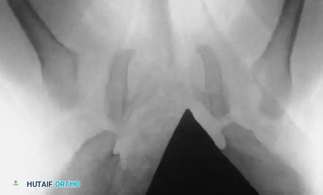
For suspected posterior hip dislocation, after patient stabilization, the initial management involves obtaining an AP pelvis X-ray and a lateral hip X-ray. This is critical *before* emergent closed reduction to rule out associated fractures (e.g., femoral head, acetabular wall), which could complicate reduction or necessitate open reduction, preventing further complications.

Orthopedic Board Prep MCQs: High-Yield Trauma & Dislocation Questions

Comprehensive 100-Question Exam


00:00

Start Quiz

Question 1

An 18-year-old male rugby player is brought to the emergency department after falling on his shoulder during a tackle. He complains of severe pain at the base of his neck, difficulty swallowing, and a feeling of shortness of breath. On examination, the medial end of his right clavicle is not palpable, and there is a visible depression at the sternoclavicular joint. He is hemodynamically stable. What is the most appropriate next step in management?


Explanation

This patient presents with a posterior sternoclavicular (SC) joint dislocation, which is a life-threatening injury due to the proximity of the great vessels, trachea, and esophagus. Because he is hemodynamically stable but symptomatic (dysphagia, dyspnea indicating mediastinal compression), a CT of the chest with IV contrast (or CT angiogram) is the most critical next step. It accurately defines the displacement and evaluates for occult vascular or visceral injuries. Closed reduction should only be attempted after advanced imaging, ideally in the operating room with a cardiothoracic surgeon on standby.

Question 2

A 30-year-old male is brought to the trauma bay after a high-speed motorcycle collision. He has a grossly deformed left knee. Radiographs reveal an anterior knee dislocation. The dislocation is urgently reduced. Post-reduction, he has a palpable dorsalis pedis pulse, but his Ankle-Brachial Index (ABI) is measured at 0.85. What is the most appropriate next step in management?


Explanation

Knee dislocations carry a high risk of popliteal artery injury. Current guidelines recommend measuring the Ankle-Brachial Index (ABI) after reduction of all knee dislocations. If the ABI is >0.9, serial examinations are generally safe. However, an ABI <0.9 or asymmetric pulses indicates a high likelihood of vascular compromise and necessitates advanced imaging, most commonly CT angiography (CTA), to locate the level and extent of the vascular injury before surgical intervention.

Question 3

A 45-year-old female presents after falling on an outstretched hand. Radiographs reveal a posterolateral elbow dislocation, a displaced radial head fracture, and a Type II coronoid fracture. Operative intervention is planned. To optimize biomechanical stability, what is the generally accepted surgical sequence for repairing the 'terrible triad' of the elbow?


Explanation

The 'terrible triad' of the elbow consists of an elbow dislocation, a radial head fracture, and a coronoid process fracture. The standard surgical sequence works deep to superficial and anterior to posterior/lateral: (1) Fixation of the coronoid process to restore anterior stability, (2) Fixation or replacement of the radial head to restore the anterior radiocapitellar buttress, and (3) Repair of the lateral collateral ligament (specifically the lateral ulnar collateral ligament) complex. The MCL is typically only explored if the elbow remains unstable after the first three steps.

Question 4

A 25-year-old male driver involved in a head-on motor vehicle collision presents with severe hip pain. Radiographs reveal a posterior dislocation of the right hip. Prior to reduction, a detailed neurologic exam notes inability to extend the right great toe and decreased sensation over the dorsal first web space. The hip is successfully reduced via closed means under conscious sedation. Post-reduction, the neurologic deficit remains unchanged. What is the most appropriate management of the neurologic deficit?


Explanation

Sciatic nerve injury (most commonly the peroneal division) occurs in 10-20% of posterior hip dislocations. If the neurologic deficit is present BEFORE reduction and persists post-reduction, the standard of care is observation, as the injury is usually a neuropraxia from the initial stretch, and most patients recover spontaneously. An AFO helps prevent equinus contracture and assists with ambulation. Surgical exploration is indicated only if a new nerve palsy develops AFTER reduction (suggesting iatrogenic entrapment of the nerve or a bone fragment) or if an incarcerated fragment is seen on post-reduction CT.

Question 5

A patient sustains a posterior hip dislocation with an associated fracture of the femoral head. CT scan reveals that the fracture involves the portion of the femoral head inferior to the fovea capitis. According to the Pipkin classification, what type of fracture is this?


Explanation

The Pipkin classification describes femoral head fractures associated with posterior hip dislocations. Type I is a fracture of the femoral head inferior to the fovea capitis (non-weight-bearing portion). Type II is a fracture superior to the fovea capitis (weight-bearing portion). Type III is a Type I or II fracture associated with a femoral neck fracture. Type IV is a Type I or II fracture associated with an acetabular rim fracture.

Question 6

A 22-year-old male athlete presents with recurrent anterior shoulder dislocations. Pre-operative imaging and 3D CT reconstruction indicate an engaging Hill-Sachs lesion and a 26% anterior glenoid bone loss. What is the most appropriate definitive surgical management?


Explanation

In the setting of recurrent anterior shoulder instability with significant anterior glenoid bone loss (>20-25%), soft tissue stabilization alone (Bankart repair) is associated with an unacceptably high failure rate. Bony augmentation is required. The Latarjet procedure (transfer of the coracoid process with the attached conjoint tendon to the anterior glenoid) provides a 'triple blocking' effect (bone, sling, and capsule) and is the gold standard for significant glenoid bone loss.

Question 7

A 30-year-old construction worker falls from scaffolding, landing on his extended, ulnar-deviated wrist. Lateral radiographs of the wrist demonstrate that the lunate maintains its normal articulation with the distal radius, but the capitate is dorsally displaced relative to the lunate. What is the most likely diagnosis?


Explanation

This describes a perilunate dislocation. On a lateral radiograph, the lunate maintains its 'teacup' articulation with the distal radius, but the capitate is dislocated dorsally out of the teacup. In contrast, a lunate dislocation (the end stage of perilunate instability) occurs when the lunate is tipped off the radius (volarly), appearing like a 'spilled teacup,' while the capitate remains aligned with the radius.

Question 8

A 60-year-old male slips on ice and grabs a railing to break his fall, sustaining a forceful hyperabduction injury to his shoulder. He presents to the ER with his arm locked in 120 degrees of abduction and his elbow flexed, with his hand resting near his head. What is the most commonly associated nerve injury with this specific type of dislocation?


Explanation

The clinical presentation (arm locked in extreme abduction/flexion) is pathognomonic for luxatio erecta (inferior shoulder dislocation). This severe injury has a high rate of associated complications. The axillary nerve is the most commonly injured nerve (up to 60% of cases) due to traction as the humeral head is forced inferiorly into the axilla. There is also a significant risk of axillary artery injury and massive rotator cuff tears.

Question 9

A 25-year-old cyclist is struck by a vehicle and lands directly on the acromion of his shoulder. Radiographs reveal an acromioclavicular (AC) joint injury. The distal clavicle is displaced 200% superiorly relative to the acromion, and the coracoclavicular distance is more than double the contralateral side. According to the Rockwood classification, what type of injury is this?


Explanation

The Rockwood classification of AC joint injuries is based on the direction and degree of clavicular displacement. Type III involves 25-100% superior displacement. Type V involves 100-300% superior displacement of the distal clavicle, accompanied by severe disruption of the deltotrapezial fascia. Type IV is posterior displacement into or through the trapezius. Type VI is inferior displacement (subcoracoid or subacromial).

Question 10

A 22-year-old collegiate football player experiences a severe axial load on a plantarflexed foot. He complains of intense midfoot pain and inability to bear weight. An anteroposterior (AP) radiograph demonstrates the 'fleck sign' in the first intermetatarsal space. This bony avulsion historically represents the attachment site of the Lisfranc ligament to which of the following structures?


Explanation

The Lisfranc ligament is a stout, interosseous ligament that connects the lateral aspect of the medial cuneiform to the medial aspect of the base of the second metatarsal. There is no ligamentous connection between the first and second metatarsals. The 'fleck sign' is pathognomonic for a Lisfranc injury and represents a bony avulsion fracture typically from the base of the second metatarsal, though it can occasionally avulse from the medial cuneiform.

Question 11

A 35-year-old male sustains a high-energy traumatic knee dislocation. On examination in the trauma bay, the knee is locked in a slightly flexed position, and there is a distinct transverse furrow or 'puckering' of the skin over the medial joint line (the 'dimple sign'). An attempted closed reduction is unsuccessful. What anatomic structure is primarily responsible for blocking the reduction?


Explanation

The 'dimple sign' represents a posterolateral knee dislocation in which the medial femoral condyle buttonholes through the anterior medial capsule and retinaculum. The intact capsule prevents the condyle from returning to its anatomical position, rendering the dislocation irreducible by closed means. It requires immediate open reduction. Applying excessive force during closed reduction attempts can cause skin necrosis or further soft tissue damage.

Question 12

A 28-year-old male sustains a lateral subtalar dislocation after a severe inversion and plantarflexion injury. Attempted closed reduction in the emergency department is unsuccessful, requiring operative intervention. What is the most common anatomical structure that blocks the reduction of a lateral subtalar dislocation?


Explanation

Subtalar dislocations are classified by the direction the foot moves relative to the talus. Medial subtalar dislocations are the most common (up to 80%) and when irreducible, are typically blocked by the extensor retinaculum, extensor digitorum brevis, or talonavicular capsule. Lateral subtalar dislocations (where the foot moves laterally) are less common but are frequently irreducible because the posterior tibial tendon (PTT) becomes interposed in the talonavicular joint, blocking reduction.

Question 13

A 40-year-old female presents after falling from a height. She sustains a comminuted, unsalvageable radial head fracture, a longitudinal tear of the interosseous membrane, and dislocation of the distal radioulnar joint (DRUJ). Which of the following is the most appropriate management of the proximal radius in this specific clinical entity?


Explanation

This patient has an Essex-Lopresti fracture-dislocation (radial head fracture, interosseous membrane disruption, and DRUJ dislocation). Because the interosseous membrane is torn, the radiocapitellar joint becomes the primary restraint to proximal migration of the radius. Therefore, excision of the radial head is absolutely contraindicated, as it will lead to rapid proximal migration of the radius and ulnocarpal impaction. Immediate radial head arthroplasty is essential to restore longitudinal stability to the forearm.

Question 14

A 6-year-old boy sustains a traumatic posterior hip dislocation while playing. What is the most critical modifiable factor in preventing the development of avascular necrosis (AVN) of the femoral head in this patient?


Explanation

Avascular necrosis (AVN) of the femoral head is the most devastating complication of traumatic hip dislocations in pediatric patients. The single most important modifiable factor to reduce the incidence of AVN is the time to reduction. Reduction within 6 hours significantly lowers the risk of AVN. While age and energy mechanism affect risk, they are not modifiable. Immobilization modality does not directly alter AVN rates.

Question 15

A 30-year-old male is brought to the trauma center after a diving accident. He is intubated, sedated, and paralyzed on arrival. Lateral cervical radiographs reveal a bilateral facet dislocation at C5-C6 with 50% anterior subluxation. His hemodynamic status is stable. What is the most appropriate next step in the management of his cervical spine injury?


Explanation

In a patient with a cervical facet dislocation who cannot participate in a reliable neurologic examination (e.g., intubated, comatose, or paralyzed), an MRI of the cervical spine must be obtained before any reduction attempts (closed or open). This is to evaluate for a concurrent cervical disc herniation, which occurs in up to 50% of facet dislocations. Reducing the spine without removing a herniated disc can draw the disc material into the spinal canal, leading to an iatrogenic spinal cord injury. If the patient were awake and cooperative, an awake closed traction reduction with serial neurologic exams would be indicated.

Question 16

A 45-year-old male sustains a 'floating shoulder' injury (ipsilateral midshaft clavicle fracture and scapular neck fracture) following a motorcycle collision. Which of the following radiographic parameters is the most recognized indication for operative fixation of this injury?


Explanation

A 'floating shoulder' results from ipsilateral fractures of the clavicle and scapular neck, disrupting the superior shoulder suspensory complex. While many can be treated non-operatively, significant displacement warrants surgery to prevent long-term shoulder dysfunction. The glenopolar angle (GPA) assesses the rotational displacement of the glenoid. A normal GPA is 30-45 degrees. A GPA of less than 20-22 degrees indicates severe inferomedial tilt of the glenoid and is a strong indication for surgical fixation (usually starting with the clavicle).

Question 17

A 16-year-old female dancer experiences a primary lateral patellar dislocation. Which of the following ligamentous structures is most likely to be injured and represents the primary soft-tissue restraint to lateral patellar translation at 20 degrees of knee flexion?


Explanation

The medial patellofemoral ligament (MPFL) is the primary static stabilizer against lateral patellar translation, providing 50-60% of the restraining force, particularly in early flexion (0-30 degrees) before the patella engages fully in the trochlear groove. It is torn in nearly 100% of acute lateral patellar dislocations, typically avulsing from its femoral attachment (near the adductor tubercle) or its patellar attachment.

Question 18

A 28-year-old male presents with chronic wrist pain and a 'clunking' sensation. Radiographs demonstrate a 'Terry Thomas' sign with a widened scapholunate interval. If isolated repair is considered, which portion of the scapholunate interosseous ligament (SLIL) is biomechanically the most critical for carpal stability?


Explanation

The scapholunate interosseous ligament (SLIL) is composed of three distinct anatomical zones: dorsal, proximal (membranous), and volar. The dorsal portion is the thickest, strongest, and biomechanically most important restraint against scapholunate dissociation. In contrast, for the lunotriquetral (LT) ligament, the volar portion is the thickest and most critical for stability.

Question 19

A 32-year-old male sustains a Galeazzi fracture-dislocation. He undergoes open reduction and internal fixation of the radial shaft. Intra-operatively, the distal radioulnar joint (DRUJ) remains unstable in supination. Which of the following characteristics of the radius fracture most significantly increases the risk of post-fixation DRUJ instability?


Explanation

Galeazzi fractures are fractures of the distal third of the radial shaft with associated disruption of the DRUJ. Bado and others have classified these based on distance from the articular surface. Fractures occurring within 7.5 cm of the distal radial articular surface (Type I) have a significantly higher rate of DRUJ instability (up to 55%) following anatomic fixation of the radius compared to fractures >7.5 cm proximal to the joint (Type II, ~6% instability rate).

Question 20

A 24-year-old gymnast sustains a rare pure tibiotalar (ankle) dislocation without any associated fractures of the malleoli or talus. What is the most common direction of a pure ankle dislocation, and what specific foot positioning during the traumatic axial load strongly predisposes to this injury pattern?


Explanation

Pure tibiotalar dislocations (without fracture) are exceedingly rare due to the inherent bony stability of the ankle mortise. The most common direction for a pure ankle dislocation is posteromedial. This injury classically occurs from a high-energy axial load applied to a foot that is maximally plantarflexed and inverted. In this position, the narrow posterior portion of the talar dome is positioned in the mortise, rendering the joint anatomically at its least stable state.

Question 21

An 18-year-old male rugby player presents with anterior chest pain, dysphagia, and a sensation of choking after a direct blow to the anteromedial shoulder. Clinical examination reveals the arm is held in an adducted and flexed position, and there is a palpable void adjacent to the sternum. Based on the patient's age and clinical presentation, what is the most likely true anatomical pathology?


Explanation

The medial clavicular physis is the last physis in the body to fuse, typically closing between the ages of 20 and 25. Therefore, injuries in patients under age 25 that clinically appear to be sternoclavicular dislocations are almost always Salter-Harris fractures through the medial physis. Posterior displacement is a medical emergency due to the proximity of the trachea, esophagus, and great vessels, requiring reduction in the operating room with thoracic surgery standby.

Question 22

A 32-year-old male is brought to the trauma bay after a high-speed motor vehicle collision. He sustained an obvious left knee dislocation that was immediately reduced in the emergency department. Post-reduction, he has palpable and symmetric dorsalis pedis and posterior tibial pulses. The Ankle-Brachial Index (ABI) on the affected limb is 0.85. What is the most appropriate next step in management?


Explanation

In the setting of a knee dislocation, vascular injury (specifically to the popliteal artery) is a major concern. Even in the presence of palpable pulses, an ABI of less than 0.90 is highly sensitive for arterial injury and mandates further advanced vascular imaging, most commonly a CT angiogram. Immediate surgical exploration is reserved for hard signs of vascular injury (e.g., absent pulses, expanding hematoma, pulsatile bleeding).

Question 23

A 30-year-old female sustains a lateral subtalar dislocation after a fall from a height. Closed reduction under conscious sedation in the emergency department is unsuccessful. Which of the following anatomical structures is most likely interposing and preventing closed reduction?


Explanation

Subtalar dislocations are classified by the direction of the foot relative to the talus. Medial dislocations are more common and, when irreducible, are typically blocked by the extensor retinaculum, extensor digitorum brevis, or the talonavicular joint capsule. Lateral dislocations, though less common, are more likely to be irreducible and are classically blocked by the interposition of the posterior tibial tendon.

Question 24

A 45-year-old male sustains a 'terrible triad' injury of the elbow (elbow dislocation, radial head fracture, and coronoid fracture). The coronoid fracture is a Type II according to Regan and Morrey, and the radial head is comminuted. When proceeding with operative management, what is the classic and most biomechanically sound sequence of reconstruction?


Explanation

The classic sequence for reconstructing a terrible triad injury of the elbow proceeds from deep to superficial and medial to lateral: 1) Fixation of the coronoid process, 2) Repair or replacement of the radial head, and 3) Repair of the lateral collateral ligament complex (LUCL). The medial collateral ligament is only repaired if the elbow remains unstable after the primary triad of structures is restored.

Question 25

According to the Mayfield classification of progressive perilunate instability, Stage III represents the disruption of which of the following ligaments?


Explanation

Mayfield described a progressive, four-stage sequence of perilunate instability based on a perilunar pattern of energy transmission. Stage I is scapholunate dissociation; Stage II involves the capitolunate articulation with disruption through the space of Poirier; Stage III is disruption of the lunotriquetral articulation (perilunate dislocation); Stage IV represents failure of the dorsal radiocarpal ligament, allowing the lunate to dislocate completely (usually volarly into the carpal tunnel).

Question 26

A 35-year-old male is brought in following a severe motor vehicle collision. He is intubated, sedated, and obtunded upon arrival. Radiographs and CT of the cervical spine reveal a bilateral facet dislocation at C5-C6. What is the most appropriate next step in the management of his cervical spine injury prior to attempted reduction?


Explanation

In an obtunded or unexaminable patient with a cervical facet dislocation, an MRI of the cervical spine is mandatory prior to any reduction maneuvers (closed or open). This is to evaluate for a herniated cervical disc, which could be drawn into the spinal canal during reduction, leading to catastrophic spinal cord transection. Awake, neurologically intact patients can undergo closed traction reduction prior to MRI.

Question 27

A 24-year-old male manual laborer presents with recurrent anterior shoulder instability. A 3D CT scan reveals 28% glenoid bone loss and a large, engaging Hill-Sachs lesion. Which of the following surgical procedures is the most appropriate definitive management?


Explanation

Critical glenoid bone loss in anterior shoulder instability is generally accepted to be >20-25%. Soft tissue procedures (Bankart repair, with or without remplissage) have a high failure rate when significant glenoid bone loss is present. The Latarjet procedure (transfer of the coracoid process to the anterior glenoid) provides both a bony block and a dynamic sling effect (via the conjoined tendon) and is the gold standard for instability with >25% glenoid bone loss.

Question 28

A 6-year-old boy presents with a Bado Type I Monteggia fracture-dislocation (anterior dislocation of the radial head with fracture of the ulnar diaphysis). In the operating room, an anatomic closed reduction of the ulnar shaft is achieved and confirmed under fluoroscopy; however, the radial head remains persistently dislocated. What is the most likely cause of this persistent radial head dislocation?


Explanation

The most common overall cause of persistent radial head dislocation in a Monteggia injury is failure to achieve an anatomic reduction of the ulna (ulnar malreduction or length discrepancy). However, the question specifies that anatomic reduction of the ulna was achieved. When the ulna is perfectly out-to-length and the radial head still will not reduce, the most common blocking structure is interposition of the torn annular ligament or joint capsule.

Question 29

A 40-year-old male is involved in a dashboard injury and sustains a posterior hip dislocation. Post-reduction CT scan reveals a fracture of the femoral head with the fracture line extending cephalad to the fovea capitis, with a 2.5 mm step-off of the articular surface. According to the Pipkin classification, what is the stage and appropriate management?


Explanation

Pipkin classification defines femoral head fractures associated with posterior hip dislocations. Pipkin I fractures occur caudad to the fovea capitis (non-weight bearing portion); Pipkin II fractures occur cephalad to the fovea capitis (weight-bearing portion). A step-off of >1 mm in a Pipkin II fracture necessitates Open Reduction and Internal Fixation (ORIF) to restore joint congruity and reduce post-traumatic arthritis risk. Pipkin III includes a femoral neck fracture, and Pipkin IV includes an acetabular fracture.

Question 30

A 50-year-old male arrives at the trauma bay in hemorrhagic shock following an anteroposterior compression (APC III) pelvic ring injury. Emergency medical services placed a commercial pelvic binder in the field. Upon evaluation, to maximize the mechanical closure of the pelvic ring and tamponade the presacral venous plexus bleeding, the pelvic binder must be accurately centered over which anatomic landmark?


Explanation

Pelvic binders are critical in the acute management of mechanically unstable, open-book pelvic ring injuries (APC II/III) to reduce pelvic volume and tamponade venous bleeding. To be effective, the binder must be placed directly over the greater trochanters. Placement too high (e.g., over the iliac crests) is a common error and may paradoxically open the pelvis further or fail to reduce the volume adequately.

Question 31

In multiligamentous knee injuries (knee dislocations), the popliteal artery is at extremely high risk for intimal tear or transection due to its anatomic tethering points. Which of the following correctly identifies the proximal and distal tethering sites of the popliteal artery?


Explanation

The popliteal artery is highly susceptible to traction injury during knee dislocations because it is firmly fixed anatomically. Proximally, it is tethered as it exits the adductor hiatus (opening in the adductor magnus). Distally, it is tethered as it passes deep to the tendinous arch of the soleus muscle (soleal arch) before bifurcating.

Question 32

A 26-year-old male boxer sustains a Bennett fracture-dislocation of the thumb base. The main metacarpal shaft is displaced proximally, dorsally, and radially by the deforming pull of the abductor pollicis longus (APL). However, a small volar-ulnar beak fragment remains anatomically located. Which ligament maintains the position of this volar-ulnar fragment?


Explanation

In a Bennett fracture, the shaft of the first metacarpal is dislocated proximally, dorsally, and radially by the pull of the APL, extensor pollicis longus, and extensor pollicis brevis. The small volar-ulnar lip fragment remains in its anatomic position, secured to the trapezium by the strong anterior oblique ligament (AOL).

Question 33

A 30-year-old male with a comminuted tibial shaft fracture complains of severe, unrelenting pain out of proportion to the injury. His blood pressure is 105/65 mmHg. Intracompartmental pressure testing of the anterior compartment yields a value of 40 mmHg. What is the calculated Delta P, and what is the most appropriate management?


Explanation

Delta P is calculated as Diastolic Blood Pressure minus Intracompartmental Pressure (Delta P = DBP - ICP). In this patient, 65 mmHg - 40 mmHg = 25 mmHg. A Delta P of less than 30 mmHg is an absolute indication for immediate four-compartment fasciotomy of the lower leg to prevent irreversible muscle and nerve necrosis associated with acute compartment syndrome.

Question 34

A 45-year-old male sustains a twisting injury to his midfoot. Anteroposterior radiographs demonstrate a 'fleck sign' in the first intermetatarsal space. This pathognomonic finding represents an avulsion of the Lisfranc ligament. What are the correct anatomical attachment sites of the intact Lisfranc ligament?


Explanation

The Lisfranc ligament is a large, strong interosseous ligament that connects the lateral aspect of the medial cuneiform to the medial base of the second metatarsal. It is the primary stabilizer of the midfoot arch, as there is no intermetatarsal ligament connecting the first and second metatarsal bases. The 'fleck sign' represents an avulsion fracture of this ligament's attachment, most commonly off the base of the second metatarsal.

Question 35

During intraoperative evaluation of ankle syndesmotic instability (the 'Cotton test'), a lateral force is applied to the fibula using a bone hook. Which of the following ligaments provides the primary resistance to lateral displacement of the fibula, functioning as the strongest component of the syndesmotic complex?


Explanation

The syndesmotic complex consists of the AITFL, PITFL, interosseous ligament/membrane, and the transverse ligament. Biomechanical studies have demonstrated that the posterior inferior tibiofibular ligament (PITFL) is the strongest component of the syndesmosis, contributing approximately 42% of the total resistance to lateral fibular displacement.

Question 36

A 25-year-old male is evaluated in the trauma bay after a high-speed rollover collision. Lateral cervical spine radiography reveals a Basion-Dental Interval (BDI) of 14 mm. Based on this diagnosis, which of the following interventions is strictly CONTRAINDICATED in the immediate management of this patient?


Explanation

A Basion-Dental Interval (BDI) > 12 mm is diagnostic of occipitocervical dissociation (craniocervical dissociation), a highly unstable injury disrupting the tectorial membrane and alar ligaments. Cervical traction is absolutely contraindicated as it can cause fatal over-distraction, leading to severe brainstem and spinal cord stretch injuries. Initial management includes rigid cervical collar immobilization followed by definitive occipitocervical fusion.

Question 37

A 35-year-old motorcycle accident victim presents with a flail upper extremity, ipsilateral clavicle fracture, and absent radial pulse. You suspect scapulothoracic dissociation. Which of the following radiographic measurements is most reliably used to diagnose this condition on a non-rotated AP chest radiograph?


Explanation

Scapulothoracic dissociation is defined by complete disruption of the scapulothoracic articulation with lateral displacement of the scapula, often accompanied by severe brachial plexus and subclavian vessel injury. The radiographic diagnosis is made using the scapular index, which is the ratio of the distance from the thoracic midline to the medial border of the scapula on the injured side compared to the normal side. A ratio of > 1.07 is highly specific for the diagnosis.

Question 38

A 16-year-old female sustains a first-time lateral patellar dislocation, which reduces spontaneously. Subsequent MRI evaluates the medial patellofemoral ligament (MPFL). Where is the most common anatomic location of an MPFL tear in the setting of acute patellar dislocation?


Explanation

The Medial Patellofemoral Ligament (MPFL) is the primary restraint to lateral patellar translation at 0-30 degrees of flexion. In an acute lateral patellar dislocation, the MPFL tears in >90% of cases. The most common location of the tear is at its femoral origin (near Schöttle's point, situated between the medial epicondyle and adductor tubercle).

Question 39

A 35-year-old construction worker is extricated after being trapped beneath concrete rubble for 8 hours. He has bilateral severe crush injuries to the lower extremities. Laboratory tests reveal significant myoglobinuria. Alongside aggressive isotonic intravenous fluid resuscitation, which of the following medications is most appropriate to specifically prevent the precipitation of myoglobin in the renal tubules?


Explanation

In crush syndrome, massive muscle breakdown leads to rhabdomyolysis and myoglobinuria. Myoglobin is highly nephrotoxic, particularly in acidic environments where it precipitates and causes acute tubular necrosis. Aggressive IV hydration is the primary treatment, but the addition of IV sodium bicarbonate is recommended to alkalinize the urine (target pH > 6.5), which significantly prevents myoglobin precipitation. Calcium, insulin/dextrose, and sodium polystyrene sulfonate are used to treat hyperkalemia, not specifically to protect renal tubules from myoglobin.

Question 40

A 28-year-old female sustains a Galeazzi fracture-dislocation. After Open Reduction and Internal Fixation (ORIF) of the radial shaft, the distal radioulnar joint (DRUJ) is noted to be unstable dorsally when evaluated. In what forearm position should the arm be splinted postoperatively to maximize DRUJ stability, and what anatomical structure is primarily tensioned in this position?


Explanation

In a Galeazzi fracture, most DRUJ dislocations are volar (ulnar head is volar to the radius) and are most stable in supination. Supination confers stability by moving the radius relative to the ulna to reduce the joint, tensioning the intact palmar radioulnar ligament, and relaxing the deforming force of the pronator quadratus. Note: If the ulnar head dislocates dorsally, stability is usually achieved in pronation. However, standard teaching states that DRUJ volar dislocations (classic Galeazzi) require immobilization in supination to tighten the palmar radioulnar ligament.

Question 41

A 25-year-old male sustains a multiligamentous knee injury following a high-speed motorcycle collision. His knee was visibly dislocated at the scene but reduced by paramedics. In the emergency department, his Ankle-Brachial Index (ABI) is measured at 0.85. His foot is warm and well-perfused, and he has palpable but slightly diminished distal pulses compared to the contralateral side. What is the most appropriate next step in management?


Explanation

An Ankle-Brachial Index (ABI) < 0.90 or asymmetry in pulses after a knee dislocation is highly suspicious for a vascular injury. The current standard of care dictates that patients with an ABI < 0.90 should undergo advanced imaging, typically CT angiography (CTA), to delineate the vascular anatomy and rule out intimal tears or occlusions. Immediate surgical exploration is reserved for 'hard signs' of arterial injury (e.g., active pulsatile bleeding, expanding hematoma, absent pulse with ischemia). If the ABI is > 0.90, serial observation is appropriate.

Question 42

A 38-year-old female presents in hemorrhagic shock following a crush injury to the pelvis. Radiographs demonstrate a vertical shear pelvic ring disruption with marked displacement of the sacroiliac joint. Despite the application of a pelvic binder and massive transfusion protocol, she remains hemodynamically unstable. If arterial bleeding is contributing to her shock, which artery is most likely injured in the posterior aspect of this pelvic ring disruption?


Explanation

While venous bleeding (from the presacral venous plexus) and cancellous bone bleeding are the most common sources of hemorrhage in pelvic fractures overall, arterial bleeding can be catastrophic. The superior gluteal artery is the most frequently injured artery in posterior pelvic ring disruptions, particularly those involving sacral fractures or major sacroiliac joint disruptions. The internal pudendal and obturator arteries are more commonly injured in anterior ring disruptions (e.g., rami fractures).

Question 43

A 30-year-old man falls from a height and sustains a Hawkins Type III fracture of the talar neck. Which of the following accurately describes the displacement pattern and the approximate associated risk of avascular necrosis (AVN) of the talar body?


Explanation

The Hawkins classification describes talar neck fractures. Type I is non-displaced (0-15% AVN risk). Type II involves subluxation or dislocation of the subtalar joint (20-50% AVN risk). Type III involves dislocation of both the subtalar and tibiotalar joints, and the risk of AVN is exceedingly high, approaching 90-100% due to the disruption of the major blood supply to the talar body (artery of the tarsal canal, deltoid artery, and superior capsular network). Type IV adds talonavicular dislocation.

Question 44

When evaluating a proximal humerus fracture for the risk of developing avascular necrosis (AVN) of the humeral head, Hertel described specific radiographic criteria that predict ischemia. Which of the following findings is the most reliable predictor of subsequent ischemia?


Explanation

Hertel's criteria for predicting ischemia in proximal humerus fractures identified that a calcar length (metaphyseal extension of the humeral head fragment) of less than 8 mm, combined with disruption of the medial hinge, are the most reliable predictors of humeral head ischemia and subsequent AVN. A calcar length > 8 mm and an intact medial hinge are protective, as they preserve the intraosseous blood supply from the anterior and posterior circumflex humeral arteries.

Question 45

Surgical management of a 'Terrible Triad' injury of the elbow (elbow dislocation, radial head fracture, coronoid fracture) typically follows a systematic approach to restore joint stability. What is the standard, most widely accepted sequence of structural repair/fixation during the operation?


Explanation

The standard surgical algorithm for a terrible triad injury utilizes a deep-to-superficial (inside-out) approach. Fixation begins with the deepest anterior structure, the coronoid (or anterior capsule repair if the fragment is too small). Next, the radial head is either fixed (ORIF) or replaced, restoring the anterior/lateral bony column. Finally, the lateral collateral ligament (LCL) complex is repaired to the lateral epicondyle. MCL repair or cross-pinning/hinged ex-fix is reserved for residual instability after the primary lateral-sided and bony repairs.

Question 46

A 45-year-old female falls on an outstretched hand and sustains a capitellum fracture. CT imaging demonstrates a coronal shear fracture that involves the capitellum and the lateral ridge of the trochlea, with extensive posterior articular comminution. Based on the Dubberley classification, this is a Type 3B fracture. What does the 'B' designation specifically indicate in this classification?


Explanation

The Dubberley classification of capitellar/trochlear shear fractures categorizes based on anatomical involvement: Type 1 (capitellum only), Type 2 (capitellum + lateral trochlear ridge), Type 3 (capitellum + entire trochlea). The modifiers 'A' and 'B' refer to the absence or presence of posterior radiocapitellar comminution, respectively. A Type 'B' fracture is critical because posterior comminution means there is no posterior buttress, making isolated anterior-to-posterior screw fixation prone to failure; these often require a posterior surgical approach and structural grafting or arthroplasty.

Question 47

A 28-year-old man sustains an isolated, closed medial subtalar dislocation while playing basketball. The head of the talus is palpable laterally. An attempt at closed reduction in the emergency department is unsuccessful. Which anatomic structure is most commonly interposed and blocking reduction in a medial subtalar dislocation?


Explanation

Subtalar dislocations are most commonly medial (85%), where the foot is displaced medially and the talar head is prominent laterally. Irreducible medial dislocations are typically blocked by lateral structures that become interposed over the talar head, most commonly the extensor digitorum brevis (EDB) muscle, the extensor retinaculum, or the talonavicular joint capsule. In contrast, lateral subtalar dislocations are often blocked by medial structures, specifically the posterior tibial tendon (PTT) or the flexor hallucis longus (FHL).

Question 48

A 25-year-old male is admitted to the ICU with a severely comminuted tibia fracture. He is sedated and intubated. An intracompartmental pressure monitor is placed in the anterior compartment of his leg, yielding a pressure of 45 mmHg. His systemic blood pressure is 110/65 mmHg. What is the calculated Delta P, and what is the indicated management?


Explanation

Acute compartment syndrome is a surgical emergency. The definitive diagnostic metric is the Delta P, which is calculated as the Diastolic Blood Pressure minus the Intracompartmental Pressure. In this case, 65 mmHg - 45 mmHg = 20 mmHg. A Delta P of less than 30 mmHg (some sources cite <20-30 mmHg) is widely accepted as an absolute indication for emergent fasciotomy due to inadequate tissue perfusion. Since the Delta P is 20 mmHg, immediate four-compartment fasciotomy is indicated.

Question 49

During the surgical planning for a complex pilon fracture, the surgeon identifies an avulsed bone fragment from the anterolateral aspect of the distal tibia. This is classically known as the Chaput fragment. Which syndesmotic ligament is attached to this specific fragment?


Explanation

The Chaput fragment is an avulsion of the anterolateral distal tibia, which serves as the tibial attachment site for the Anterior Inferior Tibiofibular Ligament (AITFL). Correspondingly, the fibular avulsion of the AITFL is the Wagstaffe-Le Fort fragment. The Volkmann fragment is the posterolateral distal tibia avulsion, where the Posterior Inferior Tibiofibular Ligament (PITFL) attaches.

Question 50

A 35-year-old male sustains a posterior wall acetabular fracture following a motor vehicle collision. The hip was reduced in the ED. Which of the following radiographic or intraoperative findings is considered an absolute indication for operative fixation (ORIF) of the posterior wall?


Explanation

Absolute indications for ORIF of a posterior wall acetabular fracture include hip joint instability (often defined as >20-50% wall involvement, or dynamic instability on Exam Under Anesthesia), an irreducible fracture-dislocation, intra-articular incarcerated fragments, and marginal impaction of the articular surface. Marginal impaction cannot be reduced closed and requires surgical elevation, bone grafting, and fixation to restore the articular congruity and prevent rapid post-traumatic arthrosis. A wall size <20% is typically stable and can often be treated non-operatively if congruent.

Question 51

A 27-year-old construction worker sustains a Galeazzi fracture (fracture of the distal third of the radial shaft with associated distal radioulnar joint (DRUJ) disruption). Following anatomic open reduction and internal fixation of the radius with a volar plate, the surgeon must assess the DRUJ. Which fracture characteristic is most predictive of persistent DRUJ instability requiring intraoperative stabilization?


Explanation

Galeazzi fractures located within 7.5 cm of the radiocarpal joint have a significantly higher rate of persistent DRUJ instability following anatomic fixation of the radius compared to those located > 7.5 cm proximal. This proximity to the joint is strongly associated with severe disruption of the triangular fibrocartilage complex (TFCC) and supporting radioulnar ligaments, often necessitating DRUJ pinning or direct TFCC repair.

Question 52

A 6-year-old boy presents with a displaced Gartland Type III supracondylar humerus fracture. On initial presentation, his hand is pink, but the radial pulse is absent. The patient is taken emergently to the OR. After anatomic closed reduction and percutaneous pinning, the hand remains pink, warm, and well-perfused (capillary refill < 2 seconds), but the radial pulse remains absent by Doppler. What is the most appropriate next step in management?


Explanation

A 'pulseless, pink' hand following reduction and pinning of a pediatric supracondylar humerus fracture suggests adequate collateral circulation despite probable brachial artery spasm, kinking, or intimal injury. Current guidelines recommend close observation and hospital admission for a pulseless but well-perfused (pink) hand after definitive fracture stabilization. Routine vascular exploration is not indicated unless the hand becomes poorly perfused (white, cool, capillary refill > 3 seconds), in which case open exploration of the artery would be warranted.

Question 53

A 19-year-old male football player sustains a traumatic posterior sternoclavicular (SC) joint dislocation. He complains of dysphagia, neck pressure, and mild shortness of breath. Closed reduction is planned in the operating room. What is the most critical logistical preparatory step prior to attempting the closed reduction?


Explanation

Posterior sternoclavicular dislocations are orthopedic emergencies due to the proximity of critical mediastinal structures, including the trachea, esophagus, and great vessels (subclavian artery/vein, brachiocephalic vein). Closed reduction carries a risk of catastrophic vascular injury or displacement if a great vessel has been compromised or pseudoaneurysm is present. Therefore, it is absolutely critical to have a thoracic surgeon or vascular surgeon present or immediately available in the operating room before attempting reduction.

Question 54

According to Mayfield's progressive stages of perilunate instability, which of the following sequential ligamentous failures represents Stage III?


Explanation

Mayfield described four sequential stages of perilunate instability resulting from wrist hyperextension, ulnar deviation, and intercarpal supination. Stage I is scapholunate dissociation. Stage II involves disruption of the capitolunate joint (dorsal displacement of the capitate). Stage III involves lunotriquetral dissociation (tearing of the LT ligament). Stage IV results in dorsal radiocarpal ligament failure and volar dislocation of the lunate into the carpal tunnel.

Question 55

A 42-year-old farmer sustains an open midshaft tibia fracture (Gustilo-Anderson Type IIIA) after his leg is caught in a tractor. The wound is heavily contaminated with soil and organic material. In addition to prompt surgical debridement, which intravenous antibiotic regimen is most appropriate according to standard trauma guidelines?


Explanation

For Gustilo-Anderson Type III open fractures, standard prophylaxis includes a first-generation cephalosporin (for Gram-positive coverage) and an aminoglycoside (for extended Gram-negative coverage). In cases of farm-related injuries, highly contaminated soil injuries, or potential bowel contamination, there is a high risk of anaerobic infection, specifically Clostridium perfringens. Therefore, high-dose penicillin is added to the regimen to provide necessary anaerobic coverage.

Question 56

A 40-year-old male sustains a Schatzker Type IV (medial) tibial plateau fracture following a high-energy motor vehicle collision. Due to the mechanism and specific fracture pattern, which critical anatomic structure is at the highest risk of injury and must be meticulously evaluated?


Explanation

A Schatzker Type IV fracture involves the medial tibial plateau. Unlike lateral plateau fractures (Schatzker I-III) which are often lower energy, medial plateau fractures typically result from high-energy varus forces. They are clinically treated as knee dislocation equivalents. Because the popliteal artery is tethered proximally at the adductor hiatus and distally at the soleus arch, the severe hyperextension/varus mechanism places the popliteal artery at extreme risk for traction injury, intimal tear, or transection.

Question 57

In young adults with femoral neck fractures, the Pauwels classification is frequently used to assess biomechanical stability. Which feature of a Pauwels Type III fracture is most directly responsible for its high rates of nonunion and osteonecrosis?


Explanation

The Pauwels classification is based on the angle of the fracture line relative to the horizontal plane. Type I is <30 degrees, Type II is 30-50 degrees, and Type III is >50 degrees (highly vertical). A vertical fracture line converts the weight-bearing loads across the hip joint into sheer forces rather than compressive forces. This high shear environment leads to extreme instability, making it difficult to achieve stable internal fixation, thereby significantly increasing the risk of mechanical failure, nonunion, and subsequent osteonecrosis.

Question 58

A 25-year-old male is brought to the trauma bay after sustaining a low-velocity civilian gunshot wound to the mid-thigh. Radiographs show a comminuted midshaft femur fracture. The bullet passed 'through and through'. The patient is neurovascularly intact with no expanding hematoma, and soft tissues are relatively clean. What is the standard operative management for this injury?


Explanation

Civilian, low-velocity gunshot wounds resulting in femur fractures generally behave like closed fractures regarding infection risk. Standard of care includes local superficial wound debridement/irrigation in the ER, tetanus prophylaxis, appropriate short-term IV antibiotics, and acute intramedullary nailing. Formal operative debridement of the entire bullet tract is unnecessary and may increase morbidity, provided there is no severe contamination (e.g., shotgun wadding), vascular compromise, or intra-articular bullet retention.

Question 59

A 22-year-old professional basketball player presents with lateral foot pain after a cutting maneuver. Radiographs demonstrate an acute, non-displaced Zone 2 fracture of the proximal fifth metatarsal (Jones fracture). To minimize the risk of nonunion and allow for the fastest, most reliable return to elite play, what is the recommended treatment?


Explanation

A Jones fracture occurs in Zone 2 of the proximal fifth metatarsal (the metaphyseal-diaphyseal junction), a vascular watershed area with a high propensity for delayed union or nonunion. While non-operative management (non-weight bearing cast) can be successful in the general population, intramedullary screw fixation is recommended for elite athletes. Surgical fixation significantly decreases the time to union, decreases the nonunion rate, and allows for a faster and more predictable return to sport.

Question 60

A 35-year-old female sustains a purely ligamentous Lisfranc injury with complete disruption of the Lisfranc ligament complex and resultant dorsal displacement of the first and second metatarsals. Based on prospective randomized evidence, which treatment modality provides the best long-term functional outcomes and lowest reoperation rate for a purely ligamentous injury?


Explanation

Historically, ORIF was the standard for all Lisfranc injuries. However, landmark prospective randomized studies (e.g., Ly and Coetzee) demonstrated that for purely ligamentous Lisfranc injuries, primary arthrodesis of the medial 2 or 3 tarsometatarsal joints results in significantly better functional outcomes, less pain, and a lower reoperation rate (avoiding the need for hardware removal and salvaging subsequent post-traumatic arthritis) compared to ORIF. Bony avulsion injuries may still be treated effectively with ORIF.

Question 61

A 45-year-old male is brought to the trauma bay after a crush injury. He is hypotensive with a blood pressure of 80/50 mmHg. An AP pelvis radiograph reveals an 'open book' (APC III) pelvic ring injury. A pelvic binder is requested. To biomechanically optimize the closure of the pelvic ring and control hemorrhage, at what anatomical level should the binder be centered?


Explanation

Pelvic binders should be centered directly over the greater trochanters to effectively close the pelvic ring and reduce pelvic volume. Placement over the iliac crests can paradoxically open the pelvis further in certain fracture patterns.

Question 62

A 28-year-old male sustains a posterior hip dislocation following a motor vehicle collision. Closed reduction is successful. Post-reduction CT reveals a Pipkin Type I fracture (femoral head fracture inferior to the fovea capitis) with a 1 mm step-off, a concentrically reduced joint, and no intra-articular loose bodies. What is the most appropriate next step in management?


Explanation

A Pipkin Type I involves the non-weight-bearing portion of the femoral head (below the fovea). If anatomically reduced (less than 1-2 mm step-off) and stable without loose bodies, conservative management yields excellent outcomes.

Question 63

A 35-year-old female undergoes open reduction and internal fixation for a Hawkins Type II talar neck fracture. At her 8-week follow-up, an AP ankle radiograph reveals a distinct band of subchondral radiolucency in the talar dome. What does this radiographic finding signify?


Explanation

This finding describes the Hawkins sign, which is subchondral osteopenia observed 6-8 weeks post-injury. It indicates intact vascularity and bone resorption, making avascular necrosis highly unlikely.

Question 64

A 24-year-old male presents with severe wrist pain after a fall onto an outstretched hand. Radiographs show that the lunate is displaced volarly into the carpal tunnel, and the capitate is situated dorsal to the lunate. According to Mayfield's progressive stages of perilunate instability, the failure of which ligament marks the transition to this final stage (Stage IV)?


Explanation

Mayfield Stage IV is a volar lunate dislocation, which occurs when the dorsal radiocarpal ligament tears. This allows the lunate to rotate and dislocate volarly into the carpal tunnel.

Question 65

A 30-year-old male sustains a closed, highly comminuted midshaft tibial fracture. His blood pressure is 110/70 mmHg. He reports out-of-proportion pain, and intracompartmental pressure testing of the anterior compartment yields a value of 45 mmHg. What is the most appropriate next step in management?


Explanation

Compartment syndrome is diagnosed when the Delta P (Diastolic BP minus Compartment Pressure) is less than 30 mmHg. In this case, Delta P is 25 mmHg (70 - 45), indicating immediate four-compartment fasciotomy.

Question 66

A 22-year-old male athlete presents with recurrent anterior shoulder dislocations. A 3D CT scan reveals 25% anterior glenoid bone loss and an engaging Hill-Sachs lesion, indicating an 'off-track' lesion. What is the gold standard surgical intervention to restore stability in this patient?


Explanation

For anterior shoulder instability with critical glenoid bone loss (greater than 20-25%), isolated soft tissue repairs have unacceptably high failure rates. The Latarjet procedure (coracoid transfer) is the standard of care.

Question 67

A 25-year-old female presents with midfoot pain after an axial load injury to a plantarflexed foot. Weight-bearing radiographs show 3 mm of widening between the first and second metatarsal bases. The primary ligament injured in this classic Lisfranc injury connects which two anatomical structures?


Explanation

The Lisfranc ligament is a strong interosseous ligament that runs from the lateral aspect of the medial cuneiform to the medial base of the second metatarsal. It is critical for midfoot stability.

Question 68

A 40-year-old male sustains a closed, distal-third spiral fracture of the humeral shaft (Holstein-Lewis type). His initial neurologic exam in the emergency department is fully intact. Following a closed reduction and application of a coaptation splint, the patient is unable to actively extend his wrist or fingers. What is the most appropriate next step in management?


Explanation

A secondary radial nerve palsy that develops after a closed reduction attempt of a humerus shaft fracture is an absolute indication for surgical exploration. This is to identify and resolve potential nerve entrapment within the fracture site.

Question 69

A 70-year-old female sustains a subtrochanteric femur fracture. Preoperatively, the proximal fracture fragment is observed to be displaced into flexion, abduction, and external rotation. Which muscle is primarily responsible for the significant flexion deformity of this proximal fragment?


Explanation

In subtrochanteric fractures, the iliopsoas tendon inserts on the lesser trochanter and acts as the primary deforming force pulling the proximal fragment into flexion.

Question 70

A 35-year-old male sustains a high-energy ankle injury. CT imaging reveals a distinct fracture fragment at the anterolateral aspect of the distal tibia, commonly referred to as the Tillaux-Chaput fragment. Which ligament attaches to this specific fragment?


Explanation

The Tillaux-Chaput fragment is an avulsion of the anterolateral distal tibia. It serves as the attachment site for the Anterior Inferior Tibiofibular Ligament (AITFL).

Question 71

A 28-year-old male sustains a coronal shear fracture of the lateral femoral condyle (Hoffa fracture) following a motorcycle crash. Operative intervention is planned. What is the optimal surgical approach and mechanical fixation strategy for this fracture pattern?


Explanation

Hoffa fractures are coronal plane shear fractures usually of the lateral condyle. They are optimally fixed via a lateral approach using posterior-to-anterior (PA) lag screws, which biomechanically better resist shear and pullout forces.

Question 72

A 6-year-old boy presents with a displaced extension-type supracondylar humerus fracture. Upon initial presentation, his radial pulse is absent, but his hand is warm, pink, and well-perfused. Following successful closed reduction and percutaneous pinning in the operating room, his hand remains pink with brisk capillary refill, but the radial pulse remains absent. What is the most appropriate management?


Explanation

In a 'pulseless but pink' hand following reduction and pinning of a pediatric supracondylar fracture, observation is the recommended management. Excellent collateral circulation maintains perfusion, and the pulse typically returns within days.

Question 73

A 32-year-old male presents with persistent dorsal wrist pain after a fall. Radiographs demonstrate a widened scapholunate interval of 4 mm (the 'Terry Thomas' sign). The scapholunate interosseous ligament (SLIL) complex is disrupted. Which anatomical portion of the SLIL is the thickest and provides the primary biomechanical restraint to diastasis?


Explanation

The scapholunate interosseous ligament is composed of three regions. The dorsal band is the thickest and strongest part, serving as the primary restraint to scapholunate diastasis.

Question 74

A 26-year-old male presents with a Galeazzi fracture-dislocation. Following rigid plate fixation of the radial shaft fracture, the distal radioulnar joint (DRUJ) remains dorsally dislocated and cannot be reduced with closed manipulation. What soft tissue structure is most likely interposing and blocking the reduction?


Explanation

In an irreducible dorsal DRUJ dislocation, the ulnar head frequently buttonholes through the joint capsule. The Extensor Carpi Ulnaris (ECU) tendon is the most common anatomical structure that blocks closed reduction.

Question 75

A 45-year-old male presents with a posterior hip dislocation and an associated posterior wall acetabular fracture after a dashboard injury. The hip is closed reduced in the trauma bay. Which of the following is an absolute indication for operative fixation of this posterior wall fracture?


Explanation

Operative indications for a posterior wall acetabular fracture include a fragment size >20%, joint instability, and marginal impaction. Marginal impaction must be operatively elevated and grafted to prevent rapid post-traumatic osteoarthritis.

Question 76

A 22-year-old male undergoes surgical stabilization of a rotational ankle fracture. Intraoperative external rotation stress testing confirms syndesmotic instability. Of the ligaments comprising the syndesmotic complex, which one is anatomically the thickest and provides the greatest resistance to posterior-lateral translation of the fibula?


Explanation

The Posterior Inferior Tibiofibular Ligament (PITFL) is the strongest component of the syndesmotic complex. It provides the greatest restraint to lateral and posterior displacement of the distal fibula.

Question 77

A 35-year-old male sustains a posterior hip dislocation with an associated fracture of the femoral head that extends into the weight-bearing zone, along with an acetabular posterior wall fracture. Which Pipkin classification best describes this injury?


Explanation

Pipkin IV fractures involve a posterior hip dislocation with a femoral head fracture and an associated acetabular fracture. Management typically requires surgical fixation of both the acetabulum and femoral head to restore joint stability.

Question 78

A 42-year-old male presents to the ED with his arm locked in 120 degrees of abduction and his forearm resting on his head following a fall. He reports numbness over the lateral aspect of his shoulder. Radiographs confirm luxatio erecta. Which neurovascular structure is most commonly injured in this type of dislocation?


Explanation

Luxatio erecta is an inferior shoulder dislocation presenting with the arm locked in hyperabduction. The axillary nerve is the most commonly injured neurovascular structure due to the severe inferior displacement of the humeral head.

Question 79

A 28-year-old female sustains a severe twisting injury to her ankle. Radiographs show a fracture-dislocation with the proximal fibular shaft fragment trapped behind the posterolateral ridge of the tibia. Closed reduction in the ED is unsuccessful. What is the most likely diagnosis?


Explanation

A Bosworth fracture is a rare fracture-dislocation of the ankle where the proximal fibular fragment becomes entrapped behind the posterior tubercle of the distal tibia. It is characteristically irreducible by closed means and requires open reduction.

Question 80

A 25-year-old male falls from a height onto a hyperextended wrist. Lateral radiographs show the capitate rests dorsally to the lunate, while the lunate maintains its normal articulation with the distal radius. According to Mayfield's stages of perilunate instability, which ligamentous structure is disrupted first?


Explanation

Mayfield described a progressive, four-stage perilunate instability pattern starting radially and progressing ulnarly. Stage I involves disruption of the scapholunate interosseous ligament.

Question 81

A 22-year-old football player sustains an axial load to a plantarflexed foot. Weight-bearing radiographs show a 3 mm diastasis between the bases of the first and second metatarsals. The disrupted primary stabilizing ligament connects which two structures?


Explanation

The Lisfranc ligament is a strong interosseous ligament that connects the medial cuneiform to the base of the second metatarsal. It is the primary stabilizer of the midfoot and tarsometatarsal joint complex.

Question 82

A 30-year-old male sustains a high-energy motor vehicle collision resulting in an open talar neck fracture with complete extrusion of the talar body. What is the expected rate of avascular necrosis (AVN) for this Hawkins Type III injury?


Explanation

Hawkins Type III talar neck fractures involve complete displacement of the talar body from both the subtalar and tibiotalar joints. The risk of avascular necrosis in Type III fractures is historically reported as 75-100%.

Question 83

A 45-year-old male is involved in a high-speed rollover MVC. He complains of severe neck pain and bilateral upper extremity weakness. Cervical radiographs reveal 50% anterior translation of C5 over C6. Which mechanism of injury is most classically responsible for this specific pattern?


Explanation

Bilateral facet dislocations result from extreme hyperflexion and distraction forces. Radiographically, they are characterized by approximately 50% anterior translation of the superior vertebral body over the inferior one.

Question 84

A 34-year-old male arrives in the trauma bay in hemorrhagic shock after a crush injury to the pelvis. AP pelvis radiograph demonstrates complete disruption of the pubic symphysis (5 cm diastasis) and widened sacroiliac joints bilaterally. A pelvic binder is to be applied. What is the correct anatomical landmark for the optimal placement of the binder?


Explanation

To effectively reduce pelvic volume and control venous hemorrhage in an 'open book' pelvic fracture, a pelvic binder must be centered directly over the greater trochanters. Placement over the iliac crests is ineffective and can exacerbate the deformity.

Question 85

A 40-year-old male experiences a first-time seizure and subsequently complains of shoulder pain. Radiographs demonstrate a posterior shoulder dislocation with an anteromedial humeral head impression fracture (reverse Hill-Sachs lesion) involving 25% of the articular surface. What is the most appropriate surgical management?


Explanation

A reverse Hill-Sachs lesion involving 20-40% of the articular surface is commonly managed with the McLaughlin procedure (transfer of the subscapularis into the defect) or its modification (lesser tuberosity transfer). This prevents the defect from engaging the posterior glenoid rim.

Question 86

A 26-year-old soccer player sustains a knee injury. Physical exam reveals a positive posterior drawer test and increased external rotation at both 30 and 90 degrees of knee flexion during the Dial test compared to the contralateral side. What combined injury pattern is present?


Explanation

The Dial test evaluates posterolateral instability. Asymmetry of greater than 10 degrees of external rotation at both 30 and 90 degrees of knee flexion indicates a combined injury to both the PCL and the Posterolateral Corner (PLC).

Question 87

A 42-year-old female presents with a highly comminuted radial head fracture and distal radioulnar joint (DRUJ) instability after a fall from a height. She undergoes radial head replacement. Intraoperatively, the DRUJ remains grossly unstable. What is the most appropriate next step in management for this Essex-Lopresti injury?


Explanation

Essex-Lopresti injuries involve a radial head fracture, interosseous membrane disruption, and DRUJ dislocation. Following radial head replacement, if the DRUJ remains unstable, it should be pinned in supination to maximize stability and allow ligamentous healing.

Question 88

A 50-year-old roofer falls from a ladder, sustaining a closed, displaced intra-articular calcaneus fracture. Computed tomography (CT) is obtained. According to the Sanders classification, which anatomical structure is evaluated in the coronal plane to determine the severity and type of fracture?


Explanation

The Sanders classification for intra-articular calcaneus fractures is based on the number and location of articular fracture lines through the posterior facet as seen on coronal CT imaging.

Question 89

A 33-year-old male sustains a lateral subtalar dislocation after a fall from scaffolding. Closed reduction in the emergency department is unsuccessful. Which anatomical structure is most commonly responsible for blocking the reduction in a lateral subtalar dislocation?


Explanation

Lateral subtalar dislocations account for about 15% of subtalar dislocations and are frequently irreducible by closed means. The posterior tibial tendon often becomes entrapped in the talonavicular joint, blocking reduction.

Question 90

A 28-year-old female falls from a horse and presents with saddle anesthesia and bowel/bladder dysfunction. CT of the pelvis shows a transverse fracture through the S2 neural foramina connecting bilateral longitudinal sacral fractures. What is the most likely diagnosis?


Explanation

A U-type sacral fracture represents spinopelvic dissociation, characterized by bilateral longitudinal sacral fractures joined by a transverse fracture. Patients typically present with significant neurological deficits and require lumbopelvic fixation.

None

Clinic OS
Medically Verified Content by
Prof. Clinic OS
Consultant Orthopedic & Spine Surgeon
Chapter Index