Full Question & Answer Text (for Search Engines)
Question 1:
A 68-year-old male presents with a 2-year history of progressive right shoulder pain, pseudoparalysis (active elevation to 45 degrees, passive 90 degrees), and significant night pain refractory to NSAIDs and corticosteroid injections. Radiographs demonstrate severe glenohumeral osteoarthritis with superior migration of the humeral head and an acromiohumeral interval of 4mm. What is the most appropriate surgical intervention?
Options:
- Arthroscopic rotator cuff repair and debridement
- Anatomic total shoulder arthroplasty
- Reverse total shoulder arthroplasty
- Hemiarthroplasty
- Subacromial decompression
Correct Answer: Reverse total shoulder arthroplasty
Explanation:
This patient presents with signs and symptoms consistent with rotator cuff tear arthropathy (RCAT), characterized by chronic shoulder pain, pseudoparalysis, superior migration of the humeral head, and glenohumeral arthritis. In such cases, the deltoid muscle becomes the primary elevator of the arm, and a reverse total shoulder arthroplasty (RTSA) is indicated to medialize and distalize the center of rotation, increasing the deltoid's lever arm and restoring function. Anatomic total shoulder arthroplasty is contraindicated in the presence of an irreparable rotator cuff tear with superior migration as it relies on an intact rotator cuff for stability and function. Arthroscopic repair is not feasible given the chronic nature, pseudoparalysis, and arthritic changes. Hemiarthroplasty alone would not address the deltoid's mechanical disadvantage.
Question 2:
A 32-year-old competitive runner presents with 6 months of worsening posteromedial ankle pain, exacerbated by push-off and toe-off activities. Clinical examination reveals pain with resisted plantarflexion and inversion. MRI shows tenosynovitis and thickening of the tendon sheath. Which tendon is most likely affected?
Options:
- Achilles tendon
- Peroneus brevis tendon
- Tibialis posterior tendon
- Flexor hallucis longus tendon
- Tibialis anterior tendon
Correct Answer: Tibialis posterior tendon
Explanation:
The clinical presentation of posteromedial ankle pain, exacerbated by push-off/toe-off, and pain with resisted plantarflexion and inversion, is classic for tibialis posterior tendon dysfunction (TPTD). The tibialis posterior tendon is a primary dynamic stabilizer of the medial longitudinal arch and is crucial for plantarflexion and inversion. Tenosynovitis and thickening on MRI further support this diagnosis. Achilles tendon pain is typically posterior, peroneus brevis pain is lateral, FHL pain is posteromedial but typically associated with hallux rigidus or dance, and tibialis anterior pain is anterior.
Question 3:
A 7-year-old male falls from a tree, sustaining a supracondylar humerus fracture. On arrival, he has a pulseless but warm and pink hand. There is no evidence of motor or sensory deficit. What is the MOST appropriate initial management step?
Options:
- Immediate operative reduction and pinning
- Observation and repeat vascular assessment in 1 hour
- Gentle closed reduction and assessment of pulse
- Angiography to assess vascular compromise
- Emergent fasciotomy
Correct Answer: Gentle closed reduction and assessment of pulse
Explanation:
A pulseless but warm and pink hand with good capillary refill following a supracondylar humerus fracture in a child suggests a vascular spasm, not complete arterial transection. The most appropriate initial step is a gentle closed reduction of the fracture. If the pulse returns after reduction, the limb should be pinned. If the pulse does not return after reduction, then further vascular assessment (e.g., Doppler, potentially angiography) and possibly exploration would be warranted. Immediate angiography or fasciotomy is premature. Observation without attempting reduction delays appropriate treatment and risks worsening ischemia.
Question 4:
Which of the following surgical approaches to the hip carries the highest risk of injury to the superior gluteal nerve?
Options:
- Direct anterior approach
- Posterior approach
- Direct lateral approach (Hardinge)
- Anterolateral approach (Watson-Jones)
- Transtrochanteric approach
Correct Answer: Direct lateral approach (Hardinge)
Explanation:
The direct lateral approach (Hardinge approach) involves incising the fascia lata and splitting the gluteus medius abductor insertion, which places the superior gluteal nerve at risk. The superior gluteal nerve innervates the gluteus medius, gluteus minimus, and tensor fascia lata. Injury to this nerve can lead to a Trendelenburg gait. While all approaches have specific nerve risks, the direct lateral approach is particularly known for this risk due to its dissection plane. The posterior approach risks the sciatic nerve, the direct anterior approach risks the lateral femoral cutaneous nerve, and the anterolateral approach carries a lower risk of superior gluteal nerve injury compared to the direct lateral but still involves the abductors.
Question 5:
A 55-year-old male with a history of diabetes mellitus and chronic kidney disease presents with acute onset, severe left foot pain and swelling, warmth, and erythema, but no history of trauma. Radiographs show disorganization of the midfoot joints, fragmentation, and 'rocker-bottom' deformity. What is the most likely diagnosis?
Options:
- Gouty arthritis
- Septic arthritis
- Osteomyelitis
- Charcot arthropathy
- Cellulitis
Correct Answer: Charcot arthropathy
Explanation:
The clinical presentation (acute onset, severe pain, swelling, warmth, erythema in a diabetic patient) combined with the radiographic findings (disorganization of midfoot joints, fragmentation, rocker-bottom deformity) is highly characteristic of Charcot neuroarthropathy. This condition results from an underlying neuropathy (common in diabetes) leading to repetitive microtrauma and subsequent destructive changes in the joints. While gout, septic arthritis, osteomyelitis, and cellulitis can present with similar acute inflammatory signs, the radiographic findings of joint disorganization and fragmentation, particularly the 'rocker-bottom' deformity, are pathognomonic for Charcot arthropathy.
Question 6:
What is the most common primary malignant bone tumor in children and young adults?
Options:
- Chondrosarcoma
- Osteosarcoma
- Ewing's sarcoma
- Multiple myeloma
- Fibrosarcoma
Correct Answer: Osteosarcoma
Explanation:
Osteosarcoma is the most common primary malignant bone tumor in children and young adults, typically presenting in the metaphysis of long bones (e.g., distal femur, proximal tibia, proximal humerus). Ewing's sarcoma is the second most common, often affecting the diaphysis. Chondrosarcoma is more common in older adults, and multiple myeloma is a plasma cell dyscrasia primarily affecting older individuals. Fibrosarcoma is a rare soft tissue sarcoma that can occasionally arise from bone.
Question 7:
A 28-year-old professional basketball player sustains an acute knee injury after an awkward landing. He reports hearing a 'pop' and describes immediate swelling and difficulty bearing weight. Lachman test is positive, and there is a positive pivot shift. What is the most appropriate next step in management?
Options:
- Immediate surgical repair of the anterior cruciate ligament
- Immobilization in a brace for 6 weeks followed by physical therapy
- MRI of the knee
- Aspiration of hemarthrosis and corticosteroid injection
- Start immediate rehabilitation focusing on quadriceps strengthening
Correct Answer: MRI of the knee
Explanation:
The clinical presentation (pop, swelling, difficulty bearing weight, positive Lachman and pivot shift) is highly indicative of an ACL tear. However, the most appropriate next step is an MRI of the knee to confirm the diagnosis, evaluate for associated meniscal tears, collateral ligament injuries, and chondral damage, which will influence treatment planning. While an ACL tear is likely, imaging provides crucial information for shared decision-making regarding operative versus non-operative management, timing of surgery, and potential concomitant procedures. Immediate surgery is not typically indicated acutely due to swelling and inflammation. Immobilization alone is not definitive treatment for a young athlete. Corticosteroid injections are not indicated for acute ligamentous injury.
Question 8:
Which of the following describes the anatomical structure primarily responsible for providing a 'rotary stability' to the knee, especially against external tibial rotation in a valgus stress?
Options:
- Medial collateral ligament (MCL)
- Lateral collateral ligament (LCL)
- Posterior cruciate ligament (PCL)
- Anterior cruciate ligament (ACL)
- Posteromedial corner (PMC) structures
Correct Answer: Posteromedial corner (PMC) structures
Explanation:
The posteromedial corner (PMC) of the knee, which includes the superficial MCL, posterior oblique ligament, and semimembranosus attachments, is crucial for controlling valgus and external rotational stability, particularly at 30 degrees of knee flexion. While the MCL resists valgus stress, the complete 'rotary stability' against combined valgus and external rotation (often seen in complex knee injuries) is attributed to the integrity of the PMC. The ACL primarily resists anterior translation and internal rotation. The LCL resists varus stress. The PCL resists posterior translation.
Question 9:
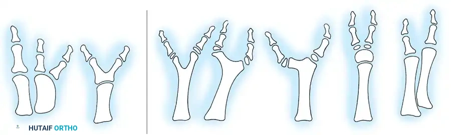
A 4-year-old child presents with a painless limp. Examination reveals limited abduction and internal rotation of the hip. Radiographs show increased density and flattening of the femoral epiphysis. What is the most likely diagnosis?
Options:
- Developmental dysplasia of the hip (DDH)
- Septic arthritis of the hip
- Slipped capital femoral epiphysis (SCFE)
- Legg-Calvé-Perthes disease
- Transient synovitis
Correct Answer: Legg-Calvé-Perthes disease
Explanation:
The clinical picture of a painless limp in a 4-year-old with limited hip abduction and internal rotation, coupled with radiographic findings of increased density (sclerosis) and flattening (fragmentation/collapse) of the femoral epiphysis, is characteristic of Legg-Calvé-Perthes disease. This condition is idiopathic avascular necrosis of the femoral head. SCFE typically occurs in older, often obese adolescents. DDH presents earlier and with different radiographic findings. Septic arthritis and transient synovitis are acute painful conditions.
Question 10:
What is the primary role of the annulus fibrosus in the intervertebral disc?
Options:
- Bear axial load
- Permit spinal flexibility
- Contain the nucleus pulposus
- Produce collagen and proteoglycans
- Serve as a shock absorber
Correct Answer: Contain the nucleus pulposus
Explanation:
The annulus fibrosus, composed of concentric lamellae of collagen fibers, primarily functions to contain the nucleus pulposus and resist tensile forces during spinal movements. While it contributes to bearing axial load and spinal flexibility, its most critical role is to encapsulate the highly hydrated nucleus pulposus. The nucleus pulposus itself acts more as the shock absorber, and the annulus maintains its position. Fibroblasts within the annulus produce collagen and proteoglycans, but this is a cellular function, not the primary structural role.
Question 11:
A 72-year-old female undergoes a total hip arthroplasty via a posterior approach. On postoperative day 2, she complains of acute, severe groin pain and inability to bear weight after attempting to reach for an object on the floor. Her leg is internally rotated, adducted, and shortened. What is the most likely diagnosis?
Options:
- Periprosthetic fracture
- Deep vein thrombosis
- Acetabular component loosening
- Posterior hip dislocation
- Infection
Correct Answer: Posterior hip dislocation
Explanation:
The presentation of sudden severe groin pain, inability to bear weight, and a classic posture of internal rotation, adduction, and shortening following a posterior approach total hip arthroplasty is pathognomonic for a posterior hip dislocation. The posterior approach is associated with a higher risk of posterior dislocation compared to anterior or direct lateral approaches due to posterior capsule compromise. While other complications are possible, this constellation of signs points directly to dislocation. Periprosthetic fracture would likely present with different deformity and radiographic findings. DVT would present with calf pain and swelling. Acetabular loosening is typically a chronic issue. Infection presents with systemic signs and progressive pain.
Question 12:
Regarding the pathogenesis of osteonecrosis of the femoral head, which of the following is considered the MOST significant initiating factor in non-traumatic cases?
Options:
- Mechanical stress
- Genetic predisposition
- Vascular compromise
- Increased intraosseous pressure
- Systemic inflammation
Correct Answer: Vascular compromise
Explanation:
Vascular compromise leading to ischemia is the most significant initiating factor in the pathogenesis of osteonecrosis of the femoral head (ONFH), whether traumatic (e.g., femoral neck fracture) or non-traumatic (e.g., steroid use, alcoholism). This compromise can be due to thrombosis, fat emboli, or direct vessel injury, ultimately leading to death of osteocytes and marrow cells. While increased intraosseous pressure can result from the ischemia and contribute to further compromise, it is typically a consequence, not the primary initiator. Mechanical stress can exacerbate the condition but isn't the primary cause. Genetic predisposition and systemic inflammation are associated factors but not the direct initiating mechanism of bone cell death.
Question 13:
A 15-year-old male presents with chronic anterior knee pain, exacerbated by squatting and prolonged sitting. Examination reveals patellar tenderness and crepitus with patellar motion. Radiographs are unremarkable. What is the most appropriate initial management?
Options:
- Arthroscopic debridement and lateral retinacular release
- Quadriceps and hamstring strengthening program
- Patellar realignment surgery
- Corticosteroid injection into the patellofemoral joint
- Activity restriction and NSAIDs only
Correct Answer: Quadriceps and hamstring strengthening program
Explanation:
This presentation is classic for patellofemoral pain syndrome (PFPS), also known as chondromalacia patellae, characterized by anterior knee pain, particularly with activities that load the patellofemoral joint. The cornerstone of initial management is a comprehensive physical therapy program focusing on quadriceps strengthening (especially vastus medialis obliquus), hamstring flexibility, and hip abductor strengthening to improve patellar tracking. Activity modification and NSAIDs can provide symptomatic relief but don't address the underlying biomechanical issues. Surgical options are reserved for cases refractory to extensive conservative management. Corticosteroid injections are generally not recommended for PFPS due to limited efficacy and potential cartilage damage.
Question 14:
Which rotator cuff muscle is primarily responsible for internal rotation of the shoulder?
Options:
- Supraspinatus
- Infraspinatus
- Teres minor
- Subscapularis
- Deltoid
Correct Answer: Subscapularis
Explanation:
The subscapularis muscle is the primary internal rotator of the shoulder and is one of the four rotator cuff muscles. The supraspinatus is responsible for abduction. The infraspinatus and teres minor are external rotators. The deltoid is a major abductor and flexor/extensor, but not a rotator cuff muscle.
Question 15:
In the management of open fractures, what is the MOST critical factor influencing the risk of infection and overall outcome?
Options:
- Timing of prophylactic antibiotics
- Type of fixation used (internal vs. external)
- Patient's age and comorbidities
- Adequacy of debridement
- Location of the fracture
Correct Answer: Adequacy of debridement
Explanation:
The adequacy and timeliness of surgical debridement are paramount in preventing infection and improving outcomes in open fractures. Debridement involves removing all non-viable tissue, foreign material, and contaminated bone, reducing the bacterial load. While prophylactic antibiotics are crucial, their effectiveness is greatly diminished if debridement is incomplete. Fixation type, patient age, comorbidities, and fracture location are important considerations but are secondary to thorough debridement in preventing infection.
Question 16:
A 45-year-old male laborer presents with gradual onset of pain, stiffness, and snapping in his right index finger, particularly worse in the morning. He notes that the finger occasionally gets 'stuck' in a flexed position and requires passive extension to straighten. What is the most likely diagnosis?
Options:
- De Quervain's tenosynovitis
- Carpal tunnel syndrome
- Flexor tendinitis (trigger finger)
- Ganglion cyst
- Rheumatoid arthritis
Correct Answer: Flexor tendinitis (trigger finger)
Explanation:
The classic symptoms of trigger finger (stenosing tenosynovitis) include pain, stiffness, and a palpable nodule or catching/locking sensation during finger flexion and extension, often worse in the morning. This is caused by inflammation and thickening of the flexor tendon sheath, particularly at the A1 pulley, leading to difficulty for the tendon to glide smoothly. De Quervain's affects the first dorsal compartment of the wrist. Carpal tunnel syndrome involves median nerve compression. Ganglion cysts are typically localized masses. Rheumatoid arthritis would involve multiple joints and systemic symptoms.
Question 17:
What is the primary function of the menisci in the knee joint?
Options:
- Provide active stability
- Produce synovial fluid
- Increase articular cartilage thickness
- Distribute compressive loads and absorb shock
- Initiate knee flexion
Correct Answer: Distribute compressive loads and absorb shock
Explanation:
The menisci are fibrocartilaginous structures within the knee joint that primarily function to distribute compressive loads evenly across the articular cartilage, thereby increasing the contact area and reducing stress on the joint surfaces. They also contribute to shock absorption, joint stability, and lubrication. They do not produce synovial fluid (that's the synovium), increase articular cartilage thickness (they sit between the cartilage surfaces), or initiate knee flexion (that's muscle action).
Question 18:
Which of the following describes a Salter-Harris Type III fracture?
Options:
- Fracture through the physis only
- Fracture through the metaphysis and physis
- Fracture through the epiphysis and physis
- Fracture through the metaphysis, physis, and epiphysis
- Crush injury to the physis
Correct Answer: Fracture through the epiphysis and physis
Explanation:
A Salter-Harris Type III fracture is an intra-articular fracture that extends from the joint surface through the epiphysis and then turns to exit through the physis (growth plate). This type of fracture can lead to growth arrest due to involvement of the germinal cells in the physis and often requires anatomical reduction to prevent articular incongruity. Type I is physis only, Type II is metaphysis and physis, Type IV is metaphysis, physis, and epiphysis, and Type V is a crush injury to the physis.
Question 19:
A 60-year-old male with a history of prostate cancer metastatic to bone presents with new onset lower back pain and bilateral leg weakness. On examination, he has decreased motor strength in both lower extremities (3/5), decreased sensation below T10, and hyperreflexia. What is the most appropriate emergent management?
Options:
- High-dose oral corticosteroids and pain management
- Urgent MRI of the spine
- Surgical decompression of the spinal cord
- Radiation therapy to the metastatic lesion
- Physical therapy for strengthening
Correct Answer: Urgent MRI of the spine
Explanation:
This patient presents with signs and symptoms highly suggestive of impending or established spinal cord compression due to metastatic disease (epidural spinal cord compression - ESCC). The most appropriate emergent step is high-dose intravenous corticosteroids (e.g., dexamethasone) to reduce peritumoral edema and preserve neurological function while further evaluation (urgent MRI) and definitive treatment planning (surgical decompression, radiation therapy) are underway. While an MRI is critical for diagnosis, steroids should not be delayed awaiting imaging if ESCC is suspected. Surgery or radiation are definitive treatments, but steroids buy time and can temporarily improve symptoms.
Question 20:
What is the primary mechanism of action of bisphosphonates in the treatment of osteoporosis?
Options:
- Increase osteoblast activity
- Inhibit osteoclast activity
- Promote calcium absorption in the gut
- Stimulate parathyroid hormone release
- Increase bone matrix mineralization
Correct Answer: Inhibit osteoclast activity
Explanation:
Bisphosphonates are potent inhibitors of osteoclast-mediated bone resorption. They bind to hydroxyapatite crystals in the bone and are internalized by osteoclasts, leading to osteoclast apoptosis and reduced bone turnover. This preserves bone mass and increases bone mineral density, thereby reducing fracture risk. They do not primarily increase osteoblast activity (though reduced resorption can indirectly lead to more effective new bone formation), promote calcium absorption, or stimulate PTH release.
Question 21:
A 38-year-old female presents with progressive pain, swelling, and crepitus in her dominant wrist. Radiographs reveal diffuse carpal collapse, scaphoid non-union, and radioscaphoid arthritis. She is a non-smoker. What is the most appropriate surgical intervention for significant pain and dysfunction?
Options:
- Scaphoid non-union repair with bone graft
- Proximal row carpectomy (PRC)
- Scapholunate ligament reconstruction
- Four-corner fusion (Scapholunate advanced collapse - SLAC wrist reconstruction)
- Total wrist arthroplasty
Correct Answer: Four-corner fusion (Scapholunate advanced collapse - SLAC wrist reconstruction)
Explanation:
The description of diffuse carpal collapse, scaphoid non-union, and radioscaphoid arthritis in a wrist indicates advanced scapholunate advanced collapse (SLAC wrist) or scaphoid non-union advanced collapse (SNAC wrist). For established arthritis of the radioscaphoid joint and midcarpal instability, a four-corner fusion (fusion of the capitate, hamate, triquetrum, and lunate) with scaphoid excision is a well-established and durable surgical option that preserves some wrist motion while alleviating pain. A proximal row carpectomy (PRC) is another option, but often contraindicated with capitolunate arthritis. Scaphoid non-union repair is not sufficient once arthritis has developed. Scapholunate reconstruction is for earlier stages before significant arthritis. Total wrist arthroplasty is typically reserved for severe, global arthritis and lower demand patients.
Question 22:
Which of the following nerves is MOST commonly injured during an anterior approach to the cervical spine (ACDF)?
Options:
- Recurrent laryngeal nerve
- Phrenic nerve
- Vagus nerve
- Hypoglossal nerve
- Spinal accessory nerve
Correct Answer: Recurrent laryngeal nerve
Explanation:
The recurrent laryngeal nerve is the most commonly injured nerve during an anterior cervical discectomy and fusion (ACDF). It supplies the intrinsic muscles of the larynx and loops around the subclavian artery (right side) or aortic arch (left side). Its proximity to the surgical field and variable course makes it vulnerable to injury during retraction, dissection, or electrocautery, leading to dysphonia (hoarseness). The phrenic nerve, vagus nerve, hypoglossal nerve, and spinal accessory nerve are also in the vicinity but are less commonly injured.
Question 23:
A 10-year-old male falls onto an outstretched hand. Radiographs show a fracture of the distal radius with dorsal displacement and angulation. There is an intact volar cortex. What type of fracture is this, based on pediatric classification?
Options:
- Greenstick fracture
- Torus fracture
- Salter-Harris Type I
- Galeazzi fracture
- Monteggia fracture
Correct Answer: Greenstick fracture
Explanation:
A greenstick fracture is an incomplete fracture of a long bone in children, characterized by a break in one side of the cortex while the opposite cortex remains intact and bent. This description of a distal radius fracture with dorsal displacement and angulation, but an intact volar cortex, perfectly fits a greenstick fracture. A torus (buckle) fracture is an incomplete fracture characterized by buckling of the cortex. Salter-Harris Type I is a complete physeal separation without metaphyseal or epiphyseal involvement. Galeazzi and Monteggia fractures are specific forearm fracture-dislocations.
Question 24:
Which cytokine is primarily responsible for promoting bone resorption by stimulating osteoclast differentiation and activation?
Options:
- Bone morphogenetic protein (BMP)
- Transforming growth factor-beta (TGF-β)
- Insulin-like growth factor 1 (IGF-1)
- Receptor activator of nuclear factor kappa-Β ligand (RANKL)
- Osteoprotegerin (OPG)
Correct Answer: Receptor activator of nuclear factor kappa-Β ligand (RANKL)
Explanation:
RANKL (Receptor Activator of Nuclear factor Kappa-Β Ligand) is a key cytokine produced by osteoblasts and stromal cells that binds to RANK receptors on pre-osteoclasts, promoting their differentiation into mature osteoclasts and activating their bone-resorbing activity. OPG (Osteoprotegerin) acts as a decoy receptor for RANKL, inhibiting its effects and thus preventing bone resorption. BMPs, TGF-β, and IGF-1 are primarily involved in bone formation and remodeling, stimulating osteoblast activity.
Question 25:
A 65-year-old female presents with persistent deep gluteal pain, worse with sitting and relieved by standing. Physical examination reveals tenderness over the sciatic notch and pain with resisted hip external rotation. What is the most likely diagnosis?
Options:
- Lumbar radiculopathy (L5-S1)
- Trochanteric bursitis
- Piriformis syndrome
- Sacroiliac joint dysfunction
- Hamstring tendinopathy
Correct Answer: Piriformis syndrome
Explanation:
Piriformis syndrome is characterized by buttock pain, often radiating down the leg (pseudoradiculopathy), caused by sciatic nerve compression or irritation by the piriformis muscle. It is typically worse with prolonged sitting and activities involving hip external rotation. Tenderness over the sciatic notch and pain with resisted hip external rotation are classic findings. Lumbar radiculopathy would usually have neurological deficits more consistent with a specific nerve root. Trochanteric bursitis causes lateral hip pain. Sacroiliac joint dysfunction would involve pain with SIJ provocative tests. Hamstring tendinopathy typically involves more distal pain and tenderness along the hamstring insertion.
Question 26:
What is the most common organism responsible for acute hematogenous osteomyelitis in otherwise healthy children?
Options:
- Escherichia coli
- Pseudomonas aeruginosa
- Staphylococcus aureus
- Streptococcus pyogenes
- Kingella kingae
Correct Answer: Staphylococcus aureus
Explanation:
Staphylococcus aureus is the most common causative organism for acute hematogenous osteomyelitis in all age groups, including otherwise healthy children. While Kingella kingae is an increasingly recognized pathogen in younger children (under 3 years old), S. aureus remains overall the predominant cause. E. coli and Pseudomonas are more common in neonates, immunocompromised individuals, or following puncture wounds (e.g., through a shoe). Streptococcus pyogenes is less common.
Question 27:
A 22-year-old collegiate athlete sustains an isolated anterior cruciate ligament (ACL) tear. He desires to return to competitive sports. What is the MOST crucial factor for a successful return to play following ACL reconstruction?
Options:
- Choice of graft (autograft vs. allograft)
- Aggressive early rehabilitation protocol
- Passing functional performance tests and adequate time from surgery
- Use of a functional knee brace post-surgery
- Surgeon's experience and volume
Correct Answer: Passing functional performance tests and adequate time from surgery
Explanation:
While all options play a role, passing objective functional performance tests (e.g., hop tests, strength testing, balance) and adequate time from surgery (typically 9-12 months for high-level athletes) are the most crucial factors indicating readiness for return to play. Premature return increases the risk of re-injury. Graft choice, while important for long-term outcomes, is less critical than achieving functional milestones. Aggressive early rehab is part of the process but needs to be balanced with biological healing. Bracing is controversial and generally not supported as the primary factor for return to play in ACL patients.
Question 28:
Which of the following is considered a 'red flag' symptom for serious spinal pathology that warrants immediate investigation?
Options:
- Chronic low back pain lasting >6 weeks
- Pain radiating below the knee
- Morning stiffness lasting <30 minutes
- New onset bowel or bladder dysfunction
- Pain worsened by activity and relieved by rest
Correct Answer: New onset bowel or bladder dysfunction
Explanation:
New onset bowel or bladder dysfunction (urinary retention with overflow incontinence, fecal incontinence) is a classic 'red flag' for cauda equina syndrome, a neurosurgical emergency requiring urgent investigation and potentially surgical decompression. Other red flags include progressive neurological deficit, unexplained weight loss, fever, night pain, and a history of cancer or immunosuppression. The other options describe common features of mechanical low back pain or radiculopathy, which, while needing evaluation, are not typically emergent 'red flags'.
Question 29:
What is the primary vascular supply to the scaphoid bone, making it susceptible to avascular necrosis after fracture?
Options:
- Radial artery, volar carpal branch
- Ulnar artery, dorsal carpal branch
- Radial artery, dorsal carpal branch
- Median artery, palmar branch
- Anterior interosseous artery
Correct Answer: Radial artery, dorsal carpal branch
Explanation:
The primary blood supply to the scaphoid bone is via branches of the radial artery, specifically the dorsal carpal branch (or dorsal carpal artery). These vessels enter the scaphoid distally and dorsally, providing a retrograde blood supply to the proximal pole. This retrograde flow makes the proximal pole particularly vulnerable to avascular necrosis (AVN) following a scaphoid waist fracture, as its blood supply can be disrupted. The volar carpal branch supplies the distal scaphoid but does not typically reach the proximal pole.
Question 30:
In the context of surgical site infection (SSI) prophylaxis, what is the generally accepted optimal timing for administering intravenous antibiotics prior to incision?
Options:
- Within 2 hours before incision
- Immediately at the time of incision
- After the incision but before wound closure
- Within 60 minutes before incision
- Postoperatively, for 24 hours
Correct Answer: Within 60 minutes before incision
Explanation:
For most surgical procedures, intravenous prophylactic antibiotics should be administered within 60 minutes prior to the surgical incision to ensure adequate tissue concentration at the time of potential contamination. For certain antibiotics (e.g., vancomycin or fluoroquinolones) with longer infusion times, administration may begin up to 120 minutes prior. Administering too early can reduce efficacy, and too late can miss the critical window of contamination. Postoperative continuation beyond 24 hours is generally not recommended for prophylaxis in clean or clean-contaminated cases.
Question 31:
A 50-year-old overweight male presents with acute onset, excruciating pain, redness, and swelling of the great toe (podagra). He reports similar episodes in the past affecting other joints. Laboratory tests show elevated serum uric acid. What is the most appropriate long-term management to prevent recurrent attacks?
Options:
- Colchicine
- Indomethacin
- Prednisone
- Allopurinol
- Aspirin
Correct Answer: Allopurinol
Explanation:
This patient presents with classic symptoms of gout. While colchicine, indomethacin, and prednisone are used for acute attack management, allopurinol is the most appropriate long-term therapy to prevent recurrent attacks. Allopurinol is a xanthine oxidase inhibitor that reduces uric acid production, thereby lowering serum uric acid levels and preventing crystal deposition. It should not be started during an acute attack. Aspirin can actually worsen gout by interfering with renal uric acid excretion at low doses.
Question 32:
What is the primary indication for surgical intervention in a patient with adult degenerative lumbar scoliosis?
Options:
- Curve magnitude >30 degrees
- Persistent low back pain refractory to conservative management
- Significant leg length discrepancy
- Progressive neurological deficit (e.g., motor weakness, cauda equina)
- Cosmetic deformity
Correct Answer: Progressive neurological deficit (e.g., motor weakness, cauda equina)
Explanation:
The primary indication for surgical intervention in adult degenerative lumbar scoliosis is progressive neurological deficit (e.g., motor weakness, radiculopathy, or cauda equina syndrome) that correlates with the deformity, or intractable pain that has failed prolonged conservative management. While curve magnitude, leg length discrepancy, and cosmetic deformity are considerations, they are typically not primary indications for surgery unless associated with significant pain or neurological compromise. Surgery for degenerative scoliosis is complex and carries significant risks, so indications are carefully considered.
Question 33:
A 25-year-old male sustains a high-energy tibial plateau fracture. He develops increasing pain, swelling, and paresthesias in his leg. On examination, the leg is tense, and passive ankle dorsiflexion elicits severe pain. Distal pulses are present. What is the most appropriate immediate action?
Options:
- Administer IV opioids and elevate the limb
- Obtain an urgent CT angiogram
- Perform compartment pressure measurements
- Apply ice packs and monitor closely
- Perform a diagnostic arthroscopy
Correct Answer: Perform compartment pressure measurements
Explanation:
This patient's symptoms (increasing pain, swelling, paresthesias, tense leg, pain with passive stretch) in the setting of a high-energy tibial plateau fracture are highly suggestive of acute compartment syndrome. Although distal pulses are present, compartment syndrome is a clinical diagnosis, and absent pulses are a late and unreliable sign. The most appropriate immediate action is to perform compartment pressure measurements to confirm the diagnosis. If pressures are elevated (typically within 30 mmHg of diastolic pressure or >30 mmHg absolute, depending on protocol), an emergent fasciotomy is indicated to prevent irreversible muscle and nerve damage. Delaying treatment for imaging or conservative measures is inappropriate and risks permanent disability.
Question 34:
Which ligament is most commonly injured in an inversion ankle sprain?
Options:
- Deltoid ligament
- Anterior talofibular ligament (ATFL)
- Posterior talofibular ligament (PTFL)
- Calcaneofibular ligament (CFL)
- Interosseous talocalcaneal ligament
Correct Answer: Anterior talofibular ligament (ATFL)
Explanation:
The anterior talofibular ligament (ATFL) is the most commonly injured ligament in an inversion ankle sprain. It is the weakest of the lateral ankle ligaments and the first to be torn. Severe inversion injuries can also involve the calcaneofibular ligament (CFL). The deltoid ligament is on the medial side and is injured in eversion sprains. The PTFL is typically injured in very severe sprains and ankle dislocations.
Question 35:
What is the classic radiographic finding associated with osteosarcoma?
Options:
- Punctuated calcifications with a 'ring and arc' pattern
- Expansile lytic lesion with a 'soap bubble' appearance
- Cortical thickening and periosteal reaction with 'onion skin' appearance
- Sunburst periosteal reaction and Codman's triangle
- Geographic lysis with sclerotic border
Correct Answer: Sunburst periosteal reaction and Codman's triangle
Explanation:
Osteosarcoma classically presents with aggressive radiographic features, including a 'sunburst' periosteal reaction (spicules of new bone radiating outwards) and Codman's triangle (triangular elevation of the periosteum at the margins of the lesion). These indicate rapid bone formation and destruction. 'Ring and arc' calcifications are characteristic of chondrosarcoma. 'Soap bubble' appearance is often seen in giant cell tumors. 'Onion skin' periosteal reaction is characteristic of Ewing's sarcoma. Geographic lysis with a sclerotic border is more indicative of a benign or slow-growing lesion.
Question 36:
A 6-month-old infant is diagnosed with developmental dysplasia of the hip (DDH). Physical examination reveals a positive Barlow test and Galeazzi sign. Radiographs show a dislocated left hip. What is the most appropriate initial treatment?
Options:
- Immediate open reduction and spica cast
- Pavlik harness
- Closed reduction under anesthesia and spica cast
- Observation with serial ultrasounds
- Referral for triple osteotomy
Correct Answer: Pavlik harness
Explanation:
For an infant diagnosed with DDH at 6 months of age, a Pavlik harness is the gold standard initial treatment for reducible hips. The Barlow test indicates reducibility. The Pavlik harness holds the hips in flexion and abduction, promoting femoral head seating and acetabular development. Open reduction or closed reduction with spica cast is typically reserved for older infants, failed harness treatment, or irreducible hips. Observation is inappropriate for a dislocated hip. Triple osteotomy is for older children with persistent dysplasia.
Question 37:
Which of the following ligaments is critical for providing primary valgus stability to the elbow joint?
Options:
- Radial collateral ligament
- Lateral ulnar collateral ligament
- Annular ligament
- Medial ulnar collateral ligament (UCL)
- Oblique cord
Correct Answer: Medial ulnar collateral ligament (UCL)
Explanation:
The Medial Ulnar Collateral Ligament (UCL), specifically its anterior bundle, is the primary static stabilizer of the elbow against valgus stress. Injuries to the UCL are common in overhead throwing athletes (e.g., 'Tommy John' injury). The radial collateral ligament complex provides varus and posterolateral rotatory stability. The annular ligament stabilizes the radial head. The oblique cord has a minor role.
Question 38:
A patient undergoing knee arthroplasty receives a neuraxial anesthetic. Which of the following potential complications of neuraxial anesthesia is correctly matched with its management?
Options:
- Hypotension: Administer IV atropine
- Spinal hematoma: Urgent MRI and surgical decompression
- Postdural puncture headache: Immediate blood transfusion
- High spinal block: Trendelenburg position and aggressive fluid bolus
- Nerve injury: Prolonged physical therapy and watchful waiting
Correct Answer: Spinal hematoma: Urgent MRI and surgical decompression
Explanation:
Spinal hematoma is a rare but devastating complication of neuraxial anesthesia, especially in patients on anticoagulants. Prompt recognition, urgent MRI for diagnosis, and emergent surgical decompression are critical to prevent permanent neurological deficits. Hypotension is managed with IV fluids and vasopressors (e.g., phenylephrine), not atropine. Postdural puncture headache is often treated with a blood patch, not transfusion. A high spinal block requires ventilatory support and hemodynamic management. Nerve injury requires thorough investigation and management, not just watchful waiting.
Question 39:
What is the most common cause of non-union in long bone fractures?
Options:
- Patient's age
- Inadequate immobilization
- Infection
- Soft tissue interposition
- Open fracture
Correct Answer: Inadequate immobilization
Explanation:
Inadequate stabilization/immobilization and insufficient blood supply (leading to inadequate biological response) are the two most common causes of non-union. While infection, soft tissue interposition, and open fractures can certainly contribute to non-union, failure to provide a stable mechanical environment for healing is a predominant factor. Age is a factor but not the primary cause of non-union.
Question 40:
A 40-year-old male presents with severe right foot pain following a fall from a ladder, landing on his heels. Radiographs reveal a comminuted fracture of the calcaneus, involving the posterior facet. What is the most appropriate initial management step (assuming no open wounds or neurovascular compromise)?
Options:
- Immediate surgical fixation
- Elevation, ice, non-weight bearing, and pain control
- Computed tomography (CT) scan of the foot
- Consultation with a spine surgeon
- Casting in a short leg walking boot
Correct Answer: Computed tomography (CT) scan of the foot
Explanation:
Following a high-energy calcaneal fracture, especially one involving the posterior facet, a CT scan of the foot is essential. It provides detailed information regarding the fracture pattern, subtalar joint involvement, degree of displacement, and comminution, which are critical for surgical planning. Additionally, calcaneal fractures resulting from axial loading often have associated injuries, particularly to the lumbar spine (up to 10-15%), so a spine assessment is important but secondary to initial local evaluation. Initial management also involves elevation, ice, and pain control, but a CT is the crucial next diagnostic step before deciding on definitive treatment (surgical vs. non-surgical).
Question 41:
Which of the following conditions is considered a contraindication to performing a total knee arthroplasty (TKA)?
Options:
- Obesity (BMI >40)
- Active knee infection
- Advanced age (>85 years)
- Diabetes mellitus
- History of prior knee surgery
Correct Answer: Active knee infection
Explanation:
Active knee infection is an absolute contraindication to total knee arthroplasty (TKA). Performing a TKA in the presence of infection would almost certainly lead to periprosthetic joint infection, which is a devastating complication. The infection must be completely eradicated and confirmed resolved before considering TKA. While obesity, advanced age, diabetes, and prior knee surgery are factors that increase surgical risk or complexity, they are relative contraindications or risk factors, not absolute contraindications. They require careful patient selection and optimization.
Question 42:
What is the primary function of the deltoid ligament complex of the ankle?
Options:
- Resist anterior translation of the talus
- Resist inversion forces
- Resist eversion forces
- Resist posterior translation of the talus
- Stabilize the distal tibiofibular syndesmosis
Correct Answer: Resist eversion forces
Explanation:
The deltoid ligament complex, located on the medial side of the ankle, is a strong, multi-component ligament that primarily resists eversion forces. It stabilizes the ankle against outward rotation and abduction of the foot. In contrast, the lateral collateral ligaments (ATFL, CFL, PTFL) resist inversion forces.
Question 43:
A 12-year-old female presents with progressive thoracic spinal deformity. Radiographs show a right thoracic curve measuring 45 degrees, with significant vertebral rotation. She is Risser 0. What is the most appropriate management?
Options:
- Observation and serial radiographs every 6 months
- Bracing with a thoracolumbosacral orthosis (TLSO)
- Posterior spinal fusion
- Anterior vertebral body tethering (VBT)
- Physical therapy and stretching exercises
Correct Answer: Posterior spinal fusion
Explanation:
For adolescent idiopathic scoliosis, a curve magnitude of 45 degrees in a skeletally immature patient (Risser 0 indicates significant growth remaining) is typically an indication for surgical intervention, most commonly posterior spinal fusion. Bracing is generally recommended for curves between 25-45 degrees in growing patients to prevent progression, but a 45-degree curve at Risser 0 has a high likelihood of progression beyond 50 degrees, warranting surgery. Observation is for curves <25 degrees. VBT is a growth modulation technique, but a 45-degree curve may be pushing its limits, and posterior fusion is more definitive for this magnitude. Physical therapy does not typically prevent progression of structural scoliosis.
Question 44:
What is the most common benign bone tumor?
Options:
- Osteoid osteoma
- Enchondroma
- Osteochondroma
- Fibrous dysplasia
- Non-ossifying fibroma
Correct Answer: Osteochondroma
Explanation:
Osteochondroma (exostosis) is by far the most common benign bone tumor, accounting for approximately 35-50% of all benign bone tumors. It is characterized by an outgrowth of bone covered by a cartilaginous cap, typically arising from the metaphysis of long bones. While other options are benign bone tumors, they are less common than osteochondromas.
Question 45:
Which metabolic bone disease is characterized by excessive bone resorption followed by disorganized and excessive bone formation, leading to enlarged and weakened bones?
Options:
- Osteoporosis
- Osteomalacia
- Paget's disease of bone
- Hyperparathyroidism
- Osteogenesis imperfecta
Correct Answer: Paget's disease of bone
Explanation:
Paget's disease of bone (osteitis deformans) is characterized by a focal disorder of bone remodeling, with greatly accelerated, chaotic bone turnover. This involves an initial osteolytic phase, followed by a mixed lytic and blastic phase, and finally a predominant sclerotic phase. The new bone formed is structurally disorganized (woven bone), leading to enlarged, weakened, and deformed bones. Osteoporosis is a decrease in bone mass. Osteomalacia is defective mineralization of new bone. Hyperparathyroidism causes increased bone resorption but not this chaotic pattern. Osteogenesis imperfecta is a genetic collagen disorder.
Question 46:
A 58-year-old female experiences sudden, sharp pain in her calf during a tennis match, followed by difficulty weight-bearing. On examination, she has a palpable gap in her Achilles tendon and a positive Thompson test. What is the most appropriate management for a healthy, active individual seeking to return to high-level activity?
Options:
- Immobilization in a plantarflexed cast for 8 weeks
- Percutaneous repair of the Achilles tendon
- Open surgical repair of the Achilles tendon
- Physical therapy focusing on eccentric loading exercises
- Steroid injection around the tendon
Correct Answer: Open surgical repair of the Achilles tendon
Explanation:
A palpable gap in the Achilles tendon and a positive Thompson test are diagnostic of an Achilles tendon rupture. For healthy, active individuals who desire to return to high-level sports, open surgical repair of the Achilles tendon is generally recommended. It provides the strongest repair, allows for anatomical reduction, and has been associated with a lower re-rupture rate compared to non-operative management or percutaneous techniques, especially in this population. Non-operative management or percutaneous repair might be considered for less active patients or those with comorbidities. Steroid injections are contraindicated due to the risk of further tendon degeneration and rupture.
Question 47:
What is the primary function of the subscapularis tendon in the shoulder?
Options:
- Abduction
- External rotation
- Internal rotation
- Adduction
- Flexion
Correct Answer: Internal rotation
Explanation:
The subscapularis is one of the four rotator cuff muscles and is the primary internal rotator of the shoulder. It also contributes to shoulder adduction. The supraspinatus is an abductor, and the infraspinatus and teres minor are external rotators. The deltoid is a major abductor.
Question 48:
A 3-year-old child presents with a painful, swollen elbow after being pulled up by the hand by an adult. The child holds the arm pronated and slightly flexed, refusing to use it. Radiographs are normal. What is the most likely diagnosis?
Options:
- Supracondylar humerus fracture
- Lateral condyle fracture
- Radial head subluxation (nursemaid's elbow)
- Olecranon fracture
- Septic elbow arthritis
Correct Answer: Radial head subluxation (nursemaid's elbow)
Explanation:
The classic presentation of a child (typically 1-4 years old) with a painful arm held in pronation and flexion after a sudden pull on the hand, with normal radiographs, is highly suggestive of a radial head subluxation, commonly known as 'nursemaid's elbow.' This occurs when the annular ligament slips over the radial head and becomes trapped in the radiohumeral joint. Fractures would typically be visible on X-ray, and septic arthritis would present with fever and more systemic signs.
Question 49:
Which of the following describes the preferred initial treatment for a displaced midshaft clavicle fracture in a healthy, active adult?
Options:
- Figure-of-eight brace
- Sling immobilization for 6 weeks
- Plate and screw osteosynthesis
- Intramedullary nailing
- Immediate physical therapy to maintain range of motion
Correct Answer: Plate and screw osteosynthesis
Explanation:
For a displaced midshaft clavicle fracture in a healthy, active adult, plate and screw osteosynthesis (open reduction internal fixation) is often the preferred treatment. While non-operative management (sling immobilization) can be successful, displaced fractures, especially with significant shortening or comminution, have a higher risk of non-union, malunion, and prolonged recovery with non-operative treatment. Surgical fixation can provide earlier return to function, improved cosmesis, and lower non-union rates in appropriately selected patients. Figure-of-eight braces have been shown to be no more effective than slings and can cause discomfort. Intramedullary nailing is less commonly used for clavicle fractures. Immediate physical therapy without stabilization is contraindicated.
Question 50:
Regarding avascular necrosis (AVN) of the femoral head, what is the MOST sensitive imaging modality for early detection?
Options:
- Plain radiographs (X-rays)
- Computed tomography (CT) scan
- Magnetic resonance imaging (MRI)
- Bone scintigraphy (bone scan)
- Ultrasound
Correct Answer: Magnetic resonance imaging (MRI)
Explanation:
Magnetic Resonance Imaging (MRI) is the most sensitive imaging modality for the early detection of avascular necrosis (AVN) of the femoral head. It can detect ischemic changes in the bone marrow before radiographic changes become apparent. Plain radiographs are often normal in the early stages and only show changes (e.g., sclerosis, crescent sign, collapse) in later stages. CT scans offer good bony detail but are less sensitive than MRI for early marrow edema. Bone scintigraphy can be sensitive but lacks specificity. Ultrasound is not typically used for AVN diagnosis.
Question 51:
What is the primary goal of surgical treatment for hallux valgus (bunion deformity)?
Options:
- Cosmetic correction of toe deformity
- Achieve a pain-free, functional foot
- Increase hallux range of motion
- Prevent recurrence of the deformity
- Reduce the intermetatarsal angle to zero
Correct Answer: Achieve a pain-free, functional foot
Explanation:
The primary goal of surgical treatment for hallux valgus is to achieve a pain-free, functional foot with good alignment. While cosmetic correction and prevention of recurrence are important secondary goals, the overarching aim is to alleviate pain, improve shoe wear, and restore normal foot mechanics. Simply reducing the intermetatarsal angle to zero is not the goal, as a slightly divergent angle is normal. Increasing range of motion is desired but secondary to pain relief and function.
Question 52:
A 60-year-old male with a history of intravenous drug use presents with acute onset fever, chills, and severe back pain localized to the lumbar spine. He has no neurological deficits. Labs show elevated ESR and CRP. What is the most likely diagnosis?
Options:
- Mechanical low back pain
- Herniated nucleus pulposus
- Spinal epidural abscess
- Vertebral osteomyelitis/Discitis
- Malignancy
Correct Answer: Vertebral osteomyelitis/Discitis
Explanation:
The patient's presentation (fever, chills, severe localized back pain, IV drug use, elevated inflammatory markers) is highly suspicious for vertebral osteomyelitis and/or discitis. IV drug use is a significant risk factor for hematogenous spread of infection to the spine. While a spinal epidural abscess is also a serious possibility and often coexists, the initial infection of the vertebral body and/or disc (osteomyelitis/discitis) is the most likely primary diagnosis. Mechanical back pain and HNP would not typically present with fever and chills. Malignancy is possible but less likely with acute fever and high inflammatory markers. Urgent MRI is needed for definitive diagnosis and differentiation.
Question 53:
Which type of collagen is primarily found in hyaline cartilage, providing its characteristic strength and resilience?
Options:
- Type I collagen
- Type II collagen
- Type III collagen
- Type IV collagen
- Type V collagen
Correct Answer: Type II collagen
Explanation:
Type II collagen is the predominant collagen found in hyaline cartilage (including articular cartilage). Its unique molecular structure and organization contribute to the tensile strength and resilience of cartilage, allowing it to withstand compressive loads. Type I collagen is found in bone, tendons, and ligaments, providing strong tensile strength. Type III collagen is found in extensible connective tissues like skin and blood vessels. Type IV collagen is found in basement membranes. Type V collagen is found in small amounts in various tissues, including cartilage.
Question 54:
A 6-month-old infant presents with a torticollis and a firm, non-tender mass in the sternocleidomastoid muscle. The child's head is tilted to one side and rotated to the opposite. What is the most likely diagnosis?
Options:
- Congenital muscular torticollis
- Klippel-Feil syndrome
- Congenital scoliosis
- Cervical dystonia
- Infectious torticollis
Correct Answer: Congenital muscular torticollis
Explanation:
Congenital muscular torticollis (CMT) is the most likely diagnosis. It typically presents in infants aged 2-4 weeks with a progressively palpable, firm, non-tender mass (pseudotumor) in the sternocleidomastoid muscle, along with a characteristic head tilt to the ipsilateral side and rotation to the contralateral side. It is thought to be due to fibrosis within the SCM. Klippel-Feil syndrome and congenital scoliosis are bony abnormalities of the spine. Cervical dystonia usually presents later in life. Infectious torticollis would typically involve pain, fever, and signs of inflammation.
Question 55:
What is the primary indication for surgical intervention in patients with carpal tunnel syndrome?
Options:
- Intermittent numbness and tingling in the median nerve distribution
- Difficulty with fine motor tasks and dropping objects
- Positive Phalen's and Tinel's signs
- Thenar muscle atrophy and persistent sensory deficit
- Night pain unresponsive to splinting
Correct Answer: Thenar muscle atrophy and persistent sensory deficit
Explanation:
While all options represent symptoms of carpal tunnel syndrome, thenar muscle atrophy and persistent sensory deficit (indicating severe nerve compression and potential irreversible damage) are considered primary indications for surgical release. These signs suggest chronic and severe median nerve compression. While other symptoms like night pain unresponsive to splinting or difficulty with fine motor tasks are strong indications for surgery, thenar atrophy signifies advanced disease. Intermittent numbness and positive provocative signs alone can often be managed conservatively initially.
Question 56:
Which type of gait is characteristic of a patient with weakness of the gluteus medius muscle?
Options:
- Foot drop gait (steppage gait)
- Antalgic gait
- Trendelenburg gait
- Cerebellar ataxic gait
- Waddling gait
Correct Answer: Trendelenburg gait
Explanation:
A Trendelenburg gait is characterized by the dropping of the contralateral hip during the stance phase of gait. This occurs due to weakness or paralysis of the ipsilateral gluteus medius and minimus muscles, which are crucial hip abductors. When the stance leg's abductors cannot stabilize the pelvis, the opposite side drops. Foot drop gait is due to dorsiflexor weakness. Antalgic gait is pain-avoiding. Cerebellar ataxic gait is uncoordinated. Waddling gait (often bilateral Trendelenburg) can be seen with bilateral abductor weakness or neuromuscular disorders.
Question 57:
What is the primary concern when managing a dislocated knee (tibiofemoral dislocation)?
Options:
- Damage to the cruciate ligaments
- Meniscal tears
- Articular cartilage damage
- Neurovascular injury, especially the popliteal artery
- Fractures of the tibial plateau
Correct Answer: Neurovascular injury, especially the popliteal artery
Explanation:
Neurovascular injury, particularly to the popliteal artery, is the MOST critical concern in a dislocated knee. The popliteal artery runs in close proximity to the posterior knee joint, and it can be stretched, kinked, or torn during dislocation, leading to limb-threatening ischemia. Immediate reduction and thorough neurovascular assessment (including ankle-brachial index - ABI, and potentially CT angiography) are paramount. While ligamentous injuries (ACL, PCL, MCL, LCL), meniscal tears, and articular cartilage damage are almost always present and require subsequent management, the vascular status dictates immediate limb salvage efforts.
Question 58:
Which zone of the physis (growth plate) is most susceptible to shear forces and is primarily responsible for longitudinal bone growth?
Options:
- Resting zone
- Proliferative zone
- Hypertrophic zone
- Calcification zone
- Resorption zone
Correct Answer: Hypertrophic zone
Explanation:
The hypertrophic zone of the physis is where chondrocytes rapidly enlarge, forming columns, and become calcified. This zone is mechanically the weakest part of the physis, making it most susceptible to shear forces and fractures (e.g., Salter-Harris fractures typically occur through this zone). The proliferative zone is where chondrocytes multiply, contributing to longitudinal growth by producing new cells. The resting zone serves as a reserve of cells. The calcification zone is where the cartilage matrix calcifies, and the resorption zone is where chondroclasts and osteoblasts invade, leading to ossification.
Question 59:
A 4-month-old infant is brought to the clinic for routine check-up. The mother notes the baby keeps the head tilted to the left. On examination, the left sternocleidomastoid muscle is palpably tight. Passive range of motion reveals limited rotation to the right and left lateral flexion. What is the most appropriate initial treatment?
Options:
- MRI of the cervical spine
- Referral for surgical release of the sternocleidomastoid
- Physical therapy focusing on stretching and strengthening exercises
- Botulinum toxin injection into the SCM
- Orthosis (collar) to correct head position
Correct Answer: Physical therapy focusing on stretching and strengthening exercises
Explanation:
This is a classic presentation of congenital muscular torticollis. The most appropriate initial treatment, especially in infants under one year of age, is conservative management with physical therapy. This involves gentle stretching exercises (passive stretching of the SCM and active range of motion) to elongate the tight muscle, along with positioning and strengthening exercises to encourage symmetrical head posture and neck muscle development. Surgery is reserved for cases that fail extensive conservative therapy (e.g., after 12-18 months of treatment). MRI might be considered if there are atypical features or persistent concerns, but not as the first step. Botulinum toxin and orthoses are less common initial treatments.
Question 60:
What is the most common direction of glenohumeral instability?
Options:
- Anterior
- Posterior
- Inferior
- Multidirectional
- Superior
Correct Answer: Anterior
Explanation:
Anterior glenohumeral instability (dislocation or subluxation) is by far the most common direction of shoulder instability, accounting for over 95% of cases. This is due to the inherent anatomy of the glenohumeral joint, with less anterior bony constraint and the typical mechanism of injury involving abduction and external rotation. Posterior and inferior instability are less common, and multidirectional instability involves instability in multiple directions.
Question 61:
In the context of bone healing, what is the sequence of events during secondary bone healing (endochondral ossification)?
Options:
- Hematoma formation → cartilage callus → bony callus → remodeling
- Hematoma formation → bony callus → cartilage callus → remodeling
- Direct osteoblast activity → woven bone → lamellar bone
- Inflammation → osteoclast activity → osteoblast activity → consolidation
- Fibrous tissue formation → cartilage formation → direct bone formation
Correct Answer: Hematoma formation → cartilage callus → bony callus → remodeling
Explanation:
Secondary bone healing, also known as indirect healing or endochondral ossification, involves a cascade of events: 1. Hematoma formation at the fracture site. 2. Inflammation and formation of a soft callus (fibrous tissue and cartilage). 3. Hard callus formation as the cartilage calcifies and is replaced by woven bone. 4. Bone remodeling, where woven bone is gradually replaced by lamellar bone, and the bone reshapes itself. The other options describe direct bone healing or incorrect sequences.
Question 62:
Which nerve is at highest risk of injury during surgical fixation of a proximal humerus fracture via a deltopectoral approach?
Options:
- Axillary nerve
- Radial nerve
- Musculocutaneous nerve
- Median nerve
- Ulnar nerve
Correct Answer: Axillary nerve
Explanation:
The axillary nerve (C5-C6) is at highest risk of injury during surgical approaches to the proximal humerus, especially the deltopectoral approach. It courses around the surgical neck of the humerus, approximately 5-7 cm distal to the acromion, making it vulnerable during plate application, screw insertion, or excessive retraction of the deltoid. Injury can lead to deltoid paralysis and sensory loss over the lateral shoulder. The radial nerve is at risk more distally in the humerus (spiral groove). The musculocutaneous, median, and ulnar nerves are generally protected by their more medial and anterior/posterior positions relative to the deltopectoral interval.
Question 63:
A 48-year-old male presents with chronic insidious bilateral heel pain, worse with the first steps in the morning and after periods of rest. Pain is localized to the plantar aspect of the heel. Physical examination reveals tenderness at the origin of the plantar fascia. What is the most likely diagnosis?
Options:
- Achilles tendinopathy
- Tarsal tunnel syndrome
- Stress fracture of the calcaneus
- Plantar fasciitis
- Fat pad atrophy
Correct Answer: Plantar fasciitis
Explanation:
The classic presentation of plantar fasciitis involves chronic insidious heel pain, primarily on the plantar aspect, that is worse with the first steps in the morning ('first step pain') and after periods of rest, improving with activity. Tenderness at the medial plantar calcaneal tuberosity (origin of the plantar fascia) is a key diagnostic finding. Achilles tendinopathy causes posterior heel pain. Tarsal tunnel syndrome would present with nerve symptoms (numbness, tingling). Calcaneal stress fractures usually involve swelling and different pain patterns. Fat pad atrophy is common in older individuals but doesn't typically present with 'first step pain' as its primary symptom.
Question 64:
What is the primary goal of conservative management for rotator cuff tendinopathy without a full-thickness tear?
Options:
- Eliminate all shoulder pain permanently
- Avoid all overhead activities indefinitely
- Improve rotator cuff strength and scapular mechanics
- Initiate corticosteroid injections early
- Prepare for inevitable surgical repair
Correct Answer: Improve rotator cuff strength and scapular mechanics
Explanation:
The primary goal of conservative management for rotator cuff tendinopathy is to improve rotator cuff strength, restore proper scapular mechanics, and optimize neuromuscular control of the shoulder. This addresses underlying biomechanical deficits that contribute to impingement and tendon overload. While pain reduction is a goal, complete elimination of pain may not always be achievable, and the focus is on functional restoration. Avoidance of overhead activities is temporary, not indefinite. Corticosteroid injections offer short-term relief but do not address the root cause and are not always initiated early. Surgical repair is typically reserved for failed conservative management or full-thickness tears.