Part of the Master Guide

Septic Arthritis in Children: Epidemiology, Diagnosis, Management & Orthopedic Considerations

Deep Surgical Site Infection & Osteomyelitis After Ankle ORIF: A Case Study

17 Apr 2026 18 min read 144 Views
Illustration of case infected ankle - Dr. Mohammed Hutaif

Key Takeaway

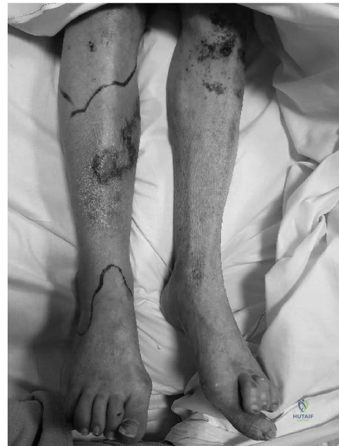
Diagnosing post-ORIF ankle infection requires observing clinical signs: pain, erythema, swelling, and purulent discharge. Key lab indicators include elevated WBC, ESR, and CRP. MRI with contrast is critical, revealing soft tissue edema, fluid collections, and marrow changes consistent with osteomyelitis. Prompt diagnosis ensures effective management and improved outcomes.

Patient Presentation and History

Our case today involves a 48-year-old male, a long-haul truck driver, who presented to the emergency department following a high-energy motor vehicle collision. He was unrestrained, resulting in significant axial loading and rotational forces to his left ankle. His medical history is significant for well-controlled Type 2 Diabetes Mellitus on oral hypoglycemics, morbid obesity (BMI 38 kg/m²), and active tobacco use (20 pack-years). He denied any prior trauma or surgical history to the affected limb.

Initial assessment revealed an open (Gustilo-Anderson Type II) left ankle fracture-dislocation. The mechanism involved a direct impact to the dashboard, leading to a comminuted distal tibia fracture extending into the ankle joint (pilon component) with an associated fibula fracture and syndesmotic disruption. Due to the open nature and significant soft tissue compromise, emergent debridement and external fixation were performed within six hours of injury. The wound was copiously irrigated with 9 liters of normal saline, debrided of all non-viable tissue, and cultures were taken. Broad-spectrum intravenous antibiotics (cefazolin and gentamicin) were initiated.

Definitive open reduction and internal fixation (ORIF) was performed eight days later, following the resolution of significant soft tissue swelling and a clean wound check. Intraoperatively, the joint was articular, congruent, and stable. A standard approach was utilized for staged fixation of the fibula with a one-third tubular plate and lag screws, followed by a medial approach for reduction and fixation of the medial malleolus, and an anterolateral approach for buttressing of the anterior pilon fragment and anti-glide plating of the anterolateral plafond. Post-operative radiographs confirmed satisfactory reduction and stable fixation. The patient was discharged home on post-operative day three with a non-weight-bearing protocol, a back slab, and a five-day course of oral cephalexin.

Approximately six weeks post-ORIF, the patient presented with increasing pain, localized erythema, swelling, and purulent discharge from the medial incision site. He also reported intermittent fevers and chills over the preceding 72 hours, significantly impacting his sleep and overall well-being.

Host Factor Analysis and Optimization Challenges

The complexity of this presentation is significantly magnified by the patient's intrinsic host factors. Utilizing the Cierny-Mader classification for osteomyelitis, this patient is definitively a Type B host (systemically and locally compromised).

The patient's active tobacco use introduces a profound local compromise. Nicotine acts as a potent vasoconstrictor, diminishing microvascular perfusion to the already tenuous angiosomes of the distal tibia and ankle. Furthermore, carbon monoxide competitively binds to hemoglobin, shifting the oxygen dissociation curve to the left and resulting in localized tissue hypoxia. This hypoxic environment impairs oxidative killing mechanisms of neutrophils, rendering the surgical site highly susceptible to bacterial colonization.

Concurrently, the patient's Type 2 Diabetes Mellitus exacerbates the risk of surgical site infection (SSI) and fracture-related infection (FRI). Hyperglycemia impairs leukocyte chemotaxis, phagocytosis, and intracellular bactericidal activity. The accumulation of advanced glycation end-products (AGEs) leads to microangiopathy, further compromising the delivery of systemic antibiotics and essential nutrients to the zone of injury. The patient's morbid obesity (BMI 38 kg/m²) adds a mechanical disadvantage, increasing the sheer stress on the soft tissue envelope and complicating the initial external fixation and subsequent internal fixation constructs.

Clinical Examination Findings

Upon re-presentation at the six-week postoperative mark, the patient was afebrile but appeared systemically unwell and fatigued.

Local Inspection and Palpation

The left ankle was notably edematous, erythematous, and warm to palpation, particularly around the medial malleolar incision. There was an obvious fluctuant area with frank purulent discharge emanating from a dehiscence of approximately 1 cm along the medial incision. The surrounding soft tissue envelope exhibited induration, and the skin appeared tense and shiny, indicative of underlying fluid accumulation and deep space infection.

Significant tenderness was elicited upon palpation around the medial malleolus and the anterior aspect of the ankle. The skin temperature was increased regionally compared to the contralateral limb. No overt crepitus was appreciated, though deep palpation was limited by patient intolerance. Examination of the remainder of the limb, including the proximal tibia and knee, was unremarkable.

Range of Motion and Neurovascular Status

Active and passive dorsiflexion and plantarflexion were severely restricted due to pain, effusion, and capsular distension. Motion was estimated at 0 to 10 degrees for dorsiflexion and 0 to 20 degrees for plantarflexion. Subtalar motion was essentially absent due to guarding and edema.

Neurological assessment revealed that sensation remained intact to light touch in all dermatomes of the foot, specifically encompassing the distributions of the superficial peroneal, deep peroneal, sural, saphenous, and tibial nerves. Motor function was intact distally, with the patient able to recruit the extensor hallucis longus (EHL) and flexor hallucis longus (FHL), albeit limited by profound pain.

Vascular assessment demonstrated palpable and strong distal pulses (dorsalis pedis and posterior tibial) bilaterally. Capillary refill was brisk (under two seconds) in all digits. There were no clinical signs of venous congestion or deep vein thrombosis (DVT), such as calf pain or palpable cords.

Laboratory Investigations

Laboratory investigations were ordered immediately upon presentation to quantify the systemic inflammatory response and establish a baseline for therapeutic monitoring.

Complete Blood Count (CBC) demonstrated a White Blood Cell count of 14.5 x 10^9/L with a significant left shift and neutrophil predominance (85% polymorphonuclear leukocytes), indicating an acute systemic response to a bacterial pathogen.

Erythrocyte Sedimentation Rate (ESR) was markedly elevated at 78 mm/hr.

C-Reactive Protein (CRP) was significantly elevated at 142 mg/L. The extreme elevation of CRP in the context of a six-week postoperative period is highly suggestive of a deep fracture-related infection rather than standard postoperative inflammation, which typically normalizes within two to three weeks following definitive fixation.

Comprehensive Metabolic Panel (CMP) revealed a blood glucose level of 195 mg/dL, highlighting suboptimal glycemic control in the acute setting, likely exacerbated by the systemic stress response to infection. Renal and hepatic function panels were within normal limits, which is critical for anticipating the administration of nephrotoxic or hepatotoxic systemic antibiotics. Pre-operative peripheral blood cultures were drawn from two distinct venipuncture sites during a febrile spike, though empirical antibiotics were withheld pending deep intraoperative tissue cultures to avoid sterilizing the samples.

Imaging and Diagnostics

The diagnostic imaging algorithm for suspected fracture-related infection mandates a multimodal approach to evaluate the integrity of the osseous structures, the status of the implanted hardware, and the extent of soft tissue involvement.

Radiographic Evaluation

Standard anteroposterior, mortise, and lateral radiographs of the left ankle were obtained. At six weeks post-ORIF, the expected radiographic progression should demonstrate early callus formation and maintenance of the articular reduction. However, the current radiographs revealed alarming pathology.

There was evidence of periosteal reaction along the medial distal tibia, adjacent to the medial malleolar hardware. Subtle radiolucencies were observed surrounding the lag screws of the medial plate, highly suggestive of osteolysis and aseptic or septic hardware loosening. The fracture lines remained distinctly visible with no evidence of bridging trabeculae, indicating an infected nonunion or delayed union. The syndesmotic interval appeared maintained, but the overall bone quality exhibited regional osteopenia, likely secondary to hyperemia and disuse.

Image

Advanced Cross Sectional Imaging

To further delineate the extent of the infection and assist in surgical templating for debridement, a Computed Tomography (CT) scan of the left ankle with metal artifact reduction sequence (MARS) was performed.

The CT scan confirmed the presence of profound osteolysis surrounding the medial malleolar screws and the distal aspect of the anterolateral plate. A focal area of cortical destruction was identified at the medial metaphyseal-diaphyseal junction, communicating directly with the soft tissue defect noted on clinical examination. Furthermore, a 1.5 cm hyperdense fragment surrounded by a radiolucent halo was identified within the medullary canal of the distal tibia, representing a true sequestrum (necrotic bone acting as a nidus for infection) with an early surrounding involucrum.

Magnetic Resonance Imaging (MRI) with and without intravenous gadolinium contrast was considered but ultimately deferred. While MRI is the gold standard for diagnosing osteomyelitis in the native bone, the presence of extensive stainless steel or titanium hardware creates significant susceptibility artifacts, limiting the evaluation of the immediately adjacent bone marrow. The combination of clinical purulence, elevated inflammatory markers, and definitive CT evidence of osteolysis and sequestrum formation provided sufficient diagnostic certainty to proceed with surgical intervention without the need for MRI.

Differential Diagnosis

The presentation of pain, swelling, erythema, and wound dehiscence at six weeks post-ORIF necessitates a structured differential diagnosis. The primary considerations revolve around infectious, mechanical, and neuropathic etiologies.

Condition Clinical Presentation Radiographic Findings Laboratory Markers Primary Differentiating Factors
Deep Fracture-Related Infection / Osteomyelitis Fever, severe localized pain, purulent discharge, wound dehiscence, systemic malaise. Osteolysis around hardware, periosteal reaction, sequestrum/involucrum formation, lack of fracture healing. Markedly elevated WBC, ESR, and CRP. Left shift on differential. Presence of frank purulence from the deep space. Confirmatory criteria met per FRI consensus guidelines.
Superficial Surgical Site Infection Erythema and warmth limited to the skin and subcutaneous tissue. Mild serous or serosanguinous drainage. Normal progression of fracture healing. Intact hardware without surrounding radiolucency. Mildly elevated or normal WBC. Mildly elevated CRP that is trending downward. Infection does not penetrate the deep fascial layers. No communication with the implanted hardware or bone.
Aseptic Hardware Loosening / Nonunion Mechanical pain exacerbated by weight-bearing or motion. Absence of systemic inflammatory signs. Radiolucent lines around screws, hardware failure (breakage), hypertrophic or atrophic nonunion signs. Normal WBC, ESR, and CRP. Lack of erythema, warmth, or purulence. Pain is strictly mechanical rather than constant/rest pain.
Charcot Neuroarthropathy (Acute Phase) Profound swelling, erythema, bounding pulses, often painless or less painful than expected for the degree of swelling. Early: may be normal. Late: joint subluxation, periarticular fragmentation, debris. Normal WBC. ESR/CRP may be mildly elevated due to localized inflammation but not systemic infection. History of profound peripheral neuropathy. Elevation of the limb typically reduces erythema (dependent rubor).

In this specific case, the presence of frank purulence communicating with the hardware, combined with the systemic inflammatory response and radiographic evidence of osteolysis, definitively points to a Deep Fracture-Related Infection (FRI) with concomitant acute-on-chronic osteomyelitis.

Surgical Decision Making and Classification

The management of a deep fracture-related infection requires a delicate balance between eradicating the pathogen, achieving skeletal stability, and preserving the soft tissue envelope.

According to the 2018 International Consensus Meeting on Fracture-Related Infection, this patient meets the confirmatory criteria for an FRI based on the presence of a sinus tract communicating with the bone/implant and the presence of frank purulence during clinical examination.

The timing of the infection is a critical determinant in the surgical algorithm. Infections presenting within the first 2 to 3 weeks post-operatively (acute early infections) can occasionally be managed with aggressive irrigation and debridement (I&D), hardware retention, and implant biofilm suppression with targeted systemic antibiotics, provided the hardware is stable and the soft tissue envelope can be closed.

However, this patient presents at six weeks post-operatively. At this stage, mature biofilms have established themselves on the surface of the titanium or stainless steel implants. Biofilms consist of complex bacterial communities encased in a self-produced extracellular polymeric substance (EPS) matrix. This glycocalyx barrier renders the enclosed bacteria (often in a metabolically inactive, sessile state) highly resistant to systemic antibiotics and host immune clearance.

Hardware Removal Justification

Given the delayed presentation (six weeks), the radiographic evidence of hardware loosening (indicating loss of absolute stability), and the presence of a necrotic sequestrum, a hardware retention strategy is contraindicated. The implants are no longer providing mechanical benefit and are acting solely as a nidus for persistent infection.

The surgical decision is therefore a staged reconstructive approach:
1. Stage 1: Aggressive oncologic debridement of all infected bone and soft tissue, complete hardware removal, obtaining targeted microbiological cultures, and dead space management with local antibiotic delivery systems (e.g., polymethylmethacrylate [PMMA] cement spacer). Skeletal stability must be re-established, typically via a spanning external fixator.
2. Stage 2: Following normalization of inflammatory markers and clinical eradication of the infection (typically 6 to 8 weeks later), definitive skeletal reconstruction. Given the articular involvement and profound bone loss anticipated from the debridement, this will likely necessitate a tibiotalocalcaneal (TTC) arthrodesis utilizing a retrograde intramedullary nail or a definitive fine-wire circular frame.

Surgical Technique and Intervention

The patient was optimized medically, with tight glycemic control protocols initiated and a strict nicotine cessation mandate enforced. The patient was taken to the operating room for Stage 1 debridement and stabilization.

Patient Positioning and Preparation

The patient was positioned supine on a radiolucent Jackson table. A bump was placed under the ipsilateral hip to correct external rotation of the lower extremity. A high thigh tourniquet was applied but not inflated initially. The decision to avoid initial tourniquet inflation is paramount in osteomyelitis surgery; it allows the surgeon to accurately assess tissue viability and bone bleeding (the "paprika sign") during the debridement phase. The entire left lower extremity, from the toes to the proximal thigh, was prepped and draped in a standard sterile fashion. An iliac crest drape was also prepared in the event that autologous cancellous bone graft was required for subsequent stages, though not intended for this acute infectious stage.

Approach and Hardware Extraction

The previous medial and anterolateral incisions were utilized. The medial incision, which exhibited the dehiscence, was excised elliptically to remove the necrotic wound edges and the epithelialized sinus tract.

Upon entering the deep fascial layers, copious purulent fluid was encountered. Before any irrigation was initiated, five distinct tissue samples were obtained from the deep peri-implant space, the medullary canal, and the surrounding soft tissues. These samples were obtained using separate, sterile instruments to prevent cross-contamination and were sent for aerobic, anaerobic, mycobacterial, and fungal cultures, as well as final histopathology.

The medial one-third tubular plate and lag screws were exposed. The screws were found to be grossly loose, lacking any cortical purchase, confirming the radiographic suspicion of septic loosening. The hardware was sequentially removed. The explanted hardware was placed in a sterile container with Ringer's lactate and sent for sonication. Sonication utilizes low-frequency ultrasound to dislodge the bacterial biofilm from the implant surface, significantly increasing the yield of subsequent microbiological cultures compared to standard tissue swabs.

The anterolateral incision was subsequently opened. While there was no frank purulence in this compartment, the tissues were highly friable, and the distal screws of the anti-glide plate demonstrated loss of purchase. This hardware was also completely removed.

Oncologic Bone Debridement

The fundamental principle of osteomyelitis surgery is to treat the infected bone with the same aggressive margins as a benign aggressive tumor.

Using a combination of sharp curettes, rongeurs, and a high-speed motorized burr under continuous saline irrigation, all necrotic, devascularized, and infected bone was meticulously resected. The previously identified sequestrum in the distal tibia was isolated and extracted. The medullary canal of the tibia was reamed using flexible reamers to remove the endosteal biofilm and infected marrow until healthy, punctate cortical bleeding was observed—the classic "paprika sign."

The articular surface of the tibial plafond, which had been breached by the initial pilon fracture and subsequent infection, was found to be completely denuded of viable cartilage. The talar dome also exhibited significant chondral necrosis. Consequently, the remaining cartilage was aggressively debrided using a curette and burr, preparing the joint for a future arthrodesis.

Dead Space Management and Local Antibiotics

Following the extensive debridement, a critical cavitary defect remained in the distal tibia and the ankle joint space. Leaving this dead space allows for hematoma accumulation, providing an ideal medium for recurrent bacterial colonization.

To manage the dead space and deliver high concentrations of local antibiotics, a custom polymethylmethacrylate (PMMA) cement spacer was fabricated. Two mixes of standard viscosity PMMA were utilized. To this, 4 grams of Vancomycin powder and 3.6 grams of Tobramycin powder were added per 40-gram bag of cement. These specific antibiotics were chosen for their broad-spectrum coverage (targeting MRSA and Pseudomonas), their heat stability during the exothermic polymerization of the PMMA, and their synergistic elution profiles.

The antibiotic-laden cement was molded into a block and inserted into the distal tibial defect and the tibiotalar joint space while in the doughy phase, ensuring it interdigitated with the surrounding bone to completely obliterate the dead space.

Skeletal Stabilization

With the internal hardware removed and the structural integrity of the distal tibia severely compromised, rigid skeletal stabilization was required to protect the soft tissues and prevent further neurovascular injury.

A multiplanar circular external fixator (Ilizarov-type frame) was constructed. Two proximal tibial rings were secured with tensioned fine wires and hydroxyapatite-coated half-pins. A foot plate was applied, securing the calcaneus and midfoot with tensioned transosseous wires. The frame was connected using threaded rods, bypassing the infected ankle zone entirely. This construct provides absolute stability, which is biologically essential for infection eradication and soft tissue healing, while allowing unobstructed access to the surgical wounds for subsequent dressing changes and wound care.

The wounds were copiously irrigated with 9 liters of normal saline utilizing low-pressure pulsatile lavage. The anterolateral wound was closed loosely over a closed suction drain. The medial wound, due to the initial dehiscence and tissue loss, could not be closed primarily without undue tension. A negative pressure wound therapy (NPWT) device was applied to the medial defect at -125 mmHg continuous pressure to manage exudate, reduce local edema, and promote the formation of healthy granulation tissue.

Post Operative Protocol and Rehabilitation

The immediate postoperative period was managed in conjunction with the Infectious Disease and Plastic Surgery services.

Systemic Antimicrobial Therapy

Based on the intraoperative cultures, which ultimately grew Methicillin-Resistant Staphylococcus aureus (MRSA) and Enterobacter cloacae, the empirical broad-spectrum antibiotics were narrowed. The patient was transitioned to intravenous Vancomycin (dosed via pharmacy to maintain trough levels between 15-20 mcg/mL) and intravenous Cefepime. A Peripherally Inserted Central Catheter (PICC) was placed for long-term administration. The planned duration of systemic intravenous antibiotic therapy is six weeks, tailored to weekly inflammatory marker trends (CRP and ESR) and renal function monitoring.

Soft Tissue Management

The negative pressure wound therapy (NPWT) dressing on the medial ankle was changed every 72 hours under sterile conditions on the ward. Following ten days of NPWT, healthy, robust granulation tissue had covered the underlying antibiotic cement spacer and exposed bone. The Plastic Surgery service was consulted, and the patient underwent a split-thickness skin graft (STSG) harvested from the ipsilateral thigh to achieve definitive soft tissue closure over the medial defect. The use of a free tissue transfer (e.g., Anterolateral Thigh flap) was avoided due to the successful granulation, though it remained a contingency plan if the local tissues failed.

Rehabilitation and Weight Bearing

The patient was maintained on a strict non-weight-bearing protocol for the left lower extremity. The circular external fixator provided sufficient stability to allow for aggressive physical therapy focusing on the proximal joints. Active and passive range of motion exercises for the knee and hip were initiated immediately to prevent contractures.

Given the non-weight-bearing status and the presence of a severe lower extremity injury, chemical venous thromboembolism (VTE) prophylaxis was initiated utilizing Low Molecular Weight Heparin (Enoxaparin 40 mg subcutaneously daily), pending regular monitoring of platelet counts to avoid heparin-induced thrombocytopenia (HIT).

Host Optimization

A rigorous host optimization protocol was enforced. The Endocrinology service managed the patient's Type 2 Diabetes, utilizing a basal-bolus insulin regimen to maintain perioperative blood glucose levels strictly between 140-180 mg/dL. The patient was enrolled in a formal smoking cessation program, and transdermal nicotine patches were utilized to manage withdrawal symptoms, recognizing that while systemic nicotine is still a vasoconstrictor, removing the carbon monoxide and particulate matter from active smoking provides a net benefit to tissue oxygenation.

Clinical Pearls and Pitfalls

The management of deep fracture-related infections, particularly in compromised hosts following high-energy pilon fractures, is fraught with complications. The following clinical pearls and pitfalls are critical for orthopedic trauma surgeons navigating these complex cases.

  • Pearl: The "Paprika Sign" is Mandatory. When debriding osteomyelitis, the visual appearance of the bone is insufficient. You must debride until you observe uniform, punctate cortical bleeding. Sclerotic, avascular bone will not deliver systemic antibiotics and will act as a permanent nidus for recurrent infection.
  • Pearl: Sonication of Explanted Hardware. Standard tissue swabs have a notoriously high false-negative rate in the presence of biofilms. Sending the explanted hardware for sonication disrupts the glycocalyx and significantly increases the diagnostic yield of the offending pathogen, allowing for highly targeted antimicrobial therapy.
  • Pearl: Heat Stability in PMMA. When creating antibiotic cement spacers, only heat-stable antibiotics in powder form should be utilized. Vancomycin, Tobramycin, and Gentamicin are standard choices. Aqueous solutions or heat-labile antibiotics (like many cephalosporins) will be denatured by the exothermic reaction of the PMMA polymerization, rendering them ineffective and compromising the structural integrity of the cement.
  • Pitfall: Premature Hardware Retention. Attempting to retain hardware in the setting of a delayed (greater than 3-4 weeks) deep infection with radiographic signs of loosening is a recipe for failure. The biofilm cannot be eradicated while the foreign body remains. Eradicate the infection first, reconstruct second.
  • Pitfall: Ignoring Host Factors. The most meticulous surgical debridement will fail if the host envelope is not optimized. Failure to strictly control hyperglycemia and enforce smoking cessation will lead to persistent microvascular compromise, impaired wound healing, and inevitable recurrence of the osteomyelitis. Treat the patient, not just the radiograph.
  • Pitfall: Inadequate Tissue Sampling. A single swab of a draining sinus is diagnostically useless and often misleading, growing only superficial skin flora. At least five distinct deep tissue samples must be taken from different areas of the wound bed using separate instruments to accurately identify the true deep pathogens.

Dr. Mohammed Hutaif
Medically Verified Content by
Prof. Dr. Mohammed Hutaif
Consultant Orthopedic & Spine Surgeon
Chapter Index