Complex Ankle Fracture Case Study: PER Stage IV & Posterior Malleolus Management
Key Takeaway
A complex ankle fracture, such as a PER Stage IV or AO/OTA 44-C3 variant, involves multi-ligamentous and multi-bony injuries, often including syndesmotic disruption and significant posterior malleolar fragments. Diagnosis relies on detailed clinical examination, standard X-rays (AP, mortise, lateral), and crucially, CT imaging for precise assessment of fracture patterns, articular involvement, and pre-operative planning.
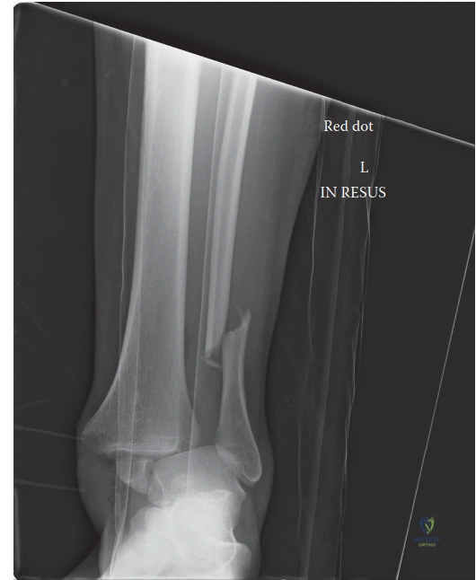
A 48-year-old construction worker presents following a 10-foot fall. He has severe ankle pain, inability to weight-bear, and significant swelling with Grade II fracture blisters. You are presented with his initial AP/Mortise/Lateral radiographs.
Describe your assessment of these radiographs and outline your immediate management plan.
Candidate: The X-ray shows an unstable ankle fracture. There is a fibular fracture, widening of the medial clear space, and a posterior malleolar fragment with talar subluxation. I would classify this as a PER IV fracture. I would splint the leg, keep him non-weight bearing, and book him for surgery once the swelling goes down.
Failure to mention the soft tissue status (Tscherne classification), omission of the patient's diabetic and smoking status which dictates the surgical timeline, and lack of systematic description of the radiographic columns (medial, lateral, posterior, syndesmotic).
A structured response is essential: "Radiographically, this is a multi-column injury: a high fibular fracture, syndesmotic diastasis, widened medial clear space, and a large posterior malleolar fragment with posterior talar subluxation. I would classify this as a Lauge-Hansen PER IV / AO 44-C3 injury. Clinically, the patient has Tscherne Grade II soft tissue injury and significant comorbidities (Diabetes, Smoking). I would perform an immediate closed reduction and application of a spanning external fixator given the soft tissue compromise. Definitive fixation is deferred until the 'wrinkle sign' is present, utilizing a prone approach to address the posterior malleolus and fibula to ensure anatomic reduction of the incisura."
You mentioned a posterior malleolar fracture. How do you determine the threshold for operative fixation of the posterior malleolus in this case, and what is your preferred surgical construct?
Candidate: We typically fix it if it's over 25% of the articular surface or if the talus is still subluxed. I would use an AP screw through a small stab incision or a posterior plate if the fragment is large.
Relying solely on the "25-33% rule." Modern teaching emphasizes that posterior malleolar fractures associated with syndesmotic instability should be fixed regardless of size, as the fragment houses the PITFL and is crucial for rotational stability.
State that the decision is now based on syndesmotic stability and articular congruency rather than just the percentage of the articular surface. I would obtain a CT scan to classify the fracture (Bartoníček-Rammelt) to assess for intercalary or 'die-punch' fragments. My preferred construct is a posterior buttress plate (anti-glide) via a prone posterolateral approach, which provides superior mechanical stability compared to AP lag screws, especially in a diabetic patient at risk of fixation failure.
The patient has significant Type 2 Diabetes and is a heavy smoker. How do these factors influence your perioperative management and surgical planning?
Candidate: These factors increase his risk of infection and wound healing problems. I would make sure his blood sugar is controlled before surgery and tell him to stop smoking.
Vague references to "controlling blood sugar" without discussing the physiological impact (microvascular compromise, collagen cross-linking) or the specific surgical strategy adjustments (avoiding hardware prominence, conservative soft tissue handling, and staged fixation).
"Diabetes and tobacco use create a high-risk environment for wound dehiscence and non-union. I would optimize the HbA1c preoperatively, strictly counsel on smoking cessation, and employ a staged approach—delaying definitive fixation until the soft tissues are pristine. During surgery, I would minimize soft tissue stripping, prefer locking plates to distribute load, and maintain a very low threshold for prophylactic antibiotic coverage. I would also have a very low threshold for using an external fixator to allow soft tissue 'resuscitation' before definitive internal fixation."
