Calcaneotibial Fusion & Talar Fractures: Surgical Guide
Key Takeaway
Calcaneotibial fusion is a powerful salvage procedure for severe talar body fractures, avascular necrosis, and end-stage arthropathy. This comprehensive guide details the anterolateral approach, talar extirpation, and rigid fixation techniques. It also explores the Blair fusion alternative for preserving limb length and midtarsal motion, alongside evidence-based management protocols for lateral process talar fractures, ensuring optimal biomechanical restoration and functional outcomes in complex hindfoot reconstruction.
Comprehensive Introduction and Patho-Epidemiology
Calcaneotibial fusion, formally known as tibiocalcaneal arthrodesis, represents a formidable and definitive salvage procedure reserved for the most severe, intractable pathologies of the hindfoot and ankle. When the talus is rendered entirely unsalvageable—whether due to highly comminuted, high-energy crush injuries, profound avascular necrosis (AVN), recalcitrant chronic osteomyelitis, or massive oncologic bone loss—standard tibiotalar arthrodesis is no longer a viable reconstructive option. In these highly complex clinical scenarios, the complete extirpation of the talar body followed by the direct coaptation and fusion of the distal tibia to the calcaneus provides a stable, plantigrade, and painless limb. This functional restoration, however, comes at the permanent cost of hindfoot kinematics and a mandatory reduction in overall limb length.
The patho-epidemiology of conditions necessitating calcaneotibial fusion is heavily skewed toward high-velocity trauma and its devastating sequelae. Talar body and neck fractures, particularly those classified as Hawkins Type III or IV, carry an extraordinarily high risk of avascular necrosis due to the tenuous, retrograde blood supply of the talus. When the delicate anastomotic sling formed by the artery of the tarsal canal and the artery of the sinus tarsi is disrupted, ischemic necrosis of the talar dome ensues. Subsequent subchondral collapse rapidly leads to end-stage post-traumatic tibiotalar and subtalar arthrosis. In cases where the necrotic segment is massive, or when secondary infection supervenes, joint-sparing procedures or standard fusions are doomed to failure, leaving talectomy and calcaneotibial fusion as the only viable alternative to major lower extremity amputation.
Beyond the realm of massive talar destruction, the orthopedic surgeon must also master the management of localized talar trauma, most notably fractures of the lateral process of the talus. Historically underdiagnosed and frequently mismanaged as severe lateral ankle sprains, these "snowboarder's fractures" carry significant morbidity if missed. The lateral process acts as a critical anatomical hub for the lateral ligamentous complex of the ankle. Failure to recognize and appropriately treat displaced lateral process fractures inevitably leads to subtalar nonunion, chronic lateral column pain, and early-onset degenerative joint disease, fundamentally altering the patient's functional trajectory.
This comprehensive masterclass delineates the intricate surgical indications, complex biomechanical considerations, and step-by-step operative techniques for standard calcaneotibial fusion. Furthermore, it explores the Blair fusion—a highly effective, height-preserving alternative—and provides an evidence-based algorithmic approach to the surgical management of lateral process talar fractures. Mastery of these techniques is essential for the advanced foot and ankle surgeon tasked with salvaging the unsalvageable limb.
Detailed Surgical Anatomy and Biomechanics
A profound understanding of the osteology, vascularity, and ligamentous anatomy of the hindfoot is paramount when undertaking a calcaneotibial fusion or addressing talar fractures. The talus is a unique osseous structure, devoid of any muscular or tendinous attachments, relying entirely on capsuloligamentous structures for its stability and vascular supply. Approximately 60% of its surface is covered by articular cartilage, leaving limited extra-articular real estate for vascular penetration. The primary blood supply arises from the posterior tibial artery via the artery of the tarsal canal, which anastomoses with the artery of the sinus tarsi (derived from the anterior tibial and peroneal arteries). Disruption of this vascular network during high-energy trauma or aggressive surgical dissection directly precipitates avascular necrosis, necessitating the salvage procedures discussed herein.
Biomechanically, the complete removal of the talus profoundly alters the functional mechanics of the lower extremity. Direct coaptation of the distal tibia to the calcaneus inherently shortens the limb by 2 to 3 centimeters, depending on the volume of necrotic bone resected and whether structural allografts are utilized to span the defect. This loss of height has cascading biomechanical consequences. Most notably, the absence of the talus shortens the moment arm of the Achilles tendon, significantly diminishing plantarflexion power and altering the gait cycle. The surgeon must meticulously balance the desire to restore limb length using massive structural grafts against the risk of catastrophic soft tissue tension.
This soft tissue tension is clinically referred to as the "accordion effect." Chronic absence, collapse, or fragmentation of the talus leads to a severe, progressive contracture of the surrounding neurovascular bundles, tendinous units, and the cutaneous envelope. Attempting to acutely restore the original limb length by jacking the calcaneus away from the tibia with a large structural graft can stretch the posterior tibial neurovascular bundle beyond its physiological limits, resulting in ischemic neuropathy or catastrophic wound dehiscence. Consequently, the surgeon must often accept a degree of limb shortening to ensure tension-free soft tissue closure and preservation of distal perfusion.
In the context of lateral process fractures, the anatomy is equally unforgiving. The lateral process of the talus is a wedge-shaped prominence that articulates with the fibula superolaterally and the calcaneus inferolaterally. It serves as the critical footprint for three major stabilizing ligaments: the lateral talofibular ligament (LTFL), the anterior talofibular ligament (ATFL), and the posterior talofibular ligament (PTFL). Despite these dense ligamentous attachments, surgical excision of a comminuted, ununited lateral process fragment does not typically induce clinically significant ankle or subtalar instability, provided the deep deltoid and syndesmotic complexes remain intact. This anatomical quirk underpins the surgical rationale for primary excision of highly comminuted lateral process fragments that defy anatomical reduction.
Exhaustive Indications and Contraindications
The decision to proceed with a calcaneotibial fusion is never taken lightly; it is a definitive, end-of-the-line procedure indicated only when catastrophic failure or absolute absence of the talar body precludes any other reconstructive effort. Severe, high-energy comminuted talar body fractures that defy open reduction and internal fixation (ORIF) due to massive articular cartilage destruction or complete extrusion represent a primary indication. In these scenarios, the native architecture is so thoroughly obliterated that attempting fixation would merely delay the inevitable onset of necrosis and infection. Similarly, advanced avascular necrosis of the talus, frequently a sequela of displaced talar neck fractures, leads to talar dome collapse and secondary pan-talar arthritis, necessitating radical resection and fusion.
Chronic osteomyelitis of the talus presents a particularly challenging indication. Intractable infections, often resulting from open fractures or failed prior osteosyntheses, require aggressive, complete, or partial talectomy to eradicate the nidus of infection. In such cases, calcaneotibial fusion may be performed in a staged manner, utilizing antibiotic-impregnated cement spacers before definitive arthrodesis. Neuropathic (Charcot) arthropathy with severe midfoot and hindfoot collapse also frequently requires rigid stabilization via tibiocalcaneal fusion to prevent ulceration and eventual amputation. Finally, the resection of primary benign aggressive or malignant tumors of the talus may leave a massive osseous void that can only be managed via this salvage pathway.
Contraindications to calcaneotibial fusion must be rigorously respected to prevent disastrous postoperative complications. Active, untreated deep infection is an absolute contraindication to single-stage fusion with internal hardware; such cases mandate a staged approach. Severe peripheral vascular disease that compromises the healing potential of the surgical incisions or the fusion mass must be optimized prior to any intervention, or alternative management (such as amputation) should be considered. Relative contraindications include profound osteoporosis, which compromises hardware purchase, and severe medical comorbidities that preclude prolonged anesthesia or the rigorous demands of postoperative non-weight-bearing rehabilitation.
| Parameter | Calcaneotibial Fusion / Talectomy | Lateral Process Talar Fractures |
|---|---|---|
| Primary Indications | Unsalvageable comminuted talar body fractures; End-stage AVN with collapse; Chronic intractable osteomyelitis; Severe Charcot neuroarthropathy; Oncologic resection. | Displaced (>2mm) Hawkins Type I fractures; Comminuted Hawkins Type II fractures; Symptomatic nonunions of the lateral process. |
| Absolute Contraindications | Active, untreated local or systemic infection (for single-stage hardware placement); Critical limb ischemia; Non-ambulatory patient status. | Undisplaced fractures amenable to casting; Active localized soft tissue infection over the lateral hindfoot. |
| Relative Contraindications | Severe osteopenia/osteoporosis (risk of hardware failure); Active smoking (high nonunion risk); Uncontrolled diabetes mellitus. | Polytrauma precluding immediate fixation; Severe peripheral neuropathy; Poor soft tissue envelope. |
| Alternative Salvage | Below-knee amputation (BKA); Blair fusion (if talar neck is viable); Custom orthotic accommodation (rarely successful long-term). | Delayed excision of symptomatic nonunion fragment; Subtalar arthrodesis for end-stage post-traumatic arthrosis. |
Pre-Operative Planning, Templating, and Patient Positioning
Meticulous preoperative planning is the cornerstone of a successful calcaneotibial fusion. The evaluation begins with high-quality, weight-bearing orthogonal radiographs of the foot and ankle to assess the degree of deformity, bone loss, and adjacent joint arthritis. However, plain radiography is insufficient for three-dimensional preoperative templating. A fine-cut Computed Tomography (CT) scan with sagittal and coronal reconstructions is mandatory to precisely quantify the volume of the talar defect, evaluate the bone stock of the distal tibia and calcaneus, and plan the trajectory for intramedullary or extramedullary fixation. In cases of suspected AVN or chronic osteomyelitis, Magnetic Resonance Imaging (MRI) with intravenous contrast is invaluable for delineating the extent of necrotic or infected bone that must be resected.
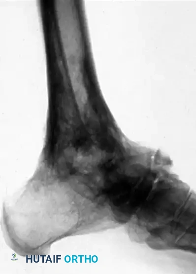
FIGURE 88-42 A: Preoperative radiograph demonstrating a severe talar body fracture with an associated fibular fracture, a classic injury pattern that, if unsalvageable, may necessitate tibiocalcaneal arthrodesis.
Preoperative templating must account for the anticipated limb length discrepancy (LLD). The surgeon must decide whether to accept the shortening—which facilitates tension-free soft tissue closure—or to utilize a structural allograft, such as a fresh-frozen femoral head, to restore height. If an allograft is planned, the templating process must precisely calculate the dimensions of the graft required to fill the void while avoiding the catastrophic "accordion effect." Hardware selection is also determined during this phase. While rigid multi-planar external fixation remains an option, particularly in cases of active infection, the modern gold standard for definitive internal fixation is a robust, retrograde tibiotalocalcaneal (TTC) intramedullary nail, supplemented by large-fragment compression screws.
Patient positioning is critical for optimal surgical exposure and intraoperative fluoroscopic imaging. The patient is typically placed in the lateral decubitus position or the supine position with a massive ipsilateral hip bump to internally rotate the leg. This provides unimpeded, direct access to the lateral hindfoot and allows for an extensile anterolateral or transfibular approach. A well-padded thigh tourniquet is applied to ensure a bloodless surgical field, though it should be deflated prior to final closure to meticulously assess distal perfusion, especially if a structural graft has been utilized to lengthen the limb.
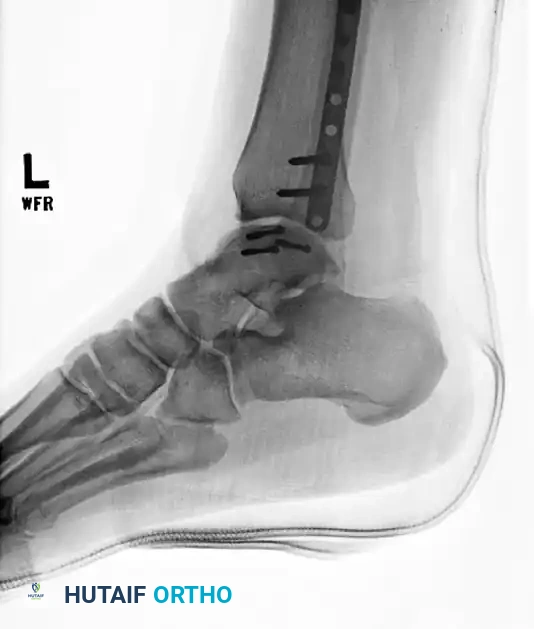
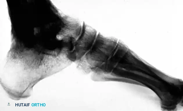
FIGURE 88-42 B: Attempted fixation of the talar body utilizing headless compression screws.
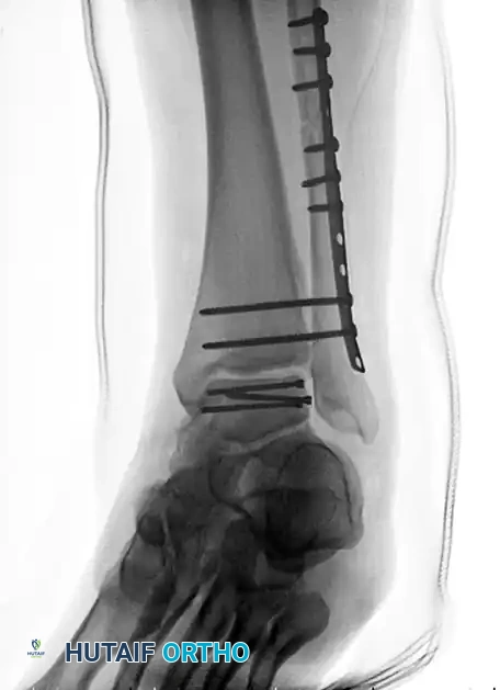
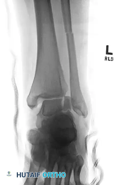
FIGURE 88-42 C: Concurrent fixation of the fibula with a neutralization plate and screws. Failure of such constructs often leads to the salvage pathways discussed herein.
The operating room setup must include a radiolucent table and a C-arm fluoroscopy unit positioned to allow seamless acquisition of both anteroposterior (AP) and lateral views of the tibia, ankle, and foot without compromising the sterile field. The surgical team must ensure that all necessary equipment, including spherical reamers, flexible osteotomes, high-speed burrs, and a comprehensive TTC nailing system, are readily available. If autogenous bone grafting is planned, the ipsilateral anterior iliac crest or proximal tibia should be prepped and draped into the sterile field.
Step-by-Step Surgical Approach and Fixation Technique
The primary objective of calcaneotibial fusion is to achieve rigid, compressive osteosynthesis between the distal tibia, the superior calcaneus, and frequently the navicular, ensuring a stable, plantigrade foot optimized for weight-bearing. This requires a systematic, meticulously executed surgical approach.
The Extensile Anterolateral Approach and Talar Extirpation
The operative field is exposed through an extensile anterolateral incision, beginning approximately 10 centimeters proximal to the ankle joint, centered over the fibula, and extending distally toward the base of the fourth metatarsal. This approach utilizes the internervous plane between the superficial peroneal nerve (supplying the anterior compartment) and the sural nerve (supplying the lateral compartment). Deep dissection requires meticulous identification and retraction of the extensor digitorum longus and peroneus tertius tendons medially. The lateral malleolus is often osteotomized and resected to serve as local autograft and to provide unhindered access to the subtalar and tibiotalar articulations.
Once the joint spaces are accessed, the fragmented, infected, or necrotic remnants of the talar body are systematically removed. In cases of comminuted fractures that have consolidated into a malunion, or when the junction of the talar body and neck remains partially intact, the surgeon must not attempt to pry the talus out en bloc, as this risks severe iatrogenic damage to the surrounding neurovascular structures. Instead, the talus should be aggressively divided with a sharp osteotome or high-speed burr into multiple fragments for safe, atraumatic piecemeal extirpation. To facilitate anterior column fusion, an osteotome is driven through the proximal aspect of the navicular to resect its proximal articular cartilage en bloc with the remaining head and neck of the talus.
Joint Preparation and Addressing the Accordion Effect
Following complete talectomy, the surgeon must meticulously prepare the remaining articular surfaces to optimize the biological environment for fusion. The articular cartilage and subchondral bone of the distal tibial plafond and the superior facet of the calcaneus are aggressively excised down to healthy, bleeding, punctate cancellous bone using a combination of osteotomes, curettes, and spherical reamers. If the lateral malleolus was preserved, its medial surface must be roughened to promote lateral gutter fusion.
Overcoming the "accordion effect" is often the most challenging aspect of the procedure. The chronic absence of the talus leads to severe soft tissue contracture. To allow the calcaneus to translate proximally and the navicular to displace posteriorly into direct contact with the anterior tibia, extensive soft tissue stripping around both the medial and lateral malleoli is mandatory. Surgical Warning: The soft tissues will fiercely resist efforts to properly appose the calcaneus to the tibia. Forcing this apposition without adequate soft tissue release or sufficient bony resection risks catastrophic skin necrosis and neurovascular compromise. It is frequently necessary to resect a larger portion of both malleoli to relieve tension on the cutaneous envelope.
Structural Grafting and Coaptation
Once adequate mobilization is achieved, the anterior tibia is denuded at the exact point of anticipated contact with the navicular to facilitate a secondary, stabilizing anterior fusion mass. If active infection has been definitively ruled out and the surgeon elects to restore limb height, a massive structural allograft—typically a fresh-frozen femoral head—is introduced. The allograft, the distal tibia, and the superior calcaneus must be meticulously contoured using matching spherical reamers. This creates a "cup and cone" geometry that ensures a perfect, flush fit, maximizing the surface area for osteoconduction and providing intrinsic biomechanical stability prior to hardware placement.
Definitive Fixation Strategies
Positioning the foot is paramount; it must be held strictly at a right angle to the leg (neutral dorsiflexion/plantarflexion) or in a maximum of 5 degrees of dorsiflexion. The hindfoot should be positioned in approximately 5 degrees of valgus, with external rotation matching the contralateral limb (typically 10 to 15 degrees). Two heavy Steinmann pins are inserted transversely through the calcaneus and tibia to provisionally hold this critical reduction.
Definitive fixation is most commonly achieved utilizing a robust, retrograde tibiotalocalcaneal (TTC) intramedullary nail. A guide wire is introduced through the plantar aspect of the calcaneus, advanced across the prepared calcaneotibial interface, and passed centrally up the tibial diaphysis under multi-planar fluoroscopic guidance. After sequential reaming, the nail is inserted and locked proximally and distally, utilizing internal compression mechanisms to maximize contact pressure across the fusion site. Alternatively, robust arthrodesis plates or multi-planar external fixators can be utilized. The navicular is subsequently fixed to the anterior tibia using a fully threaded cortical screw or a headless compression screw to enhance anterior column stability. Finally, local bone chips obtained from the malleolar resection, often augmented with orthobiologics or autogenous iliac crest bone graft, are densely packed around the osseous junctions.
The Blair Fusion Alternative
Because of the inevitable decrease in limb height and the severe rigidity of the ankle joint following standard calcaneotibial fusion, Blair introduced a highly effective, joint-preserving alternative. This technique is specifically indicated when the talar body is destroyed, but the talar head and neck remain viable, well-vascularized, and free of infection. In the Blair fusion, the comminuted or necrotic fragments of the talar body are completely excised. However, instead of dropping the tibia down to the calcaneus, a sliding cortical bone graft (typically 2 cm wide and 5 cm long) is harvested from the anterior surface of the distal tibia.
This cortical graft is translated distally and inserted directly into a meticulously prepared slot in the remnant of the head and neck of the talus. The primary goal is to obtain a solid, bridging fusion across the anterior tibia and the talar neck. Blair reported profound biomechanical advantages to this technique: the anatomical position of the foot remains unchanged, limb length is preserved, and the weight-bearing thrust is placed on undisturbed, native subtalar joint tissue. Crucially, the retained talonavicular and subtalar facets allow a compensatory "rocking motion," enabling the patient to walk with a fairly elastic gait and minimal limp.
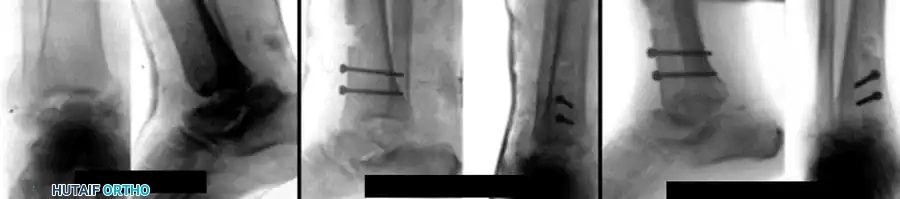
FIGURE 88-44: Results of Blair fusion. (A) Preoperative Type III fracture-dislocation of the talus with severe body comminution. (B) Immediate postoperative radiograph demonstrating the sliding anterior tibial graft slotted into the talar neck. (C) Solid fusion achieved at 3 months, preserving hindfoot height and subtalar kinematics.
Surgical Management of Lateral Process Talar Fractures
While massive talar body trauma necessitates salvage fusion, isolated fractures of the lateral process of the talus require a completely different, joint-preserving surgical algorithm. Often misdiagnosed as severe ankle sprains, these injuries demand high clinical suspicion. The classic mechanism involves axial loading, dorsiflexion, external rotation, and eversion. Hawkins classified these fractures to guide operative decision-making: Type I (large, single fragment), Type II (comminuted fragments), and Type III (nonarticular avulsions).
Operative intervention is strongly indicated for displaced Type I and Type II fractures. For Type I fractures, Open Reduction and Internal Fixation (ORIF) is performed via a small incision over the sinus tarsi. The fragment is anatomically reduced and secured using 1.5mm to 2.4mm mini-fragment headless compression screws or low-profile plates. Type II comminuted fractures, however, rarely heal anatomically and frequently lead to subtalar arthritis if internal fixation is attempted. These highly benefit from primary surgical débridement and complete excision of the comminuted fragments. Despite the removal of the ligamentous footprints, the deep deltoid and syndesmotic structures provide adequate stability, and excision yields excellent long-term functional outcomes.
Complications, Incidence Rates, and Salvage Management
Despite meticulous surgical technique, calcaneotibial fusion remains a high-risk salvage procedure with a substantial complication profile. The surgeon must be acutely aware of these risks, actively mitigate them during the perioperative period, and possess the requisite skills to manage them when they arise. Nonunion and delayed union are the most frequently encountered complications, with incidence rates reported between 15% and 30% in the literature. The risk of nonunion is exponentially higher in patients with active tobacco use, uncontrolled diabetes mellitus, profound peripheral neuropathy, or a history of prior local infection.
Infection, both superficial wound dehiscence and deep hardware-associated osteomyelitis, is another major concern. The compromised soft tissue envelope, particularly after high-energy trauma or multiple prior surgeries, is exceptionally vulnerable to breakdown. The "accordion effect," if not properly managed with adequate bone resection or soft tissue release, places immense tension on the anterior incision, leading to marginal necrosis and subsequent deep infection. Deep infections occurring before solid osseous consolidation are disastrous, often requiring complete hardware explantation, aggressive serial débridements, placement of antibiotic-impregnated cement spacers, and application of a circular external fixator (e.g., Ilizarov frame) to maintain alignment while the infection is eradicated.
Neurovascular injuries, while less common, carry profound morbidity. The sural nerve and the superficial peroneal nerve are at direct risk during the extensile anterolateral approach and during percutaneous distal locking of a TTC nail. Furthermore, acute lengthening of a chronically shortened limb using a massive structural allograft can cause a traction injury to the posterior tibial nerve or compromise the posterior tibial artery, leading to ischemic necrosis of the plantar foot. Careful intraoperative monitoring of distal perfusion and immediate adjustment of the graft size if ischemia is noted are critical preventative measures.
| Complication | Estimated Incidence | Risk Factors | Salvage / Management Strategy |
|---|---|---|---|
| Nonunion / Delayed Union | 15% - 30% | Smoking, Diabetes, AVN, Inadequate compression, Poor bone stock. | Prolonged immobilization; Bone stimulators; Revision surgery with robust bone grafting (autograft/rhBMP-2) and hardware exchange. |
| Deep Infection / Osteomyelitis | 5% - 15% | Prior open fracture, Immunosuppression, Poor soft tissue envelope. | Hardware removal; Aggressive surgical débridement; IV antibiotics; Application of circular external fixator; Staged reconstruction. |
| Wound Dehiscence / Necrosis | 10% - 20% | "Accordion effect" tension, Peripheral vascular disease, Smoking. | Local wound care; Negative pressure wound therapy (NPWT); Local rotational flaps or free tissue transfer (e.g., ALT flap). |
| Adjacent Segment Arthritis | 40% - 60% (Long-term) | Altered hindfoot kinematics, Pre-existing midtarsal joint damage. | Conservative management (custom orthotics, rocker-bottom shoes); Selective midfoot fusions (e.g., talonavicular or calcaneocuboid) if conservative measures fail. |
| Neurovascular Compromise | < 5% | Excessive acute lengthening with structural allograft, Retractor injury. | Immediate removal or downsizing of structural graft; Exploration and release of neurovascular bundles; Nerve repair if transected. |
Phased Post-Operative Rehabilitation Protocols
The postoperative rehabilitation following a calcaneotibial fusion is protracted, demanding immense patient compliance and close clinical monitoring. The protocol is strictly phased to protect the fragile biological environment required for massive bone consolidation while preventing secondary complications such as deep vein thrombosis or severe disuse osteopenia.
Phase I: Immediate Postoperative to 6-8 Weeks (Strict Non-Weight-Bearing)
Immediately following surgery, the limb is placed in a bulky, well-padded, sterile compressive dressing and a rigid posterior splint with a U-shaped stirrup to maintain neutral alignment and control edema. The patient is kept strictly non-weight-bearing. At the two-week mark, the sutures or staples are removed, provided the soft tissue envelope has healed adequately. The patient is then transitioned into a rigid, fiberglass short-leg cast. Strict non-weight-bearing must be maintained for a minimum of 6 to 8 weeks. Premature weight-bearing during this phase places catastrophic shear forces across the fusion interface, virtually guaranteeing hardware failure or nonunion.
Phase II: 6-8 Weeks to 12-16 Weeks (Progressive Weight-Bearing)
At the 6 to 8-week postoperative visit, orthogonal radiographs are obtained. If there is early radiographic evidence of bridging trabeculae across the calcaneotibial and naviculotibial interfaces, and the patient is clinically non-tender at the fusion sites, a transition to progressive weight-bearing may commence. The patient is typically placed in a removable controlled ankle motion (CAM) boot or a weight-bearing cast. Weight-bearing is advanced incrementally, starting with 25% of body weight using crutches or a walker, and progressing to full weight-bearing over the next 4 to 6 weeks. Physical therapy is initiated during this phase, focusing heavily on strengthening the proximal musculature (quadriceps, hamstrings, gluteals) and maintaining mobility in the toes and knee.
Phase III: Long-Term Maintenance and Return to Activity
Once solid osseous consolidation is definitively confirmed—typically between 12 and 16 weeks, though it may take up to 6 months in compromised hosts—the patient transitions to regular footwear. However, the limb must be protected for the next several months utilizing a custom-molded short-leg double-upright brace or an ankle-foot orthosis (AFO) with a locked ankle joint. This bracing prevents excessive shear stress on the maturing, remodeling fusion mass. Long-term, patients benefit immensely from customized shoe modifications, specifically a rigid shank and a rocker-bottom sole. Because the ankle is
Clinical & Radiographic Imaging Archive