Comprehensive Introduction and Patho-Epidemiology
Femoroacetabular impingement (FAI) represents a paradigm shift in our understanding of hip joint preservation, transitioning the orthopedic focus from end-stage arthroplasty to early biomechanical intervention. First comprehensively described by Reinhold Ganz and colleagues in the early 2000s, FAI is a dynamic, pathomechanical process characterized by abnormal morphological contact between the proximal femur and the acetabular rim. Cam impingement, the specific focus of this chapter, arises from an aspherical femoral head-neck junction. This morphological aberration—frequently described radiographically as a "pistol grip" deformity or a quantifiable reduction in the normal femoral head-neck offset—creates a space-occupying lesion that forcefully enters the joint during physiological ranges of motion.
The patho-epidemiology of cam morphology is heavily skewed toward young, active male patients, particularly those participating in sports that demand repetitive, high-impact hip flexion and internal rotation, such as ice hockey, soccer, and martial arts. Current literature suggests that the development of the cam lesion is intrinsically linked to altered mechanical loading across the proximal femoral physis during the critical stages of adolescent skeletal maturation. As the physis closes under these supraphysiologic shear forces, reactive bone formation occurs at the anterolateral head-neck junction, solidifying the aspherical deformity. While many individuals with cam morphology remain asymptomatic, the presence of this osseous prominence is a well-documented, independent risk factor for the development of symptomatic FAI and subsequent early-onset osteoarthritis.
The biomechanical consequence of this repetitive abutment is profoundly destructive to the articular ecosystem. During terminal hip flexion and internal rotation, the cam lesion is forcefully driven into the anterosuperior acetabulum. Unlike pincer impingement, which primarily crushes the labrum against the acetabular rim, the cam lesion acts as a cam-lobe in a mechanical engine. It generates massive shear forces directly at the chondrolabral junction. This repetitive shear stress leads to an "inside-out" delamination of the acetabular articular cartilage from the subchondral bone, often leaving the overlying labrum macroscopically intact in the early stages. Over time, this progressive chondral failure compromises the structural integrity of the labral attachment, leading to complex labral tears, loss of the hip fluid seal, and accelerated joint degeneration.
Arthroscopic osteochondroplasty has firmly established itself as the gold standard for addressing symptomatic cam morphology. The procedure has evolved from highly morbid open surgical dislocations to a minimally invasive, arthroscopic technique that demands a meticulous understanding of three-dimensional hip anatomy. The primary objective is not merely to create a perfectly spherical femoral head, but to precisely restore a functional, impingement-free range of motion while meticulously preserving the delicate lateral retinacular vessels that supply the femoral head. Mastery of this procedure requires a systematic approach to both the central and peripheral compartments of the hip joint, coupled with rigorous preoperative planning and dynamic intraoperative assessment.
Detailed Surgical Anatomy and Biomechanics
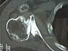
A profound mastery of the osseous, capsuloligamentous, and vascular anatomy of the hip is the absolute prerequisite for safe and effective arthroscopic cam resection. The proximal femur's osseous anatomy is defined by the spherical femoral head transitioning into the cylindrical femoral neck. In the normal hip, this transition creates a concave offset that allows the femur to clear the acetabular rim during deep flexion. In cam morphology, this concavity is lost, predominantly in the anterosuperior and anterior quadrants. The severity of this osseous buildup is quantified by the alpha angle, a radiographic measurement that identifies the exact point where the femoral head deviates from a perfect sphere. Understanding the precise topographic distribution of the cam lesion is critical, as it dictates the spatial boundaries of the arthroscopic osteochondroplasty.
The capsuloligamentous complex of the hip is a robust, dynamic stabilizer that must be carefully managed during arthroscopy. The anterior capsule is reinforced by the massive iliofemoral ligament (the Y ligament of Bigelow), which originates from the anterior inferior iliac spine (AIIS) and inserts along the intertrochanteric line. This ligament is the primary restraint to hip extension and external rotation. Deep to the longitudinal fibers of the capsule lies the zona orbicularis, a distinct collar of circumferential fibers that encircles the narrowest portion of the femoral neck. The zona orbicularis acts as a critical locking mechanism, resisting joint distraction and contributing significantly to the hip's inherent stability. During the surgical approach, particularly when performing a T-capsulotomy, the surgeon must respect these structures to prevent iatrogenic microinstability or frank subluxation.
The vascular anatomy of the proximal femur is arguably the most critical anatomical consideration during cam resection, as iatrogenic injury can lead to catastrophic avascular necrosis (AVN) of the femoral head. The primary blood supply to the femoral head is derived from the medial circumflex femoral artery (MCFA). The MCFA courses posteriorly around the femoral neck and gives rise to the lateral epiphyseal artery, which subsequently branches into the superior retinacular vessels. These delicate vessels pierce the joint capsule near the superior base of the femoral neck and run subsynovially along the posterosuperior and lateral aspects of the neck before entering the femoral head. The absolute "no-go" zone during osteochondroplasty is the superior-lateral quadrant of the femoral neck. Resection must remain strictly anterior to the lateral retinacular vessels to ensure the vascular integrity of the femoral head is preserved.
Biomechanically, the hip joint functions as a highly constrained ball-and-socket mechanism dependent on the delicate interplay between osseous morphology and the soft-tissue fluid seal. The acetabular labrum deepens the socket by 21%, increases the articular surface area, and maintains a pressurized intra-articular fluid seal that is vital for cartilage nutrition and load distribution. When a cam lesion violates this seal during the FADIR (Flexion, Adduction, Internal Rotation) maneuver, it dramatically increases the contact pressures within the anterosuperior quadrant. The arthroscopic intervention must therefore not only eliminate the osseous conflict but also restore the labral seal. Failure to recognize and respect these intricate biomechanical relationships will inevitably lead to suboptimal surgical outcomes and persistent postoperative pain.
Exhaustive Indications and Contraindications
Patient selection is the cornerstone of successful hip preservation surgery. The diagnosis of cam-type FAI is a triad requiring a characteristic clinical history, reproducible physical examination findings, and corroborating advanced imaging. Patients typically present with an insidious onset of deep, aching anterior groin pain that is mechanically exacerbated by prolonged sitting, driving, deep squatting, or pivoting movements. The classic "C-sign" is frequently observed, wherein the patient cups their hand over the greater trochanter with the thumb positioned posteriorly and the fingers wrapping anteriorly into the groin, precisely localizing the deep intra-articular pathology. Mechanical symptoms such as catching, clicking, or a sensation of the hip "giving way" are highly indicative of associated labral tearing or unstable chondral flaps resulting from the cam impingement.
The physical examination must be rigorous and systematic. The anterior impingement test (FADIR maneuver) is the most sensitive clinical indicator for anterior FAI and concomitant labral pathology. By passively moving the hip into 90 degrees of flexion, followed by adduction and internal rotation, the examiner forces the anterolateral cam deformity directly into the anterosuperior acetabular rim, sharply reproducing the patient's characteristic groin pain. The FABER (Flexion, Abduction, External Rotation) test is utilized to assess for concurrent sacroiliac joint pathology, iliopsoas tightness, or lateral rim impingement. Additionally, a comprehensive assessment of hip abductor strength, core stability, and generalized ligamentous laxity (Beighton score) is mandatory, as underlying microinstability or muscular dysfunction can profoundly influence the surgical outcome and rehabilitation trajectory.
Advanced imaging is non-negotiable for preoperative templating and confirming the diagnosis. Standard weight-bearing anteroposterior (AP) pelvis, Dunn 45-degree, and cross-table lateral radiographs are utilized to evaluate the joint space, assess pelvic tilt, and measure the alpha angle. An alpha angle exceeding 50 to 55 degrees on the lateral projections is widely accepted as indicative of cam morphology. Magnetic Resonance Arthrogram (MRA) is the imaging modality of choice for evaluating the soft-tissue envelope. MRA provides unparalleled resolution of the acetabular labrum, allowing for the classification of tears and the detection of subtle chondral delamination at the chondrolabral junction. Furthermore, 3D Computed Tomography (CT) reconstructions are increasingly utilized by high-volume hip preservation surgeons to precisely map the three-dimensional volume and spatial orientation of the cam lesion, facilitating highly accurate preoperative virtual resections.
Despite the high success rates of arthroscopic osteochondroplasty, strict adherence to contraindications is vital to prevent surgical failures and rapid progression to total hip arthroplasty (THA). Advanced osteoarthritis is the most absolute contraindication; patients with less than 2 mm of remaining joint space or a Tönnis grade of 2 or higher experience uniformly poor outcomes following hip arthroscopy. Similarly, severe acetabular dysplasia (Lateral Center Edge Angle < 20 degrees) is a contraindication for isolated cam resection, as the cam lesion often acts as a secondary stabilizer in the dysplastic hip; removing it without addressing the underlying lack of acetabular coverage can precipitate catastrophic rapid-onset instability and joint subluxation.
| Category | Specific Parameter | Clinical Significance and Rationale |
|---|---|---|
| Indications | Symptomatic FAI failing > 3 months conservative tx | Confirms mechanical nature of pain unresponsive to PT/NSAIDs. |
| Indications | Alpha Angle > 55° on Dunn/Lateral views | Quantifies the presence of a pathological cam morphology. |
| Indications | Concomitant symptomatic labral tear | Requires central compartment access and repair alongside resection. |
| Indications | Joint space > 2mm (Tönnis Grade 0 or 1) | Ensures sufficient articular cartilage remains for joint preservation. |
| Contraindications | Advanced Osteoarthritis (Tönnis Grade 2 or 3) | High failure rate; patient will likely require Total Hip Arthroplasty. |
| Contraindications | Severe Acetabular Dysplasia (LCEA < 20°) | Cam lesion provides stability; resection causes iatrogenic subluxation. |
| Contraindications | Severe Obesity (BMI > 35-40) | Technical impossibility of safe portal placement and adequate traction. |
| Contraindications | Active joint infection or severe medical comorbidities | Standard surgical and anesthetic contraindications apply. |
Pre-Operative Planning, Templating, and Patient Positioning
Preoperative planning for arthroscopic cam resection requires a meticulous, multi-modal approach to ensure precise execution and avoid catastrophic complications. The surgeon must synthesize data from plain radiographs, MRI, and 3D CT scans to construct a mental, and often digital, model of the patient's specific pathoanatomy. Templating involves measuring the maximal depth and arc of the cam lesion to determine the exact volume of bone that must be resected. A critical biomechanical threshold must be respected: resection of more than 30% of the femoral neck diameter creates a dangerous stress riser, exponentially increasing the risk of a postoperative femoral neck fracture. Therefore, the surgeon must pre-calculate the safe depth of resection and identify anatomical landmarks—such as the medial synovial fold and the lateral retinacular vessels—that will serve as the absolute boundaries during the arthroscopic procedure.
Patient positioning and operating room setup form the critical foundation of a successful hip arthroscopy. The procedure is most commonly performed with the patient in the supine position on a specialized, radiolucent orthopedic traction table. The setup requires meticulous attention to detail to prevent devastating traction-related neuropraxias and soft-tissue injuries. A well-padded, oversized perineal post is positioned eccentrically against the medial thigh of the operative leg. This eccentric placement lateralizes the vector of traction, directing the force toward the medial thigh rather than compressing the delicate pudendal nerve against the pubic symphysis. The patient's torso and upper extremities are carefully padded and secured to prevent shifting during the application of massive traction forces.
The application of traction is a highly controlled, step-wise process. Gross traction is applied to the operative extremity to distract the hip joint by approximately 10 to 15 millimeters. This distraction is necessary to break the powerful suction seal of the joint and allow safe introduction of arthroscopic instruments into the central compartment without causing iatrogenic scuffing of the femoral head or acetabular cartilage. The distraction is continuously monitored and confirmed via an intraoperative fluoroscopic C-arm, which is positioned perpendicularly to the patient to allow for rapid acquisition of both AP and lateral imaging. Simultaneous counter-traction is applied to the non-operative leg, which is placed in slight abduction, to stabilize the pelvis and prevent pelvic tilt during the procedure.
A strict, non-negotiable temporal limit governs the use of traction. To prevent irreversible ischemic injury to the pudendal, sciatic, and femoral nerves, continuous traction must never exceed 120 minutes. High-volume hip arthroscopists typically complete the central compartment work (diagnostic sweep, labral repair, pincer resection) well within 45 to 60 minutes. Once the central compartment pathology is addressed, traction must be completely released. The transition to the peripheral compartment for the cam resection is performed entirely without traction, relying instead on dynamic hip flexion and capsular distension to visualize the femoral head-neck junction. This strict adherence to traction protocols is the single most effective measure for eliminating traction-related neurological complications.
Step-by-Step Surgical Approach and Fixation Technique
The surgical approach to the hip joint demands extreme precision in portal placement to avoid the complex neurovascular structures surrounding the joint. The procedure typically utilizes two or three primary portals. The Anterolateral (AL) portal is established first under direct fluoroscopic guidance. A spinal needle is introduced 1 cm anterior and 1 cm superior to the tip of the greater trochanter, directing the trajectory toward the superior-lateral aspect of the femoral neck. Once the capsule is breached and the joint is vented, a nitinol guidewire is advanced, followed by a dilator and a 5.0 mm arthroscopic cannula. The Mid-Anterior Portal (MAP) is then established under direct arthroscopic visualization from the AL portal. The MAP is located approximately 5 to 7 cm distal and slightly anterior to the AL portal, carefully avoiding the lateral femoral cutaneous nerve (LFCN) by remaining lateral to the sartorius muscle. A Distal Anterolateral Accessory (DALA) portal is frequently added to provide the optimal trajectory for the motorized burr during the osteochondroplasty.
The operation commences in the central compartment under traction. The surgeon performs a systematic diagnostic sweep, evaluating the ligamentum teres, the acetabular fossa, the integrity of the articular cartilage, and the acetabular labrum. Any central compartment pathology must be definitively addressed before proceeding to the cam lesion. This typically involves resecting any pincer morphology (acetabular rim trimming) using a 5.5 mm motorized burr, followed by labral repair or reconstruction using knotless suture anchors. If chondral delamination is present, a microfracture or chondroplasty is performed. Once the central compartment is fully restored and the labral seal is re-established, the surgeon prepares for the critical transition to the peripheral compartment.
Transitioning to the peripheral compartment begins with the complete release of traction. The operative leg is removed from the traction boot, and the hip is dynamically flexed to approximately 45 degrees. This specific degree of flexion serves a dual biomechanical purpose: it relaxes the robust iliofemoral ligament, allowing the anterior capsule to distend maximally with fluid pressure, and it rotates the anterior and anterolateral cam deformity directly into the arthroscopic field of view. To achieve adequate visualization of the expansive cam lesion, an interportal capsulotomy is performed, connecting the AL and MAP portals. In cases of massive cam lesions extending far distally down the femoral neck, a T-capsulotomy may be required. The vertical limb of the T-capsulotomy is created parallel to the femoral neck, but the surgeon must exercise extreme caution not to extend this cut too far distally, which risks severing the zona orbicularis, or too far laterally, which jeopardizes the lateral retinacular vessels.
The arthroscopic osteochondroplasty is the defining maneuver of the procedure. The arthroscope (typically a 70-degree lens for a wider field of view) is positioned in the MAP, looking back toward the femoral neck. A 5.5 mm hooded arthroscopic burr is introduced through the DALA portal. The surgeon first delineates the strict anatomical boundaries of the resection: the medial synovial fold marks the medial/inferior limit, the articular cartilage margin marks the proximal limit, and most importantly, the lateral retinacular vessels mark the absolute lateral/superior boundary. Resection begins at the apex of the cam deformity, meticulously burring away the aspherical bone to recreate a smooth, concave contour that tapers seamlessly into the intertrochanteric line.
Intraoperative dynamic assessment and fluoroscopic confirmation are mandatory to validate the adequacy of the resection. The surgeon utilizes the C-arm to obtain dynamic AP, Dunn, and cross-table lateral views, confirming the restoration of a normal alpha angle (< 50 degrees) and verifying that the resection depth has not violated the 30% rule. Following fluoroscopic clearance, the arthroscope is left in the joint while an assistant takes the hip through a full, dynamic FADIR range of motion. The surgeon directly visualizes the newly contoured head-neck junction entering the acetabulum. There must be a smooth, gliding articulation with absolutely no residual osseous abutment against the repaired labrum. Finally, the procedure concludes with meticulous capsular closure. Using specialized arthroscopic suture shuttling devices, the capsulotomy is closed with high-strength, non-absorbable sutures to restore the biomechanical tension of the iliofemoral ligament and prevent postoperative iatrogenic microinstability.
Complications, Incidence Rates, and Salvage Management
While arthroscopic management of cam FAI is highly successful in properly selected patients, the learning curve is steep, and the potential for devastating complications is significant. The most frequent complication, and the leading cause of revision hip arthroscopy, is inadequate resection of the cam lesion. Under-resection typically occurs at the distal-lateral extent of the cam deformity, an area that is notoriously difficult to visualize and access arthroscopically. When residual cam morphology remains, the patient will continue to experience mechanical impingement, leading to rapid failure of the labral repair and persistent postoperative groin pain. Salvage management for under-resection requires a revision arthroscopy to complete the osteochondroplasty, often necessitating the use of a 70-degree arthroscope and accessory distal portals to access the residual bone.
Conversely, over-resection of the cam lesion carries catastrophic biomechanical consequences. If the surgeon violates the 30% rule—resecting more than 30% of the cross-sectional diameter of the femoral neck—the structural integrity of the proximal femur is severely compromised. This creates a massive stress riser that predisposes the patient to a postoperative femoral neck fracture, particularly during the early rehabilitation phase when the bone is actively remodeling. The incidence of this complication is rare (< 1%) but devastating. Salvage management of a post-arthroscopy femoral neck fracture requires immediate open reduction and internal fixation (ORIF) with cannulated screws or a dynamic hip screw, and in older patients or cases of delayed presentation, it may necessitate conversion to a total hip arthroplasty.
Avascular necrosis (AVN) of the femoral head is the most feared biological complication of cam resection. It is entirely iatrogenic, resulting from direct mechanical injury or thermal necrosis to the lateral retinacular vessels during superior-lateral osteochondroplasty. These vessels are the terminal blood supply to the femoral head, and their destruction leads to irreversible osteocyte death and subsequent structural collapse of the articular surface. To prevent this, surgeons must maintain a strict margin of safety, staying entirely anterior to the retinacular vessels. If AVN occurs, salvage options are severely limited; core decompression may be attempted in very early stages, but the vast majority of these patients will rapidly progress to requiring a total hip arthroplasty.
Iatrogenic instability is an increasingly recognized complication arising from improper management of the hip capsule. The routine use of extensive, unrepaired T-capsulotomies destroys the stabilizing function of the iliofemoral ligament. In patients with underlying borderline dysplasia, generalized ligamentous laxity, or increased femoral anteversion, failure to close the capsule can result in anterior microinstability, persistent pain, and in severe cases, frank anterior subluxation of the femoral head. Prevention dictates routine, watertight capsular closure using high-strength sutures. Salvage for iatrogenic instability requires revision arthroscopy for capsular plication or, in cases of severe capsular deficiency, open capsular reconstruction using allograft tissue.
| Complication | Estimated Incidence | Etiology / Risk Factors | Salvage / Management Strategy |
|---|---|---|---|
| Inadequate Resection | 5.0% - 10.0% | Poor visualization; failure to access distal/lateral cam extent. | Revision arthroscopy for completion osteochondroplasty. |
| Over-Resection / Fracture | < 1.0% | Resecting > 30% of femoral neck diameter; early weight-bearing. | Immediate ORIF (cannulated screws) or THA if displaced/older. |
| Avascular Necrosis (AVN) | < 0.5% | Direct injury to lateral retinacular vessels during superior burring. | Core decompression (early) or Total Hip Arthroplasty (late). |
| Iatrogenic Instability | 1.0% - 2.0% | Unrepaired T-capsulotomy; underlying borderline dysplasia. | Revision arthroscopy for capsular plication or open reconstruction. |
| Pudendal Neuropraxia | 1.0% - 5.0% | Prolonged traction (> 2 hours); inadequate perineal post padding. | Observation; typically resolves spontaneously within 3-6 months. |
| Heterotopic Ossification | 1.0% - 3.0% | Bone debris left in capsule; failure to use NSAID prophylaxis. | NSAIDs (Naproxen/Indomethacin) x 3 weeks; surgical excision if mature/symptomatic. |
Phased Post-Operative Rehabilitation Protocols
Rehabilitation following arthroscopic cam resection is a highly structured, phased process that is equally as critical to the final clinical outcome as the surgical execution itself. The biological healing timeline of the osteochondroplasty site, the labral repair, and the capsular closure dictates the pace of progression. The rehabilitation protocol must strike a delicate balance between protecting the vulnerable surgical repairs and preventing the formation of intra-articular adhesions. Communication between the orthopedic surgeon and the physical therapy team must be seamless, with clear parameters established for weight-bearing status, range of motion limitations, and functional milestones.
Phase 1: Protection and Early Motion (Weeks 0-4)
The primary objective of the initial phase is the absolute protection of the femoral neck and the capsular closure. Due to the focal weakness created by the osteochondroplasty, patients are strictly limited to flat-foot touch weight-bearing (approximately 20 lbs of force) using bilateral crutches for the first 3 to 4 weeks. This minimizes the bending moments across the femoral neck, mitigating the risk of stress fractures. To prevent intra-articular adhesions and promote cartilage nutrition, passive motion is initiated on postoperative day one. This is achieved via a Continuous Passive Motion (CPM) machine or stationary biking with zero resistance. Crucially, active hip flexion is strictly prohibited during this phase to prevent severe iliopsoas tendinitis, a common and highly recalcitrant postoperative complication. Furthermore, external rotation and extension are limited to 0 degrees to protect the anterior capsular closure from tension failure.
Phase 2: Gait Normalization and Strengthening (Weeks 4-8)
As the osteochondroplasty site begins to consolidate and the capsular tissues heal, the patient is gradually transitioned to full weight-bearing. The focus shifts toward weaning off crutches and normalizing the gait pattern, ensuring the elimination of any compensatory Trendelenburg mechanics. Strengthening protocols are initiated with a heavy emphasis on isometric and concentric activation of the gluteus medius, gluteus maximus, and deep core musculature. Quadriceps and hamstring strengthening are also incorporated in closed-kinetic chain environments. Aquatic therapy is highly beneficial during this phase, provided the arthroscopic portal incisions are completely healed, as the buoyancy allows for functional movement patterns with reduced joint loading.
Phase 3: Advanced Strengthening and Neuromuscular Control (Weeks 8-12)
By the third month, the biological repairs are robust, and the rehabilitation intensifies to focus on advanced strengthening and dynamic pelvic control. The physical therapist introduces eccentric loading protocols to build muscular endurance and deceleration capacity. Closed kinetic chain exercises, such as single-leg squats, lunges, and step-downs, are utilized to challenge the patient's proprioception and dynamic stability. Core stability is progressed to include multi-planar movements, ensuring the pelvis remains a stable platform for the lower extremity. The patient must demonstrate symmetrical strength and a pain-free, full range of motion before progressing to the final phase.
Phase 4: Return to Sport and High-Level Function (Months 3-6)
The final phase bridges the gap between clinical rehabilitation and the specific demands of the patient's athletic endeavors. This phase introduces progressive plyometric training, agility drills, and multi-directional cutting maneuvers. The intensity and volume of the exercises are incrementally increased to simulate the metabolic and mechanical demands of the sport. Clearance for a full return to unrestricted athletic competition is not based on a strict timeline, but rather on the successful completion of rigorous functional testing. The patient must pass a battery of tests, including single-leg hop tests, agility T-tests, and sport-specific simulations, demonstrating > 90% limb symmetry index (LSI) and absolute confidence in the operative hip. Typically, full return to sport is achieved between 4 to 6 months postoperatively.
Summary of Landmark Literature and Clinical Guidelines
The evolution of arthroscopic management for cam FAI is deeply rooted in a robust and rapidly expanding body of orthopedic literature. The foundational concepts were established by Reinhold Ganz and his colleagues in their seminal 2003 paper published in Clinical Orthopaedics and Related Research (CORR), which first articulated the pathomechanical theory of FAI as a primary etiology for early-onset hip osteoarthritis. Ganz's work shifted the global orthopedic paradigm, demonstrating that correcting the osseous morphology could halt the progression of chondrolabral destruction. Initially, these corrections were performed via open surgical hip dislocation, a highly morbid procedure. The transition to arthroscopic techniques was pioneered by surgeons such as Philippon and Byrd, who demonstrated that arthroscopic osteochondroplasty could achieve equivalent morphological correction with significantly reduced morbidity and faster rehabilitation.
In recent years, high-level, randomized controlled trials have solidified the superiority of surgical intervention over conservative management for symptomatic FAI. The UK FASHIoN (Femoroacetabular Impingement Trial) and the FAIT (Femoroacetabular Impingement Trial) are landmark multicenter studies that directly compared hip arthroscopy to specialized physiotherapist-led conservative care. Both trials demonstrated clinically significant and statistically superior improvements in patient-reported outcome measures (such as the iHOT-33 and NAHS scores) at 12 and 24 months in the surgical cohorts. These trials have fundamentally shaped international clinical guidelines, establishing arthroscopic osteochondroplasty as the definitive standard of care for patients with symptomatic cam morphology who have failed an initial trial of conservative management.
Long-term survivorship data for hip arthroscopy continues to mature, providing critical insights into patient selection and surgical efficacy. Studies tracking outcomes at 10 to 15 years postoperatively indicate that arthroscopic cam resection provides durable pain relief and functional improvement in the vast majority of patients. However, the literature also clearly delineates the predictors of failure. Patients with preoperative joint space narrowing (< 2 mm), advanced age at the time of surgery,
