INTRODUCTION TO THE SCIATIC NERVE APPROACH
The sciatic nerve is the largest and longest single nerve in the human body, providing critical motor innervation to the posterior compartment of the thigh and all compartments of the leg and foot, alongside extensive sensory coverage. Surgical exposure of the sciatic nerve is a formidable undertaking, typically indicated for severe traumatic lacerations, high-velocity gunshot wounds, complex pelvic or femoral fractures with associated nerve entrapment, iatrogenic injuries (such as those following total hip arthroplasty), and the resection of peripheral nerve sheath tumors.
The approach to the sciatic nerve allows for continuous, extensile exposure from its emergence at the greater sciatic notch down to its bifurcation into the tibial and common peroneal nerves within the popliteal fossa. Mastery of this surgical anatomy is paramount for the orthopedic surgeon, as the margin for error is minimal, and the functional consequences of inadequate repair or iatrogenic damage are devastating.
💡 Clinical Pearl: The Critical Limit of Delay
According to Zachary’s established principles of nerve regeneration, useful motor and sensory recovery can only be anticipated if a sciatic nerve injured high in the thigh or buttock is sutured before 12 to 15 months post-injury. Beyond this critical limit, irreversible motor endplate degradation and muscle atrophy render primary neurorrhaphy or grafting functionally futile.
SURGICAL ANATOMY AND BIOMECHANICS
Origin and Proximal Course
The sciatic nerve arises from the lumbosacral plexus (L4-S3). It exits the pelvis through the greater sciatic foramen, typically emerging inferior to the piriformis muscle. However, anatomical variations exist (e.g., the nerve or its peroneal division piercing or exiting superior to the piriformis). It lies deep to the gluteus maximus, resting on the posterior surface of the ischium and the short external rotators (superior gemellus, obturator internus, inferior gemellus, and quadratus femoris).
The Thigh and Bifurcation
As it descends into the posterior thigh, the sciatic nerve is crossed obliquely by the long head of the biceps femoris. It lies on the posterior surface of the adductor magnus. The nerve receives its primary vascular supply from the arteria comitans nervi ischiadici, a branch of the inferior gluteal artery. In the distal third of the thigh, typically near the apex of the popliteal fossa, it bifurcates into the tibial nerve (medial) and the common peroneal nerve (lateral).
Biomechanics of Nerve Excursion
The sciatic nerve is subjected to significant excursion during normal joint motion. Hip flexion combined with knee extension places maximal longitudinal tension on the nerve. Conversely, hip hyperextension and knee flexion maximally relax the nerve. This biomechanical principle is the cornerstone of managing large nerve defects during surgical repair.
PREOPERATIVE PLANNING AND POSITIONING
Indications for Exploration
- Open Trauma: Sharp lacerations or penetrating trauma with immediate loss of sciatic nerve function.
- Closed Trauma: Failure of clinical or electromyographic (EMG) recovery after 3 to 6 months of observation following a closed stretch injury or fracture.
- Iatrogenic Injury: Suspected entrapment by orthopedic hardware or suture following pelvic or hip surgery.
- Neoplasms: Schwannomas, neurofibromas, or malignant peripheral nerve sheath tumors (MPNSTs).
Patient Positioning
The patient is placed in the prone position on a radiolucent Jackson table or standard operating table with longitudinal chest and pelvic rolls to allow free abdominal excursion and decrease epidural venous pressure.
* The operative lower extremity must be prepped and draped freely to allow for intraoperative manipulation (hip extension and knee flexion) to assess nerve tension and facilitate gap closure.
* A sterile tourniquet may be applied to the proximal thigh if only the distal aspect of the nerve is being explored, though it is generally avoided in proximal exposures to prevent hindrance of the incision and to allow for continuous assessment of tissue perfusion.
* Anesthesia Warning: Long-acting neuromuscular blocking agents must be strictly avoided to permit intraoperative nerve stimulation and continuous intraoperative neuromonitoring (CIONM).
SURGICAL TECHNIQUE: STEP-BY-STEP EXPOSURE
The sciatic nerve may be exposed easily from its emergence from the sciatic notch to the point of its division into the tibial and peroneal nerves in the popliteal fossa. The approach is highly modular and can be tailored to the specific level of injury.
1. Proximal Approach (Gluteal Region and Sciatic Notch)
For injuries near the sciatic notch, extensile exposure of the gluteal region is required.
The Incision:
Begin the incision at the posterior superior iliac spine (PSIS). Carry it diagonally, distally, and laterally in the direction of the fibers of the gluteus maximus to a point approximately 2.5 cm medial to the greater trochanter. From this point, curve the incision medially, distal to the gluteal fold, as far as the midpoint of the fold. Finally, extend the incision distalward along the posterior aspect of the thigh to a point 10 cm proximal to the skin creases of the popliteal fossa.
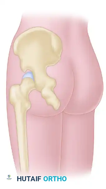
FIGURE 62-42: Skin incision for the approach to the proximal portion of the sciatic nerve extends from the posterior superior iliac spine to the trochanter and is curved distally along the posterior surface of the thigh.
Deep Dissection:
1. Fascial Incision: Deepen the proximal part of the incision through the thick gluteal fascia.
2. Muscle Splitting: Bluntly separate the coarse fibers of the gluteus maximus muscle along their length, extending as far laterally as the greater trochanter. Meticulous hemostasis is required here due to the rich vascularity of the gluteal muscle belly.
3. Distal Release: Incise the fascia of the thigh longitudinally down to the gluteal fold. To achieve full mobilization of the gluteus maximus, detach the insertion of its distal fibers from the iliotibial band.
4. Muscle Reflection: The gluteus maximus muscle, along with its neurovascular pedicle (inferior gluteal nerve and artery), may now be reflected medially. This maneuver unroofs the subgluteal space, exposing the sciatic nerve as far proximally as the piriformis muscle.
🔪 Surgical Warning: Managing the Sciatic Notch
If the injury extends directly into the greater sciatic foramen, standard soft tissue retraction will be insufficient. You must sacrifice the piriformis (tenotomize and reflect it) to expose the nerve as it emerges from the notch. If even better exposure of the nerve within the intrapelvic portion of the sciatic notch is necessary, use a Kerrison rongeur or Leksell rongeur to carefully remove a portion of the posterior sacrum and greater sciatic notch.
2. Mid-Thigh Approach
When the injury to the nerve is more distal to the sciatic notch, the incision over the buttock is made correspondingly more distal. For injuries isolated to the thigh, begin the incision at the gluteal fold and continue it distally along the midline of the posterior aspect of the thigh to a point 10 cm proximal to the knee.
Deep Dissection:
1. Fascial Opening: Open the deep fascia of the thigh longitudinally, strictly in line with the skin incision.
2. Nerve Protection: Crucial Step: Identify and protect the posterior femoral cutaneous nerve, which runs just deep to the deep fascia, often adhering to its undersurface. Iatrogenic injury to this nerve results in painful neuromas and distressing posterior thigh numbness.
3. Muscle Retraction: In the proximal thigh, identify the long head of the biceps femoris. Retract it medially to expose the underlying areolar tissue.
4. Nerve Identification: Identify the sciatic nerve in the depths of the wound, resting on the adductor magnus. Distally, trace the nerve beneath the biceps femoris to its point of bifurcation.
Alternative: When the lesion is located strictly in the middle third of the thigh, a lateral or posterolateral approach may be preferable to avoid the direct posterior scar, depending on concurrent femoral fracture patterns.
3. Distal Approach (Popliteal Fossa and Bifurcation)
Approaching the distal sciatic nerve and its bifurcation requires careful planning to avoid crossing the flexion crease of the knee, which can lead to severe hypertrophic scarring and flexion contractures.
The Mayfield Modification:
For more extensile exposure of the distal nerve branches, utilize the Mayfield modification.
* Peroneal Nerve Injury: Curve the distal end of the incision to the lateral aspect of the knee. Pass the incision distally along its course around the neck of the fibula.
* Tibial Nerve Injury: Curve the incision medially and then extend it a few centimeters distally along the medial aspect of the proximal leg.
💡 Clinical Pearl: Advantages of the Mayfield Incision
These curved incisions offer two distinct advantages:
1. They do not cross the transverse skin folds of the popliteal fossa perpendicularly; consequently, debilitating flexion contractures and ulcerating scars are significantly less likely.
2. Closing the wound is mechanically easier when the knee is flexed, as the curved flaps accommodate the bunched skin of the flexed popliteal fossa without excessive tension.
METHODS OF CLOSING GAPS IN THE SCIATIC NERVE
One of the most challenging aspects of sciatic nerve surgery is managing segmental defects following trauma or neuroma resection. Primary end-to-end neurorrhaphy without tension is the gold standard, but gaps frequently preclude this.
Joint Positioning and Mobilization
Extensive mobilization of the nerve is the first step. By mobilizing the nerve extensively—including neurolysis of its two divisions well into the leg—and manipulating the adjacent joints, massive gaps can be overcome.
* Flexing the knee to 90 degrees and hyperextending the hip allows for the closure of a gap of up to 15 cm.
* The repair must be performed with epineural sutures (e.g., 8-0 or 9-0 nylon) under microscopic magnification.
The Concomitant Femur Fracture
A highly specific and critical scenario arises when a patient presents with a fractured femur and a divided sciatic nerve.
🚨 Surgical Pitfall: Timing with Femur Fractures
When the femur has been fractured and the sciatic nerve divided, it is absolutely critical—even in the presence of draining sinuses or external fixators—to operate on the nerve before the femur has united.
Why? Aside from the detrimental effect of time on the nerve ends (Wallerian degeneration) and target muscles (fibrosis), the knee will inevitably stiffen during femoral fracture healing. If the knee stiffens in extension, it becomes impossible to flex it sufficiently to close large nerve defects. The window of opportunity for primary repair is lost.
Resecting Nerve Grafts vs. Neurolysis
When primary repair is impossible despite maximal mobilization and joint positioning, autologous nerve grafting (typically using the sural nerve) is required. Cable grafting is utilized to match the large cross-sectional area of the sciatic nerve.
Historical outcome data highlights the variability in recovery based on the required procedure:
* Neurolysis Outcomes: Of lesions treated by neurolysis alone, outcomes are generally favorable (e.g., historical cohorts show 5 excellent with complete muscle recovery, 7 good, and 1 poor).
* Grafting Outcomes: Of lesions treated by grafting, results are more guarded but still viable (e.g., 4 excellent, 4 good, and 1 poor). It is widely accepted that the tibial division recovers better than the peroneal division following grafting, largely due to the peroneal nerve's distinct fascicular topography and tighter epineural constraints.
POSTOPERATIVE PROTOCOL AND REHABILITATION
The postoperative management is just as critical as the surgical repair, particularly when joint positioning was used to close a nerve gap.
- Immobilization: If the hip was extended and the knee flexed to achieve a tension-free repair, the patient must be immobilized in a hip spica cast or a custom rigid orthosis in this exact position before waking from anesthesia.
- Duration of Immobilization: The immobilized position is maintained for 3 to 4 weeks to allow the epineural repair to gain tensile strength.
- Gradual Extension: After 4 weeks, the cast is removed, and a hinged brace is applied. The knee is gradually extended by 10 to 15 degrees per week. Rapid extension will rupture the repair or cause severe traction ischemia to the regenerating nerve.
- Physical Therapy: Once full joint extension is achieved (usually around 8-10 weeks postoperatively), aggressive physical therapy commences to restore joint mobility and maintain muscle bulk. Galvanic stimulation of denervated muscles may be considered, though evidence is mixed.
- Monitoring: Clinical recovery is monitored via advancing Tinel's sign and serial EMGs, keeping in mind that nerve regeneration occurs at a rate of approximately 1 mm per day.
CONCLUSION
The surgical approach to the sciatic nerve requires profound anatomical knowledge and meticulous surgical technique. From the proximal reflection of the gluteus maximus to the strategic curving of popliteal incisions, every step is designed to maximize exposure while minimizing iatrogenic morbidity. By adhering to strict principles of tension-free repair, utilizing joint positioning to close massive gaps, and respecting the critical time limits for intervention, the orthopedic surgeon can optimize the chances of functional recovery in these devastating injuries.
