Amputation for Chronic Osteomyelitis: Indications, Surgical Strategies, and Outcomes
Key Takeaway
Amputation remains a definitive, life-saving intervention for refractory chronic osteomyelitis, particularly when complicated by malignant transformation, severe arterial insufficiency, or profound neurovascular compromise. This surgical guide details the critical indications, preoperative optimization, and step-by-step operative techniques required to achieve a functional, infection-free residual limb. Mastery of these principles ensures optimal prosthetic fitting and restores patient mobility when limb salvage is no longer viable.
Comprehensive Introduction and Patho-Epidemiology
While the contemporary orthopaedic armamentarium heavily favors limb salvage through radical debridement, local antibiotic delivery systems, and complex soft-tissue reconstruction, amputation remains a critical, definitive treatment modality for chronic osteomyelitis. Amputation is performed infrequently for osteomyelitis in the modern era; however, in carefully selected patients, this definitive form of treatment is vastly preferable to a protracted cycle of multiple failed operations that are unlikely to eradicate the infection. The decision to amputate is rarely straightforward and requires a nuanced understanding of the patient's physiologic reserve, the anatomical extent of the disease, and the functional potential of the limb. In the context of the Cierny-Mader classification, amputation is most frequently indicated for Type IV (diffuse) osteomyelitis in a C-class host—a patient whose systemic or local comorbidities make the metabolic cost of limb salvage greater than the potential benefit.
The pathophysiology of chronic osteomyelitis is characterized by the presence of non-viable, avascular bone (the sequestrum) surrounded by a reactive envelope of new bone (the involucrum). Pathogenic microorganisms, most commonly Staphylococcus aureus and Pseudomonas aeruginosa, adhere to the necrotic bone and orthopaedic implants, elaborating a complex exopolysaccharide glycocalyx. This biofilm acts as an impenetrable barrier to both host immune cells and systemic antimicrobial agents. The bacteria within the biofilm enter a metabolically dormant, sessile state, rendering them highly resistant to cell-wall-active antibiotics. Consequently, the eradication of chronic osteomyelitis is fundamentally a surgical problem; antibiotics serve only an adjunctive role. When the anatomical extent of the sequestrum is so massive that complete extirpation would leave a skeletal defect incapable of supporting functional reconstruction, amputation becomes the most biologically sound intervention.
The metabolic toll of chronic, unrelenting bone infection cannot be overstated. Patients with decades of active osteomyelitis frequently exist in a state of chronic systemic inflammatory response syndrome (SIRS). This manifests as profound catabolism, chronic anemia of inflammation, hypoalbuminemia, and progressive immunosuppression. Furthermore, the continuous antigenic stimulation can lead to secondary systemic amyloidosis (AA amyloidosis), precipitating irreversible renal failure and hepatic dysfunction. In these severely compromised individuals, the metabolic burden of enduring multiple staged reconstructive procedures—such as Ilizarov bone transport or free tissue transfer—often exceeds their physiological reserve. Here, amputation is not a failure of orthopaedic surgery, but rather a life-saving, systemic resuscitation measure that rapidly eliminates the septic burden and reverses the catabolic cascade.
Epidemiologically, the incidence of amputation for chronic osteomyelitis is inextricably linked to the prevalence of severe peripheral neuropathy and peripheral arterial disease, most notably in the diabetic population. However, post-traumatic chronic osteomyelitis—often stemming from high-energy open fractures (Gustilo-Anderson Type III)—constitutes a significant cohort, particularly in young, otherwise healthy individuals who have endured years of reconstructive failures. A rare but critical epidemiological subset includes patients who develop malignant transformation within a chronic osteomyelitis sinus tract. This phenomenon, known as Marjolin's ulcer, occurs in approximately 0.2% to 1.6% of chronic osteomyelitis cases and fundamentally alters the treatment paradigm, mandating immediate and aggressive oncologic amputation to prevent metastatic dissemination.
Detailed Surgical Anatomy and Biomechanics
A profound mastery of cross-sectional and vascular anatomy is paramount when planning an amputation for chronic osteomyelitis. In the lower extremity, the fundamental principle is to preserve maximal limb length while ensuring the amputation level is executed through definitively uninfected, well-perfused tissue. The vascular anatomy must be meticulously evaluated, as chronic infection often induces local endarteritis and microvascular thrombosis. The concept of angiosomes—three-dimensional blocks of tissue supplied by specific source arteries—dictates flap design. For a transtibial amputation, the posterior flap is primarily supplied by the sural and muscular branches of the popliteal and posterior tibial arteries. If preoperative angiography reveals occlusion of the posterior tibial artery, a standard long posterior flap may undergo ischemic necrosis, necessitating alternative flap designs such as sagittal (skew) flaps or a more proximal transfemoral amputation.
The muscular anatomy forms the foundation of a dynamic, robust residual limb. The surgical manipulation of muscle during amputation directly dictates the functional outcome. In a transtibial amputation, the massive medial and lateral heads of the gastrocnemius muscle are utilized as a myofasciocutaneous flap. This muscle must be stabilized via myodesis (suturing muscle fascia directly to drill holes in the bone) or myoplasty (suturing opposing muscles over the bone end). Myodesis is biomechanically superior as it anchors the muscle to the skeleton, preventing the muscle from retracting and atrophying. This skeletal stabilization maintains the length-tension relationship of the musculature, enhances venous return by preserving the peripheral muscle pump, and provides a distal cushion of dynamic tissue that can tolerate the shear forces generated within a prosthetic socket. In transfemoral amputations, adductor magnus myodesis is absolutely critical to prevent the femur from resting in an abducted position, which drastically increases the energy expenditure of ambulation.
Neurologic anatomy requires equally meticulous attention to prevent the debilitating complication of terminal neuromas. The major peripheral nerves—the sciatic, tibial, common peroneal, and sural nerves—must be identified, isolated, and managed with precision. A severed nerve will inevitably form a neuroma as regenerating axons attempt to bridge the gap. The surgical goal is to ensure this neuroma forms deep within a well-vascularized muscle belly, far proximal to the weight-bearing surface of the residual limb and the rigid brim of the prosthetic socket. Contemporary techniques, such as Targeted Muscle Reinnervation (TMR), involve transferring the transected nerve stump into a nearby motor nerve branch of a redundant muscle. This provides the regenerating axons with a physiological target, significantly reducing the incidence of both symptomatic neuromas and phantom limb pain.
The biomechanics of the residual limb dictate the patient's ultimate ambulatory potential and energy expenditure. The metabolic cost of walking is inversely proportional to the length of the preserved limb and the number of preserved joints. A unilateral transtibial amputation increases the metabolic cost of ambulation by 10% to 25% compared to a non-amputee, whereas a transfemoral amputation increases energy expenditure by a staggering 60% to 70%. Therefore, preserving the knee joint is of paramount importance, provided the proximal tibia is oncologically and biologically sound. Furthermore, the residual bone must be meticulously contoured to distribute forces evenly within a modern total surface-bearing prosthetic socket. Sharp cortical edges, particularly the anterior crest of the tibia, act as stress risers that will inevitably lead to focal pressure necrosis, recurrent ulceration, and catastrophic secondary infection of the residual limb.
Exhaustive Indications and Contraindications
The transition from a limb salvage pathway to an amputation pathway is one of the most complex paradigms in orthopaedic surgery. It requires shifting the objective from preserving anatomical structure to optimizing systemic health and functional mobility. The decision is dictated by specific, evidence-based clinical thresholds. The concept of the "reconstructive ladder" is often replaced by the "reconstructive elevator," where the surgeon bypasses intermediate, high-risk salvage procedures in favor of immediate, definitive amputation when the biological or mechanical reality of the limb precludes a functional outcome. The goal is the creation of a dynamic, painless, and highly functional residual limb optimized for modern prosthetic wear, rather than the retention of a painful, non-functional, and chronically infected appendage.
Absolute indications for amputation in the setting of chronic osteomyelitis are clear and unforgiving. The foremost among these is malignant transformation, classically a well-differentiated Squamous Cell Carcinoma (SCC) arising from the epithelialized sinus tract (Marjolin’s Ulcer). Local resection or limb salvage in the presence of a Marjolin’s ulcer carries an unacceptably high rate of local recurrence and metastatic spread, particularly to regional lymph nodes. Amputation is the most reliable and oncologically sound means of treatment. Another absolute indication is irreversible neurovascular compromise. A sensate, motor-intact limb is a prerequisite for successful salvage. Profound neuropathy (e.g., complete sciatic nerve transection) resulting in an anesthetic, flail extremity inevitably leads to recurrent ulceration, Charcot arthropathy, and intractable infection. Finally, life-threatening sepsis originating from an acute-on-chronic exacerbation in a physiologically exhausted host (the C-host) mandates emergent, life-saving guillotine amputation for rapid source control.
Relative indications encompass scenarios where limb salvage is technically feasible but biologically or functionally ill-advised. Intractable, chronic pain that is refractory to multimodal analgesia and severely limits the patient's quality of life is a profound driver for amputation. Massive segmental bone loss that would require years of Ilizarov bone transport—especially in a patient with poor psychological coping mechanisms or inadequate social support—often tips the scale toward amputation. Furthermore, severe, rigid joint contractures (e.g., fixed equinovarus or knee flexion contractures exceeding 40 degrees) render the limb non-functional as a biomechanical lever. In these cases, even if the osteomyelitis is successfully eradicated, the limb remains a mechanical liability, and prosthetic rehabilitation following amputation will yield a vastly superior functional outcome.
Contraindications to amputation are exceedingly rare but must be recognized. In patients with terminal illnesses or profound physiological decline where surgical intervention will not alter the clinical trajectory or improve quality of life, palliative care and suppressive antibiotics are indicated over the trauma of a major amputation. Additionally, in a completely non-ambulatory, bed-bound patient where the chronic osteomyelitis is painless, heavily encapsulated, and not causing systemic toxicity, the risk of perioperative mortality from a major amputation may outweigh the benefits. In such scenarios, chronic suppressive antimicrobial therapy and meticulous local wound care represent the most prudent course of action.
| Category | Specific Clinical Scenarios | Rationale for Decision |
|---|---|---|
| Absolute Indications | Malignant Transformation (Marjolin's Ulcer) | High risk of local recurrence and metastasis with limb salvage; requires oncologic margins. |
| Unsalvageable Neurovascular Compromise | Anesthetic, ischemic limb will inevitably ulcerate and reinfect; lacks the biology to heal. | |
| Refractory Life-Threatening Sepsis | Emergent source control required to reverse systemic inflammatory response and prevent mortality. | |
| Relative Indications | Massive Segmental Bone Loss | Reconstructive burden (e.g., prolonged Ilizarov transport) exceeds patient's physiological/psychological reserve. |
| Severe Rigid Joint Contractures (>40° knee flexion) | Limb is mechanically non-functional; prosthetic rehabilitation offers superior ambulation. | |
| Intractable Chronic Pain | Decades of chronic infection lead to central sensitization; amputation removes the peripheral nociceptive generator. | |
| Contraindications | Terminal Illness / End of Life | Surgery will not improve quality of life or alter the ultimate clinical trajectory; palliative care preferred. |
| Painless Infection in Bed-Bound Patient | Surgical risk outweighs benefit; infection can be managed with chronic suppressive antibiotics and wound care. |
Pre-Operative Planning, Templating, and Patient Positioning
The preoperative preparation for an amputation in the setting of chronic osteomyelitis demands exhaustive, multidisciplinary coordination. The surgical team must collaborate closely with infectious disease specialists, vascular surgeons, anesthesiologists, prosthetists, and psychiatric professionals. The foremost technical objective is precise vascular mapping. Because chronic infection induces profound microvascular changes, palpable pulses alone are insufficient to guarantee flap viability. Objective non-invasive vascular studies are mandatory. Transcutaneous oxygen tension (TcPO2) mapping is the gold standard; a TcPO2 greater than 30 to 40 mmHg at the planned level of amputation is generally required to ensure primary healing of the surgical flaps. If the TcPO2 is marginal, formal CT angiography or conventional catheter angiography is indicated to delineate the arterial tree and assess the feasibility of preoperative endovascular revascularization to optimize inflow.
Advanced cross-sectional imaging is equally critical for templating the exact anatomical level of bone resection. Magnetic Resonance Imaging (MRI) with and without intravenous contrast is the modality of choice for delineating the proximal extent of intramedullary and extramedullary infection. The amputation level must be templated through definitively healthy, uninfected bone and soft tissue. The surgeon must identify the "skip lesions" common in chronic osteomyelitis, where isolated pockets of intramedullary infection exist proximal to the primary nidus. When templating a transtibial amputation, the ideal bone length is typically 12.5 to 15 cm distal to the medial joint line of the knee. This length provides an optimal lever arm for the prosthesis while allowing sufficient room within the socket for modern shock-absorbing pylons and energy-storing prosthetic feet.
Nutritional and medical optimization is a non-negotiable prerequisite for definitive closure. Chronic osteomyelitis induces a profound catabolic state, depleting the body's protein stores and impairing cellular immunity. Preoperative laboratory evaluation must include serum albumin, prealbumin, total lymphocyte count, and a comprehensive metabolic panel. An albumin level below 3.0 g/dL or a total lymphocyte count below 1,500/mm³ drastically increases the risk of postoperative wound dehiscence and stump infection. Aggressive nutritional supplementation, either enteral or parenteral, must be instituted. Furthermore, strict glycemic control (HbA1c < 7.0%) is paramount, as hyperglycemia directly impairs leukocyte phagocytosis and fibroblast proliferation, dooming the surgical flaps to failure.
Patient positioning and anesthetic management require meticulous attention to detail. The patient is typically positioned supine on a radiolucent operating table. A bump is placed under the ipsilateral hip to internally rotate the leg to a neutral position, facilitating access to the lateral and posterior compartments. While a pneumatic tourniquet is routinely applied proximally, its inflation is highly controversial in vascularly compromised patients, as tourniquet ischemia can exacerbate local tissue hypoxia and complicate the intraoperative assessment of tissue bleeding and viability. If utilized for blood conservation, the tourniquet must be deflated prior to definitive closure to ensure meticulous hemostasis. The surgical prep and drape must be extensive, extending well proximal to the planned amputation level to allow for proximal extension of the incision should intraoperative findings dictate a higher level of resection.
Step-by-Step Surgical Approach and Fixation Technique
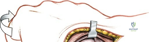
In the setting of active, purulent osteomyelitis or acute exacerbations of chronic disease, a single-stage amputation with primary closure is fraught with catastrophic failure rates. The standard of care mandates a two-stage surgical approach. The objective of the first stage, the Open (Guillotine) Amputation, is rapid source control and the elimination of the septic burden without closing potentially contaminated tissue. The incision is planned distal to the definitive amputation level. The skin, fascia, and muscle are divided in a slightly conical fashion to allow for tissue retraction. The bone is transected 2 to 3 cm proximal to the soft tissue margin using an oscillating saw. Crucially, continuous chilled saline irrigation must be applied directly to the saw blade to prevent thermal necrosis of the bone, which would otherwise act as a nidus for recurrent osteomyelitis. Major vessels are individually isolated, clamped, and double-ligated with non-absorbable suture. The wound is left entirely open and managed with negative pressure wound therapy (NPWT) or packed with antibiotic-impregnated polymethylmethacrylate (PMMA) beads to deliver massive local concentrations of antibiotics.
Once the systemic infection has resolved, the patient's catabolic state is reversed, and the local wound bed demonstrates healthy, uninfected granulation tissue (typically 7 to 14 days later), the patient is returned to the operating room for Stage 2: Definitive Closure and Stump Reconstruction. The open wound is aggressively irrigated with pulsatile lavage, and any residual necrotic tissue is sharply excised. The bone is then shortened to the definitive templated level. In a transtibial amputation, the anterior cortex of the tibia is meticulously beveled at a 45-degree angle to eliminate the sharp anterior crest. The fibula is resected 1 to 2 cm proximal to the tibial cut, and its lateral border is gently contoured. This fibular shortening is critical to prevent distal fibular pain and lateral compartment impingement during prosthetic loading.
The soft-tissue reconstruction relies heavily on precise myodesis. To provide a robust, dynamic soft-tissue envelope, the muscle fascia must be anchored to the skeleton. In a standard posterior flap transtibial amputation, the thick fascia of the gastrocnemius muscle is drawn anteriorly over the beveled distal tibia. Drill holes are placed through the anterior cortex of the tibia, approximately 1 cm proximal to the distal end. Heavy, non-absorbable sutures (e.g., #2 FiberWire) are passed through the drill holes and woven through the gastrocnemius fascia using a Krackow or locking stitch technique. The muscle is tensioned anatomically and secured to the bone. This myodesis stabilizes the bone within the soft tissue envelope, prevents the tibia from "pistoning" through the skin, and optimizes the length-tension relationship of the remaining musculature.
The final stage is meticulous skin closure. The skin flaps are approximated without tension using deep dermal absorbable sutures and superficial non-absorbable monofilament sutures or surgical staples. The creation of a smooth, cylindrical, or slightly conical stump profile is essential; therefore, any medial or lateral "dog-ears" must be meticulously excised. Deep closed-suction drains are placed beneath the myodesis flap and brought out through separate stab incisions proximally to prevent hematoma formation, which serves as a potent culture medium for residual bacteria. The incision is dressed with non-adherent gauze, and a rigid or semi-rigid postoperative dressing is applied immediately in the operating room to control edema, protect the wound from trauma, and prevent joint contractures.
Complications, Incidence Rates, and Salvage Management
Despite meticulous surgical technique and rigorous preoperative optimization, amputations performed for chronic osteomyelitis carry a distinct and formidable complication profile. The most dreaded complication is recurrent infection within the residual limb. This typically stems from inadequate proximal resection margins, retained microscopic necrotic bone, or contamination of the surgical flaps during the initial debridement. The incidence of recurrent infection ranges from 5% to 15%, heavily dependent on the host's immune status and the virulence of the pathogen. Management requires immediate return to the operating room for aggressive surgical debridement. Suppressive antibiotics alone are universally ineffective. If the bone is involved, revision to a higher anatomical level (e.g., converting a transtibial to a transfemoral amputation) is often mandatory to achieve definitive source control.
Wound dehiscence and flap necrosis represent another significant mode of failure, occurring in 10% to 20% of cases, particularly in diabetic patients with severe microvascular disease. Dehiscence is often secondary to premature suture removal, excessive flap tension over poorly contoured bone, or unrecognized arterial insufficiency. Minor marginal necrosis can often be managed conservatively with prolonged Negative Pressure Wound Therapy (NPWT) and aggressive local wound care. However, full-thickness necrosis exposing the underlying bone or myodesis requires urgent surgical intervention. Salvage management involves wedge resection of the necrotic tissue, bone shortening, and primary re-closure. In severe cases of vascular failure, proximal revision amputation is the only viable salvage strategy.
Neuropathic complications, specifically phantom limb pain and symptomatic terminal neuromas, are profoundly debilitating and occur in up to 30% to 50% of amputees. Phantom limb pain is driven by central cortical reorganization and peripheral nerve sensitization resulting from decades of chronic pain associated with the osteomyelitis. Management is initially pharmacological, utilizing gabapentinoids, tricyclic antidepressants, and NMDA receptor antagonists. Symptomatic neuromas present as exquisitely tender, focal nodules at the distal aspect of the residual limb that prevent prosthetic wear. Surgical salvage involves excision of the neuroma, proximal traction neurectomy, and burying the nerve stump deep into a proximal muscle belly or directly into the medullary canal of the bone. Targeted Muscle Reinnervation (TMR) is increasingly utilized as both a prophylactic and salvage procedure with excellent clinical outcomes.
Joint contractures are a ubiquitous mechanical complication that can render a perfectly healed residual limb functionally useless. Knee flexion contractures are particularly common following transtibial amputations, driven by the unopposed pull of the hamstring musculature and the patient's tendency to rest with the knee flexed to reduce tension on the surgical incision. Hip flexion and abduction contractures plague transfemoral amputees. Prevention is paramount and involves the use of immediate rigid postoperative dressings, early aggressive physical therapy, and strict avoidance of prolonged wheelchair sitting. Once a severe, rigid contracture is established (>20 degrees of knee flexion), prosthetic fitting becomes biomechanically impossible. Salvage management involves aggressive serial casting and dynamic splinting; rarely, surgical release of the posterior capsule and hamstring lengthening is required.
| Complication | Estimated Incidence | Etiology / Risk Factors | Salvage Management Strategy |
|---|---|---|---|
| Recurrent Infection | 5% - 15% | Retained necrotic bone, inadequate margins, virulent biofilm (e.g., MRSA). | Aggressive I&D, removal of infected bone, revision to a higher amputation level, targeted IV antibiotics. |
| Wound Dehiscence / Necrosis | 10% - 20% | Microvascular disease, excessive flap tension, premature suture removal, malnutrition. | NPWT for superficial defects; surgical wedge resection and bone shortening for full-thickness necrosis. |
| Symptomatic Neuroma | 15% - 30% | Inadequate proximal nerve retraction, nerve tethering to scar tissue or bone. | Surgical excision, proximal traction neurectomy, burying in deep muscle, or Targeted Muscle Reinnervation (TMR). |
| Joint Contracture | 20% - 40% | Poor positioning, unopposed muscle pull, lack of early physical therapy. | Aggressive physical therapy, dynamic splinting, serial casting; rarely requires surgical capsular release. |
Phased Post-Operative Rehabilitation Protocols
The postoperative rehabilitation following amputation for chronic osteomyelitis is a highly structured, phased process that demands as much clinical rigor as the surgical procedure itself. The immediate postoperative phase (Days 1 to 14) is focused on wound protection, edema control, and the prevention of joint contractures. An Immediate Postoperative Prosthesis (IPOP) or a rigid cast dressing is highly recommended. This rigid environment prevents postoperative edema, protects the vulnerable surgical flaps from accidental trauma, and holds the knee in full extension to prevent flexion contractures. Pain management is multimodal, utilizing regional nerve blocks (e.g., continuous popliteal or femoral nerve catheters), systemic non-opioid adjuncts, and judicious opioid utilization. Aggressive physical therapy is initiated on postoperative day one, focusing on upper extremity strengthening, core stability, and preservation of the contralateral limb, which is often equally compromised in diabetic or vasculopathic patients.
The subacute phase (Weeks 3 to 6) marks the transition from acute surgical recovery to residual limb maturation. Suture or staple removal is typically delayed in this patient population, often occurring at 3 to 4 weeks postoperatively, to ensure robust dermal healing in the setting of vascular compromise. Once the wound is completely healed and structurally sound, compressive shrinker socks are applied. These garments are critical for reducing residual limb volume, shaping the stump into a cylindrical or conical profile, and desensitizing the skin. Desensitization techniques, including gentle massage and tapping, are taught to the patient to mitigate hyperalgesia and prepare the limb for the tactile feedback of a prosthetic socket. Range of motion exercises are intensified, and the patient is transitioned to independent transfers and single-limb ambulation using a walker or crutches.
The prosthetic fitting phase (Weeks 6 to 10) commences once the residual limb volume has stabilized and the surgical incision demonstrates mature, robust tensile strength. The prosthetist casts the residual limb to fabricate a preparatory (temporary) prosthesis. Modern prosthetic philosophy favors a Total Surface Bearing (TSB) socket design over the traditional Patellar Tendon Bearing (PTB) socket. The TSB socket, utilized in conjunction with a silicone or urethane liner and a pin-lock or suction suspension system, distributes weight-bearing forces globally across the entire surface area of the residual limb. This global pressure distribution is critical in patients with a history of osteomyelitis, as it minimizes focal shear stresses that could lead to skin breakdown and recurrent infection.
Advanced rehabilitation and community reintegration represent the final and longest phase of the protocol. Gait training with the preparatory prosthesis focuses on achieving a symmetrical, energy-efficient gait pattern. The patient is taught to manage the dynamic volume changes of the residual limb by adding or removing prosthetic socks. Because the metabolic cost of ambulation is significantly increased, endurance training and energy conservation strategies are paramount. Psychological support is a continuous requirement, as the patient must navigate the profound alterations in body image, chronic pain management, and the emotional trauma of losing a limb to a chronic, decades-long disease process. Definitive (permanent) prosthetic fabrication typically occurs 6 to 12 months postoperatively, once the residual limb has undergone complete atrophy and volumetric stabilization.
Summary of Landmark Literature and Clinical Guidelines
The surgical management of chronic osteomyelitis and the indications for amputation are heavily informed by decades of landmark orthopaedic literature and evolving clinical guidelines. The foundation of modern treatment is rooted in the Cierny-Mader classification system, first published in 1985. Cierny and Mader revolutionized the approach to osteomyelitis by emphasizing that the host's physiological status (A, B, or C host) is a more critical determinant of treatment success than the specific anatomical extent of the bone lesion. Their landmark validation studies demonstrated that in C-class hosts (patients with profound systemic or local compromise), attempts at complex limb salvage for Type IV (diffuse) osteomyelitis resulted in unacceptable morbidity and mortality rates, thereby establishing amputation as the definitive standard of care for this specific cohort.
The literature surrounding Marjolin's ulcer provides unequivocal guidance on the necessity of amputation in the setting of malignant transformation. Landmark retrospective reviews, such as those by Hahn et al. and Chalya et al., have consistently demonstrated that Squamous Cell Carcinoma arising in a chronic osteomyelitis sinus tract is highly aggressive, with a propensity for early lymphatic metastasis. These studies validate that local wide excision or limb-sparing procedures in the presence of a Marjolin's ulcer yield unacceptably high local recurrence rates (often exceeding 40%). Consequently, contemporary oncologic and orthopaedic guidelines mandate proximal amputation—often requiring joint disarticulation or amputation at the next proximal anatomical segment—to achieve definitive oncologic margins and maximize patient survival.
Biomechanical and physiological guidelines heavily influence the selection of the amputation level. The seminal work by Waters et al. on the energy expenditure of amputee gait remains the cornerstone of surgical planning. Waters demonstrated through rigorous oxygen consumption studies that the metabolic cost of walking increases exponentially with the loss of the knee joint. A transtibial amputee requires only 10% to 25% more energy to ambulate than a non-amputee, whereas a transfemoral amputee requires up to 70% more energy. In the physiologically exhausted patient with chronic osteomyelitis, this difference in energy expenditure often dictates the difference between a patient who achieves community ambulation and one who is permanently confined to a wheelchair. This literature universally supports the mandate to preserve the knee joint whenever oncologically and biologically feasible.
Contemporary antimicrobial guidelines, published jointly by the Infectious Diseases Society of America (IDSA) and the American Academy of Orthopaedic Surgeons (AAOS), dictate postoperative medical management. The literature clearly delineates that if an amputation is performed with definitively negative, uninfected bone and soft-tissue margins, a short postoperative course of prophylactic antibiotics (24 to 48 hours) is sufficient. Prolonged systemic antibiotics do not prevent stump infection in cases of clean margins. Conversely, if the amputation is performed through a zone of reactive edema, or if residual microscopic disease is suspected but cannot be resected due to anatomical constraints, the guidelines mandate a 4-to-6-week course of targeted intravenous or highly bioavailable oral antibiotics to suppress residual bacterial burden and allow the surgical flaps to heal.
