7 Effective Ways to Fix Your Finger Sprain Fast (With Pictures)
Introduction & Epidemiology
Finger sprains represent a spectrum of ligamentous injuries affecting the small joints of the hand, primarily the interphalangeal (IP) and metacarpophalangeal (MCP) joints. While often perceived as minor, these injuries can lead to significant pain, functional impairment, and long-term instability or stiffness if not accurately diagnosed and appropriately managed. Given the critical role of precise digit function in daily activities and occupational demands, a meticulous approach to evaluation and treatment is paramount.
Epidemiologically, finger sprains are among the most common hand injuries, frequently observed in contact sports (e.g., basketball, football, rugby) and falls onto an outstretched hand. The proximal interphalangeal (PIP) joint is the most commonly injured digit joint due to its frequent exposure to axial loading and hyperextension forces. The ulnar collateral ligament (UCL) of the thumb MCP joint, often referred to as "skier's thumb" or "gamekeeper's thumb," is another frequently encountered and clinically significant ligamentous injury. Distal interphalangeal (DIP) joint sprains are less common but can also result in pain and instability.
Accurate assessment of a finger sprain involves discerning between a simple, stable ligamentous strain and a more complex, potentially unstable rupture requiring operative intervention. The initial clinical presentation often includes localized tenderness, edema, ecchymosis, and pain with attempted movement. Functional deficits can range from mild discomfort to complete inability to grasp or pinch. Early and precise evaluation of injury severity is critical for guiding appropriate management strategies and optimizing patient outcomes.
Surgical Anatomy & Biomechanics
A thorough understanding of the intricate anatomy and biomechanics of the finger joints is fundamental to managing ligamentous injuries. Each IP and MCP joint is stabilized by a complex network of structures, including the collateral ligaments, volar plate, and joint capsule.
Metacarpophalangeal (MCP) Joint Anatomy & Biomechanics
The MCP joints are diarthrodial condyloid joints, allowing flexion/extension, abduction/adduction, and circumduction.
*
Collateral Ligaments:
Consist of two main components:
*
Proper Collateral Ligament (PCL):
Originates from the metacarpal head and inserts onto the base of the proximal phalanx. These ligaments are taut in flexion and lax in extension, a crucial biomechanical feature for grip strength and stability.
*
Accessory Collateral Ligament (ACL):
Originates from the metacarpal head, volar to the PCL, and inserts onto the volar plate and tendon sheath. These are taut in extension and lax in flexion.
*
Volar Plate:
A strong fibrocartilaginous structure on the palmar aspect of the joint, protecting against hyperextension. It is firmly attached to the proximal phalanx and loosely to the metacarpal.
*
Joint Capsule:
Surrounds the entire joint, providing additional stability.
Thumb MCP Joint (UCL): The ulnar collateral ligament of the thumb MCP joint is particularly vital for pinch strength and stability. A complete rupture, especially with interposition of the adductor pollicis aponeurosis (Stener lesion), necessitates surgical repair.
Proximal Interphalangeal (PIP) Joint Anatomy & Biomechanics
The PIP joints are ginglymus (hinge) joints, primarily permitting flexion and extension. These joints are inherently less stable than MCP joints due to their smaller articular surfaces and more constrained motion.
*
Collateral Ligaments:
Also composed of proper and accessory components, similar to the MCP joints, but their tensioning characteristics are different. At the PIP joint, both proper and accessory collateral ligaments are generally taut throughout the range of motion, providing consistent stability. The radial collateral ligament (RCL) and ulnar collateral ligament (UCL) are distinct and critical.
*
Volar Plate:
A robust, quadrangular fibrocartilaginous structure that reinforces the joint capsule volarly, preventing hyperextension. It proximally attaches to the middle phalanx and distally to the proximal phalanx, with check-rein ligaments contributing to its stability. Avulsion fractures of the volar plate are common with hyperextension injuries.
*
Central Slip of the Extensor Mechanism:
Distal to the PIP joint, the central slip of the extensor tendon attaches to the dorsal base of the middle phalanx, extending the PIP joint. Injury here can lead to a boutonnière deformity.
*
Joint Capsule:
Encircles the joint, contributing to stability.
Distal Interphalangeal (DIP) Joint Anatomy & Biomechanics
The DIP joints are also hinge joints, allowing flexion and extension.
*
Collateral Ligaments:
Similar to the PIP joints, providing lateral stability.
*
Volar Plate:
Smaller and less robust than at the PIP joint, but serves a similar hyperextension-blocking function.
*
Terminal Extensor Tendon:
Attaches to the dorsal base of the distal phalanx, extending the DIP joint. Injury can lead to a mallet finger deformity.
Understanding the specific ligament involved, the mechanism of injury, and the resulting instability pattern is paramount for correct diagnosis and appropriate treatment planning.
Indications & Contraindications
The decision-making process for managing finger sprains hinges on the degree of ligamentous injury and the resulting joint stability. A thorough clinical examination, including stress testing under fluoroscopy or regional anesthesia if necessary, coupled with appropriate imaging, guides this determination.
Diagnostic Evaluation
- History: Mechanism of injury (e.g., hyperextension, varus/valgus stress, axial load), immediate symptoms, functional deficits.
-
Physical Examination:
- Inspection: Edema, ecchymosis, deformity.
- Palpation: Localized tenderness over collateral ligaments, volar plate, or central slip.
- Range of Motion: Active and passive flexion/extension. Pain with motion.
- Stability Testing: Varus and valgus stress testing at 0° and 30° of flexion (to isolate proper collateral ligaments by relaxing accessory ligaments and volar plate). Laxity compared to the contralateral digit indicates ligamentous compromise. Gross instability suggests a complete rupture.
-
Imaging:
- Plain Radiographs: AP, lateral, and oblique views are essential to rule out associated fractures, avulsion fractures (e.g., volar plate avulsion, collateral ligament avulsion), dislocations, or pre-existing arthrosis.
- Stress Radiographs: Performed if clinical stress testing is equivocal or to quantify instability, particularly for thumb MCP UCL injuries (comparison to contralateral thumb is crucial).
- MRI: Reserved for cases where soft tissue injury (e.g., Stener lesion, central slip rupture, severe volar plate injury) is suspected, and clinical examination or plain radiographs are insufficient for definitive diagnosis or surgical planning.
Indications for Operative vs. Non-Operative Management
The core principle is to restore stability and function. Non-operative management is indicated for stable injuries, while operative intervention is reserved for unstable injuries, complete ruptures with specific characteristics, or chronic instability.
Table 1: Operative vs. Non-Operative Indications for Finger Sprains
| Indication Category | Non-Operative Management | Operative Management |
|---|---|---|
| Ligamentous Injury | Stable Grade I & II sprains (partial tears without significant instability) | Grade III sprains (complete rupture with gross instability) |
| Ligamentous avulsion fractures with significant displacement or joint incongruity | ||
| Specific Cases: | ||
| * Thumb MCP UCL: Complete rupture with a Stener lesion (interposition of adductor aponeurosis) | ||
| * PIP Collateral Ligament: Gross instability (e.g., >20-30° laxity on stress testing compared to contralateral digit) | ||
| * PIP Volar Plate: Large avulsion fracture (>20-30% articular surface), chronic instability, irreducible dislocation, subluxation | ||
| Associated Injuries | Minor, stable avulsion fractures (e.g., <10% articular surface, stable) | Irreducible dislocations |
| Persistent subluxation after reduction | ||
| Significant associated articular fractures causing joint incongruity | ||
| Chronic Conditions | Stable, well-compensated chronic instability | Chronic instability causing pain, recurrent subluxation, or functional impairment despite non-operative measures |
| Failed non-operative management for unstable injuries |
Contraindications for Surgery:
Absolute contraindications are rare but include severe active infection, uncontrolled systemic comorbidities precluding safe anesthesia, or patient refusal. Relative contraindications may include severe soft tissue compromise, pre-existing advanced arthrosis (where arthrodesis might be a more appropriate primary procedure), or non-compliance with post-operative rehabilitation.
Pre-Operative Planning & Patient Positioning
Meticulous pre-operative planning is crucial for optimizing outcomes in finger sprain surgery.
Pre-Operative Planning
-
Detailed Review of Imaging:
Carefully assess radiographs (plain and stress views), MRI, or CT scans to precisely characterize the injury:
- Location and extent of ligamentous disruption.
- Presence and size of avulsion fractures.
- Joint congruity and stability.
- Identification of potential interposing structures (e.g., Stener lesion).
-
Surgical Strategy:
- Approach: Determine the appropriate surgical incision based on the involved joint and specific ligament (e.g., dorsal, lateral, volar).
- Repair Technique: Decide on direct repair (if tissue quality allows), augmentation (e.g., capsular flap), or reconstruction (if chronic injury or poor tissue quality).
- Fixation Method: Plan for suture anchors, transosseous sutures, pull-out wires, or internal brace augmentation.
- Graft Selection (for reconstruction): If reconstruction is anticipated, identify potential donor sites (e.g., palmaris longus, FCR, toe extensor).
- Anesthesia: Regional anesthesia (e.g., brachial plexus block) with sedation or general anesthesia. A tourniquet is routinely used for a bloodless field.
- Equipment: Ensure all necessary instruments, sutures (non-absorbable typically), anchors, and possibly mini-fragment plating systems for avulsion fractures are available.
Patient Positioning
- Supine Position: The patient is positioned supine on the operating table.
- Arm Table: The affected upper extremity is placed on a dedicated hand table, allowing for free range of motion and optimal exposure.
- Tourniquet: A pneumatic tourniquet is applied to the upper arm. The tourniquet time should be minimized.
- Preparation and Draping: The entire hand and forearm are prepped and draped in a sterile fashion, allowing access to the finger and wrist if a graft is needed.
Detailed Surgical Approach / Technique
The specific surgical technique varies depending on the joint involved (MCP vs. PIP), the specific ligament injured, and the chronicity of the injury. We will detail common approaches for acute collateral ligament repairs.
A. Thumb MCP Ulnar Collateral Ligament (UCL) Repair
The most common operative finger sprain involves the UCL of the thumb MCP joint.
1.
Incision:
A curvilinear or dorsal L-shaped incision centered over the ulnar aspect of the thumb MCP joint is preferred. Care must be taken to protect the dorsal sensory branch of the radial nerve.
2.
Exposure:
* Subcutaneous dissection to identify and protect the dorsal sensory radial nerve branches.
* Incise the fascia over the adductor pollicis muscle.
* Identify the adductor aponeurosis. If a Stener lesion is present, the aponeurosis will be interposed between the ruptured UCL and its insertion, preventing spontaneous healing. The aponeurosis is incised longitudinally to expose the deeper structures.
3.
Identification of Pathology:
* The torn ends of the UCL are identified. The rupture is typically off the distal insertion on the proximal phalanx, but mid-substance tears or proximal avulsions can occur.
* Assess the quality of the ligamentous tissue.
4.
Repair Technique:
*
Direct Repair:
For acute tears with good tissue quality, direct repair is preferred.
* If the ligament is avulsed from the bone, a small trough can be created in the bone, or one or two mini suture anchors (e.g., 1.5mm) are placed into the bone at the insertion site.
* Non-absorbable sutures are passed through the torn ligamentous tissue in a locking fashion (e.g., modified Krackow stitch) and tied securely to the suture anchors or passed transosseously.
* Tension the repair such that the joint is stable to valgus stress at both 0° and 30° flexion, without over-constraining the joint.
*
Augmentation/Reconstruction:
In cases of chronic instability, poor tissue quality, or delayed presentation, a reconstruction may be necessary using a free tendon graft (e.g., palmaris longus, FCR) or a local capsular flap. Internal brace augmentation using braided suture tape can also provide additional strength and allow for earlier rehabilitation.
5.
Capsular Closure:
The joint capsule is repaired where possible.
6.
Adductor Aponeurosis Closure:
The adductor aponeurosis is repaired over the UCL repair.
7.
Skin Closure:
Standard layered closure.
B. PIP Joint Collateral Ligament Repair
This typically involves the radial or ulnar collateral ligament of the PIP joint.
1.
Incision:
A curved dorsal-lateral or mid-lateral incision is made directly over the involved collateral ligament. Care is taken to protect the digital neurovascular bundles.
2.
Exposure:
* Subcutaneous dissection to protect the neurovascular bundle, which lies volar to the collateral ligament.
* Incise the overlying fibrous retinaculum.
* Carefully elevate the extensor tendon mechanism to expose the joint capsule and collateral ligament.
3.
Identification of Pathology:
* Locate the torn collateral ligament, usually avulsed from its distal insertion on the middle phalanx.
* Inspect for associated volar plate injury or articular damage.
4.
Repair Technique:
*
Direct Repair:
For acute avulsions, similar to the thumb UCL, one or two mini suture anchors are placed into the base of the middle phalanx at the ligament's insertion. Non-absorbable sutures are then passed through the torn ligament and tied.
*
Transosseous Sutures:
If tissue quality allows, transosseous sutures drilled through the phalanx can be used.
*
Reconstruction:
For chronic tears or severe tissue loss, reconstruction using a free tendon graft may be required. The graft is usually woven through drill holes in the proximal and middle phalanges and secured.
5.
Stability Assessment:
Verify joint stability through a full range of motion.
6.
Closure:
Repair the joint capsule, retinaculum, and skin in layers.
C. PIP Joint Volar Plate Repair/Reconstruction
Indicated for large avulsion fractures (often with >20-30% articular surface involvement), chronic instability, or irreducible dislocations.
1.
Incision:
A volar "zigzag" (Bruner) incision is often used to expose the PIP joint without creating a contracture.
2.
Exposure:
* Carefully dissect to protect the digital neurovascular bundles.
* The A2 and A3 pulleys of the flexor tendon sheath are exposed. The A3 pulley can be partially incised if necessary for access but should be preserved as much as possible to prevent bowstringing.
* The joint capsule is opened, and the volar plate is identified.
3.
Repair/Reconstruction:
*
Avulsion Fracture:
If a bony avulsion of the volar plate is present, the fragment is reduced anatomically. Fixation can be achieved with K-wires (transarticular or through the fragment), a mini-screw, or small suture anchors into the middle phalanx base.
*
Ligamentous Tear:
Direct repair of the volar plate to its insertion on the middle phalanx can be performed with suture anchors or transosseous sutures.
*
Reconstruction:
In cases of severe chronic instability or tissue loss, a volar plate reconstruction using a portion of the FDS tendon or a free tendon graft can be considered.
4.
Stability Assessment:
Confirm stable reduction through the full range of motion.
5.
Closure:
Repair the capsule and skin.
Complications & Management
Despite meticulous surgical technique, complications can arise following operative management of finger sprains. Anticipation, early recognition, and appropriate management are key to mitigating their impact.
Table 2: Common Complications, Incidence, and Salvage Strategies
| Complication | Incidence | Salvage Strategies |
|---|---|---|
| Joint Stiffness/Contracture | Common (10-30%) | Early protected range of motion protocols, aggressive hand therapy, serial splinting, dynamic/static progressive splinting. Surgical release (arthrolysis) for recalcitrant cases. |
| Persistent Pain | Variable (5-15%) | Activity modification, NSAIDs, local corticosteroid injections, physical therapy. Neuropathic pain management if nerve involvement. Revision surgery if due to hardware or malunion. |
| Infection | Low (<1-2%) | Culture-directed antibiotics. Surgical debridement, irrigation, and washout. Hardware removal if infection persists. |
| Recurrent Instability/Re-rupture | Moderate (5-10%) | Re-evaluation with stress radiographs and MRI. Revision surgery with reconstruction (e.g., tendon graft) or augmentation (e.g., internal brace). Arthrodesis for severe, irreparable cases. |
| Hardware-Related Issues | Variable | Symptomatic hardware removal (e.g., prominent suture anchors, K-wires). |
| Neurovascular Injury | Rare (<1%) | Careful dissection. Direct repair of nerve/vessel if recognized intra-operatively. Revision surgery for repair, nerve grafting, or vascular reconstruction. |
| Complex Regional Pain Syndrome (CRPS) | Low (1-5%) | Early diagnosis and multi-disciplinary management: pain clinic referral, stellate ganglion blocks, physical therapy, psychological support, pharmacotherapy. |
| Non-Union/Malunion of Avulsion Fracture | Low (<5%) | Depends on functional impact. Observation for asymptomatic cases. Revision fixation, bone grafting, or fragment excision for symptomatic non-union. |
| Flexor Tendon Adhesion/Bowstringing | Low (<2%) | Aggressive hand therapy, tenolysis if severe and resistant to therapy, pulley reconstruction. |
Post-Operative Rehabilitation Protocols
Post-operative rehabilitation is as critical as the surgery itself for optimizing functional outcomes and preventing complications such as stiffness or recurrent instability. The protocol must be tailored to the specific injury, surgical repair, and individual patient factors.
General Principles
- Protection: Protect the healing ligament or repair site from excessive stress.
- Controlled Motion: Initiate early, controlled range of motion to prevent stiffness and promote ligament healing.
- Gradual Progression: Slowly increase load and activity levels based on healing progression and clinical milestones.
- Pain and Edema Management: Crucial throughout the recovery period.
Phases of Rehabilitation
Phase 1: Immobilization and Protection (Weeks 0-3/4)
- Goal: Protect the repair, reduce pain and edema.
-
Immobilization:
The digit is typically immobilized in a custom thermoplastic orthosis (splint) or cast.
- Thumb MCP UCL: Immobilized in slight flexion (e.g., 20-30° flexion) and mild abduction, sparing the interphalangeal joint.
- PIP Collateral Ligament: Immobilized in slight flexion (e.g., 20-30° flexion).
- PIP Volar Plate: Immobilized in 0-10° extension, often allowing guarded flexion.
- Immobilization is usually maintained continuously for 3-4 weeks.
-
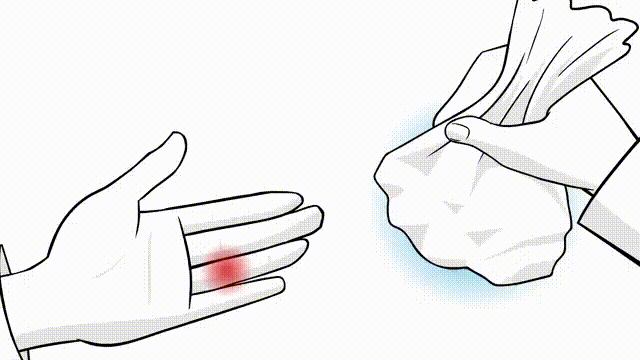
Edema Control:
- Elevation of the hand.
- Ice application (15-20 minutes, several times a day).
-
Gentle compression dressing.
-
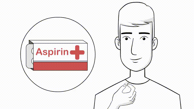
Pain Management:
-
Prescribed analgesics (e.g., NSAIDs, opioids as needed).
-
Prescribed analgesics (e.g., NSAIDs, opioids as needed).
- Patient Education: Instruct the patient on signs of complications, activity restrictions, and the importance of compliance.
- Other Joints: Maintain full range of motion of uninvolved joints (wrist, elbow, shoulder).
Phase 2: Early Protected Motion (Weeks 3/4 - 6/8)
- Goal: Gradually restore joint range of motion while protecting the healing structures.
-
Splinting:
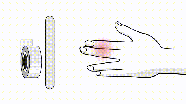
The static splint is typically replaced with a removable custom splint or buddy taping for protection during higher-risk activities. Continuous immobilization at rest.
-
Therapy:
- Initiate gentle active range of motion (AROM) exercises for the injured joint within protected arcs.
- Passive range of motion (PROM) may be initiated cautiously as tolerated, often under direct supervision by a hand therapist.
- Continue edema management.
- Light functional activities.
- Progression: The amount and type of motion are gradually increased, avoiding excessive stress in the direction of the initial injury.
Phase 3: Strengthening and Functional Integration (Weeks 6/8 - 12)
- Goal: Improve strength, endurance, and coordination. Return to more demanding activities.
- Splinting: Discontinue continuous splinting, using it only for protection during specific activities or at night if needed. Buddy taping for sports or heavy activities.
-
Therapy:
- Progressive resistive exercises (TheraBand, putty, grip strengthening tools).
- Advanced AROM and PROM exercises.
- Proprioceptive and fine motor dexterity exercises.
- Scar massage and desensitization if needed.
- Activity Modification: Gradually reintroduce specific occupational or recreational activities, with modifications as necessary.
Phase 4: Return to Activity (Weeks 12+)
- Goal: Full return to sport, work, and daily activities.
-
Criteria for Return:
- Near-normal range of motion and strength.
- Absence of pain or instability during functional tasks.
- Confidence in using the digit.
- Sport-Specific Training: Gradually progress to sport-specific drills and contact activities, often with continued buddy taping or protective splinting initially. Full return to contact sports may take 4-6 months, depending on the sport and the specific injury.
Regular communication between the surgeon and hand therapist is paramount to ensure appropriate progression and address any emerging issues.
Summary of Key Literature / Guidelines
The management of finger sprains is guided by a substantial body of literature, including clinical studies, systematic reviews, and consensus guidelines from prominent hand surgery societies. While specific recommendations may vary, several overarching principles are consistently emphasized:
-
Accurate Diagnosis is Paramount: The ability to differentiate between stable partial tears and unstable complete ruptures, especially those with interpositional tissue (e.g., Stener lesion), is the most critical determinant of successful outcome. Clinical examination and stress radiography remain the cornerstones of diagnosis. MRI is an adjunct for complex cases or equivocal findings. (e.g., Schenck RR. The thumb metacarpophalangeal joint: anatomy, pathology, and treatment of common injuries. Instr Course Lect. 1999;48:193-201. )
-
Thumb MCP UCL Injuries Require Special Attention: Due to its critical role in pinch and grip, and the high incidence of Stener lesions in complete ruptures, acute Grade III tears of the thumb UCL often warrant surgical repair. Non-operative management of unstable thumb UCL injuries carries a high risk of chronic instability and pain. (e.g., Campbell CS. Gamekeeper's thumb. J Bone Joint Surg Br. 1955;37-B(1):148-149. ; Stener B. Displacement of the ruptured ulnar collateral ligament of the metacarpo-phalangeal joint of the thumb. J Bone Joint Surg Br. 1962;44-B(4):869-879. )
-
PIP Joint Instability is Complex: PIP joint collateral ligament injuries are common. While many are amenable to non-operative treatment with buddy taping or short-term splinting, persistent instability or chronic pain may necessitate surgical repair or reconstruction. Volar plate avulsion fractures, especially those involving a significant portion of the articular surface or causing irreducible subluxation/dislocation, are indications for operative management. (e.g., Eaton RG, Littler JW. Joint injuries of the hand. Clin Orthop Relat Res. 1985 Nov;(192):22-29. )
-
Early Controlled Motion: For most ligamentous repairs in the hand, current evidence supports early controlled active and passive motion within a protected range. This approach helps prevent joint stiffness, promotes collagen alignment, and optimizes long-term functional recovery compared to prolonged rigid immobilization. (e.g., Cannon NM. Controlled mobilization in hand injuries. Hand Clin. 1990 Aug;6(3):479-487. ; Tang JB, Gu YQ, Zhang CQ. Repair and reconstruction of the collateral ligaments of the proximal interphalangeal joint. Hand Clin. 2005 May;21(2):299-308. )
-
Reconstruction for Chronic Instability: In cases of chronic instability, especially those involving significant tissue attenuation or prior failed repair, reconstructive procedures using tendon grafts (e.g., palmaris longus, FCR) are often necessary. These provide predictable stability but may have longer rehabilitation periods. The concept of an "internal brace" using suture tape augmentation is gaining traction for acute repairs and selected reconstructions, offering enhanced stability and potentially allowing for more aggressive early rehabilitation. (e.g., Franko OI, et al. Internal Brace Augmentation of Thumb Ulnar Collateral Ligament Repairs. J Hand Surg Am. 2017 Jul;42(7):527-533. )
-
Prevention of Stiffness: Stiffness remains a major complication. Factors contributing include prolonged immobilization, excessive edema, and inadequate rehabilitation. A collaborative approach involving the surgeon, patient, and certified hand therapist is essential to mitigate this risk.
In conclusion, the effective management of finger sprains requires a comprehensive understanding of anatomy, astute clinical assessment, appropriate use of diagnostic imaging, judicious application of non-operative and operative strategies, and a structured, progressive post-operative rehabilitation protocol. Adherence to these principles, informed by current literature, is critical for achieving optimal functional restoration and preventing long-term sequelae.
