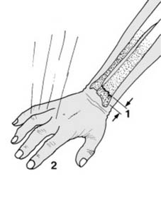
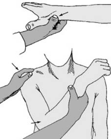
The wrist and hand 1 Colles fracture: A Colles fracture is a fracture of the radius within 2.5 cm of the wrist (1), with a characteristic deformity if displaced. It is the commonest of all fractures. It is seen mainly in middle-aged and elderly women, and osteoporosis is a frequent contributory factor. It usually results from a fall on the outstretched hand (2), and generally the distal radial fragment remains intact. (See Frame 45 et seq. for those other cases where the radiocarpal joint is involved.) 2 Displacements (a): The six characteristic features of a displaced Colles fracture (see later for details) are shown in this foreshortened view of the pronated right arm, viewed from below. The slight obliquity of impact (F) produces the two most striking features of dorsal and radial displacement (D & R) of the distal fragment. (T = triangular fibrocartilage; U = ulnar styloid; M = medial; L = lateral) 3 Displacements (b): The deformity can be followed by studying the wrist in the two planes in which the radiographs are usually taken. The impact (F) fractures the radius through the cancellous bone of the metaphysis. With greater violence the anterior periosteum tears, and the distal fragment tilts into anterior angulation (1) with loss of the 5° anterior tilt of the joint surface. 4 Displacements (c): With greater violence there is dorsal displacement of the distal fragment (2). The shaft of the radius is driven into the distal fragment leading to impaction (3). (The dotted lines indicate the position of the distal fragment prior to any displacement.) 5 Displacements (d): The altered contour of the wrist in a badly displaced Colles fracture is striking, and is referred to as a ‘dinner fork deformity’. When viewed from the side, the wrist has the same curvature as a fork, with the tines resembling the fingers. 6 Displacements (e): This radiograph shows a typical displaced Colles fracture. The anterior angulation, dorsal displacement, and impaction are obvious (deformities 1,2,3). 7 Displacements (f): In the AP plane, a small lateral component of the force of impact causes lateral (radial) displacement of the distal fragment (4). The distal fragment is attached to the ulnar styloid by the triangular fibrocartilage, and generally this leads to avulsion of the ulnar styloid. Note that in the AP projection of the normal wrist that the joint surface has a tilt of 22°. 8 Displacements (g): Sometimes the triangular fibrocartilage is torn; in either case there is disruption of the inferior radio-ulnar joint. The distal fragment tilts laterally into ulnar angulation (5) (reducing the tilt to less than 22°) and impacts. The sixth feature is a rotational or torsional deformity (6 in Frame 2), not obvious in either AP or lateral projections. 9 Displacements (h): In this AP radiograph of a Colles fracture, note the features just mentioned, i.e. radial deviation, ulnar angulation and impaction of the distal fragment. 10 Diagnosis: 1. If there is pain in the wrist and tenderness over the distal end of the radius after a fall, radiographs must be taken in every case. The site of maximum tenderness will help to differentiate fracture of the scaphoid (but see scaphoid fractures). 2. Where there is marked displacement the characteristic appearance leaves little diagnostic doubt. Note that in the normal wrist the radial styloid lies 1 cm distal to the ulnar. 11 Radiographs (a): In the majority of cases the fracture is easily identified. Sometimes it may be missed because impaction has rendered the fracture line inconspicuous. If in doubt, look at the angle between the distal end of the radius and the shaft in the lateral radiograph. Decrease to less than 0° is suggestive of fracture (but enquire about previous injury). 12 Radiographs (b): The minimally displaced fracture will also reveal itself in the lateral projection by an increase in the posterior radial concavity, often with local kinking (1) or by a separate or accompanying break in the smooth curve of the anterior surface of the radius (2). 13 Radiographs (c): In the AP view of the wrist, look for any irregularity in the smooth lateral aspect of the radius. If there is any doubtful radiographic feature suggesting fracture,…