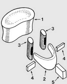
2 Anatomical features (after Kapandji1) (a): The components of a typical vertebra have a complex relationship, and can be illustrated with an exploded diagram. The elements comprise the vertebral body (1) composed of cancellous bone covered with an outer shell of cortical bone, the horseshoe-shaped neural arch (2), two articular masses or processes which take part in the facet (interarticular) joints (3) and the transverse (4) and spinous (5) processes.
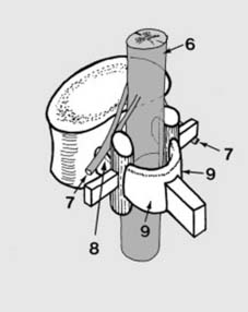
3 Anatomical features (b): When these components are brought together they form a protective bony covering for the cord (6) and the issuing nerve roots (7). The neural arch (2) is divided by the articular processes (3) into pedicles (8) and laminate (9).
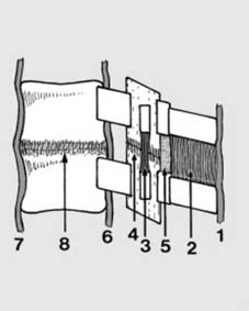
4 Anatomical features (c): The vertebrae are bound together by the following structures: The supraspinous (1), interspinous (2), intertransverse (3) and capsular ligaments (4), and the ligamentum flavum (5): together these form the so-called posterior ligament complex. Playing a less powerful but nevertheless important role are the posterior (6) and anterior (7) longitudinal ligaments and the annular ligaments (8). Trauma may result in damage to any of the bony or ligamentous structures of the spine, in isolation or in combination.
5. Assessment of spinal injuries (a): In managing any case of spinal injury it is important to determine which structures have been involved and the extent of the damage they have suffered; with this information an assessment may be made of the risks of complication. Note: 1. The history may direct you to the type of injury to suspect; 2. The clinical examination may be a valuable guide to the extent of bony and ligamentous injury (and any neurological complication); 3. Investigation by X-ray and CT scan is likely to provide the most information; and MRI scans if available may help clarify the extent of any associated soft tissue damage, especially the intervertebral discs and the posterior ligament complex.
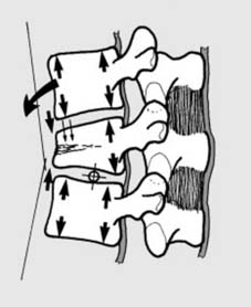
It is important to make an early assessment of the stability of the spine, i.e. to assess whether it is able to withstand stress without progressive deformity or further neurological damage. Instability may be purely mechanical (e.g. in some compression fractures where further kyphotic deformity may occur). Instability may be neurological (e.g. where shifting or further extrusion of bone fragments within the spinal canal may lead to neurological deterioration). Combined mechanical and neurological instability may be present. In assessing instability it may be helpful to regard the spine as having three main elements or columns.
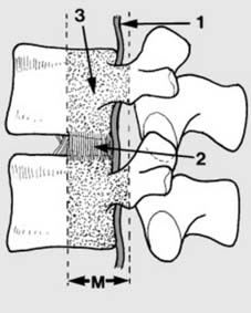
6 Assessment of spinal injuries (b): The middle column (M) consists of the posterior longitudinal ligament (1), the posterior part of the annular ligament (2), and the posterior wall of the vertebral column (3). Unstable injuries occur when damage to the middle column is combined with damage to either the anterior column or damage to the posterior column.
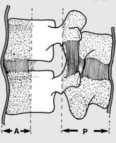
7 Assessment of spinal injuries (c): The posterior column (P), vital for stability, comprises the neural arch, the pedicles, the spinous process, and the post. ligt. complex. The anterior column (A) is formed by the anterior longitudinal ligament, and the anterior parts of the annular ligament and vertebral body. Note that failure of these columns in their role as supports can be due to bony or ligament involvement. Four categories are recognised in the Denis Classification of spinal injury:
8 Compression fractures (a): Simple compression fractures are common stable injuries involving the anterior column only. Hyperflexion of the spine round an axis passing through the disc space leads to mechanical failure of bone with either anterior (Illus.) or lateral wedging. The height of the posterior part of the vertebral body is maintained. (Note that severe wedging – e.g. more than 15–20° – often indicates damage to the other columns and a burst fracture or fracture dislocation.)
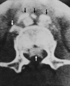
9 Burst fractures (b): Axial loading of the spine may cause failure of the anterior and middle columns. One or both end plates may be involved, and bone fragments may be extruded into the spinal canal, compromising the neurological structures and more controversially causing neurological instability. Radiographs may show the vertebral body fracture, loss of vertebral height, and in the AP, laminar fractures and separation of the pedicles. (Illus: CT scan showing displacement of bony fragments both anteriorly and posteriorly.)
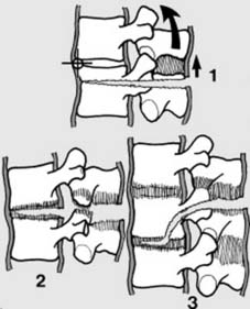
10 Seat belt type injuries (c): Rapid deceleration causes the spine to jack-knife round an axis brought forward by the lap strap part of a seatbelt, and tension forces lead to failure of the posterior and middle columns. The spine is unstable in flexion. Failure may occur entirely through bone (Chance fracture) (1), or ligaments (2), or a combination (3), involving either one (1 & 2) or two (3) levels of the spine. Injuries of this pattern may be seen in situations outside road traffic accidents, but the essential element is tension failure.
11 Fracture–dislocations (d): All 3 columns fail in these unstable injuries. Suspect if: 1. There are multiple rib or transverse process fractures (as from D1 to D8 the ribs and sternum give additional support to the spine); 2. There is a slight increase in the height of a disc or a fracture of an articular process. Types:
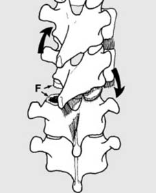
(a) Flexion–rotation fracture–dislocation. There is often a fracture of an articular process on one side (F), or a slicing fracture through a vertebral body, leading to rotation and subluxation of the spine.
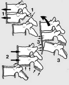
12 Fracture–dislocations (d) ctd: Shear types of fracture–dislocation: In the posteroanterior type (1) the upper segment shears forwards, often with fracture of the posterior arch at 1 or 2 levels. In the anteroposterior type (2) there is complete ligamentous disruption, but often no fracture. In the flexion–distraction type (3) there is an anterior annular tear with stripping of the anterior long. ligt. allowing anterior subluxation: this is a tension type of injury as in the seatbelt lesion.
13. Neurological examination: basic principles: Where there is evidence of a deficit, a thorough neurological examination is required on admission; this must include as a minimum:
Note also the following points:
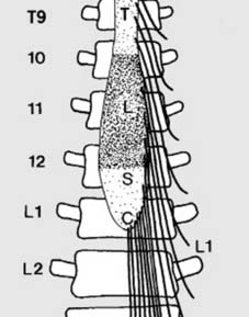
14 Anatomical features (a):Note: 1. The spinal cord ends at L1; any injury distal to this can involve the cauda but not the cord. 2. All the lumbar and sacral segments of the cord lie between T10 and L1 only. 3. Injuries at the thoracolumbar junction produce a great variety of neurological disturbances as:
NEXT
Anatomical features (b): Cervical cord
