FACTORS AFFECTING THE RATE OF HEALING OF A FRACTURE
1 TYPE OF BONE
Cancellous bone (spongy bone)
4 SEPARATION OF BONE ENDS
5 BONE LOSS
If the loss is substantial then treatment will be prolonged and difficult, and because of the mechanism of injury many of these cases have accompanying problems with skin loss, tissue contamination, and accompanying neural and vascular damage. Other factors such as the patient’s age and medical state, and other injuries will influence the management of the case. The first decision that must be made is therefore whether amputation is the best approach. This is not always easy, and the general view is that this must be decided on a completely individual assessment of the factors involved. While use of the Mangled Extremity Severity Index (see p. 51) may help in taking all the important factors into account, it has been shown to be somewhat unreliable in evaluating those cases where it indicates that amputation should be performed.
6 INFECTION
MRSA Infections
Established infections with methicillin-resistant staphylococcus aureus may pose enormous problems: for the patient, in extreme cases, with risk of life, and in others to the prolongation of treatment and inferior results; for surgeons and nursing staff, with difficulties in management; and overall, with the greatly increased cost implications. The extent of the problem and the procedures adopted to deal with it vary from country to country, but many measures are common to all. In the UK, Giannoudis and others1 suggest the following protocol for the identification of MRSA carriers, and the treatment of an established infection.
1 Strict adherence to national guidelines for screening in high-risk areas with:
2 Treatment of carriers as outlined in the national guidelines with:
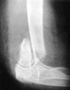
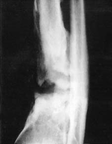
7 DISTURBANCE OF BLOOD SUPPLY
It is obvious that for the normal multiplication of bone cells and their precursors an adequate blood supply is required. Where the blood supply to an area is reduced, or where there is interference with the blood supply to both major fragments – e.g. in radionecrosis of bone – healing may be interfered with. On the other hand, reduction of the blood supply to one fragment, especially if cancellous bone is involved, may not interfere with union; indeed, in some situations it may apparently stimulate it. The most striking examples of this are fractures of the femoral neck and scaphoid, where the phenomenon of avascular necrosis is most frequently discovered in soundly united fractures. Interference with the blood supply to one fragment at the time of injury leads to immediate bone death; this is frequently followed by sound union of the fracture. Collapse of necrotic bone beyond the level of union is observed at a later date.
9 OTHER FACTORS
In closed fractures, and as a general rule, conservative methods of treatment are to be preferred wherever possible. Nevertheless, infections occurring in cases treated by internal fixation appear to be due mainly to contamination rather than an impaired immunity response, and open methods may be pursued so long as strict attention is paid to asepsis and careful tissue handling. Prophylactic antibiotics in the form of a first generation cephalosporin have been recommended by Harrison.2
In any fracture case if there is an increase in the rate of non-union, it may generally be dealt
withsuccessfully by internal fixation and bone grafting.
NEXT
COMPLICATIONS OF FRACTURES
