Wrist and Hand Anatomy
1. Osteology ( Fig. 2.8)
- Carpal bones
Ossification begins at the capitate (usually present at 1 year of age) and proceeds in a counterclockwise direction, according to posteroanterior radiographs of the right hand.
1. Several key features are important to recognize in the individual carpal bones ( Table 2.7).
- Metacarpals
- Several characteristics allow identification of individual metacarpals ( Table 2.8).
- Phalanges
Arthrology
1. Distal radioulnar joint (DRUJ) articulation (most stable in supination)
- Radiocarpal (wrist) joint
- Ellipsoid shape involving distal radius and the scaphoid, lunate, and triquetrum
- Located at the level of the crease of proximal wrist flexion
- Ligaments ( Table 2.9 ; see Fig. 2.8)
- Extrinsic ligaments bridge carpal bones to radius or metacarpals (e.g., radio-scapho-capitate); intrinsic ligaments attach carpal bones together (e.g., scapholunate).
- Palmar/volar radiocarpal ligaments are the strongest supporting structures.
Space of Poirier: central weak area in floor of carpal tunnel; implicated in volar dislocation of lunate in perilunate dislocation
1. Ligament of Testut (radioscapholunate ligament) functions as a neurovascular conduit.
- Triangular fibrocartilage complex (TFCC) ( Fig. 2.9 , Table 2.10 ): vascular supply from periphery (central portio n avascular)
- Intercarpal joints
- Proximal row
- Scaphoid linked to the lunate (via the scapholunate ligament), which is linked to the triquetrum (via the lunotriquetral ligament)
- Flexes with radial deviation and extends with ulnar deviation
- SL ligament injury: dorsal intercalated segmental instability (DISI)
- LT ligament injury: volar intercalated segmental instability (VISI)
- Dorsal intercarpal ligaments are stronger.
- Distal row
- Dorsal and palmar intercarpal ligaments connect trapezium with trapezoid, trapezoid with capitate, and capitate with hamate.
- Interosseous ligaments are much thicker in the distal row, connecting capitate and hamate (strongest), capitate and trapezoid, and trapezium and trapezoid (weakest).
- Midcarpal joint
- Transverse articulations between proximal and distal rows are reinforced by palmar and dorsal intercarpal ligaments and carpal collateral ligaments.
-
Radial ligament is stronger.


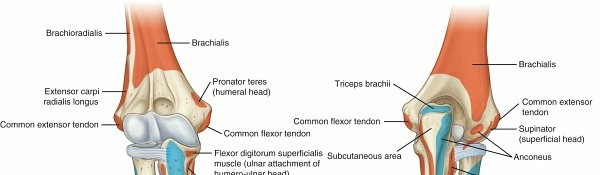
| FIG. 2.6 Muscle attachments of the forearm (anterior and posterior views). From Drake RL et al, editors:
Gray’s atlas of anatomy,
ed 2, Philadelphia, 2015, Churchill Livingstone. ### Table 2.6
Muscles of Forearm Muscle
| ** Origin
|
Insertion
|
Action
|
Innervation
|
FIG. 2.6 Muscle attachments of the forearm (anterior and posterior views). From Drake RL et al, editors:
Gray’s atlas of anatomy,
ed 2, Philadelphia, 2015, Churchill Livingstone. ### Table 2.6
Muscles of Forearm Muscle
| ** Origin
|
Insertion
|
Action
|
Innervation
|
| --- | --- | --- | --- | --- |
| Superficial Flexors Pronator teres | Medial |
| epicondyle and coronoid | Midlateral |
| radius | Pronating, flexing forearm | Median nerve Flexor carpi radialis | Medial |
| epicondyle | Second and third metacarpal bases | Flexing wrist | Median nerve Palmaris longus | Medial |
| epicondyle | Palmar |
| aponeurosis | Flexing wrist | Median nerve Flexor carpi ulnaris | Medial |
| epicondyle and posterior ulna | Pisiform | Flexing wrist | Ulnar nerve Flexor digitorum superficialis | Medial |
| epicondyle, proximal anterior ulna, | Base of middle phalanges | Flexing PIP joint | Median nerve | and anterior radius | |
| --- | --- | --- | --- | --- |
| Deep Flexors Flexor digitorum profundus | Anterior and medial ulna | Base of distal phalanges | Flexing DIP joint | Median–anterior interosseous/ulnar nerves Flexor pollicis longus | Anterior and lateral radius | Base of distal phalanges | Flexing IP joint, thumb | Median–anterior interosseous nerve Pronator quadratus | Distal ulna | Volar radius | Pronating hand | Median–anterior interosseous nerve |
| Superficial Extensors Brachioradialis | Lateral |
| supracondylar humerus | Lateral distal radius | Flexing forearm | Radial nerve Extensor carpi radialis longus | Lateral |
| supracondylar humerus | Second |
| metacarpal base | Extending wrist | Radial nerve Extensor carpi radialis brevis | Lateral |
| epicondyle of humerus | Third |
| metacarpal base | Extending wrist | Radial nerve Anconeus | Lateral |
| epicondyle of humerus | Proximal |
| dorsal ulna | Extending forearm | Radial nerve Extensor digitorum | Lateral |
| epicondyle of humerus | Extensor |
| aponeurosis | Extending digits | Radial–posterior interosseous nerve Extensor digiti minimi | Common |
| extensor tendon | Small finger extensor expansion over proximal phalanx | Extending small finger | Radial–posterior interosseous nerve Extensor carpi ulnaris | Lateral |
| epicondyle of humerus | Fifth |
| metacarpal base | Extending/adducting hand | Radial–posterior interosseous nerve |
| Deep Extensors Supinator | Lateral |
| epicondyle of humerus, ulna | Dorsolateral radius | Supinating forearm | Radial–posterior interosseous nerve Abductor | Dorsal | First | Abducting/extending | Radial–posterior pollicis longus | ulna/radius | metacarpal base | thumb | interosseous nerve | --- | --- | --- | --- | --- | Extensor pollicis brevis | Dorsal radius | Thumb |
| proximal phalanx base | Extending thumb MCP joint | Radial–posterior interosseous nerve Extensor pollicis longus | Dorsolateral ulna | Thumb dorsal phalanx base | Extending thumb IP joint | Radial–posterior interosseous nerve Extensor indicis proprius** | Dorsolateral ulna | Index finger extensor apparatus (ulnarly) | Extending index finger | Radial–posterior interosseous nerve |

- Carpometacarpal (CMC) joints
- Thumb CMC joint
- Highly mobile saddle-shaped joint
- Supported by a capsule and radial, palmar, and dorsal CMC ligaments
- Finger CMC joints
- Gliding joints with capsules, dorsal CMC ligaments (strongest), palmar CMC ligaments, and interosseous CMC ligaments
- Metacarpophalangeal (MCP) joints
- Palmar (volar plate), collateral, and deep transverse metacarpal ligaments
- Cam mechanism (collateral ligaments tighten with MCP joint flexion)
- Interphalangeal (IP) joints
-
Hinge joints with capsules and obliquely oriented collateral ligaments
1.** Muscles of the hand (** Fig. 2.10 , Table 2.11) - Extensor tendon anatomy
- Extensor compartments of wrist: formed by the extensor retinaculum over dorsal wrist ( Fig. 2.11 , Table 2.12)
- Anatomic snuffbox is bordered by tendons of the first and third dorsal wrist compartments. Extensor pollicis brevis (EPB) tendon serves as the radial snuffbox border, extensor pollicis longus (EPL) tendon as the ulnar border.
- Extensor mechanism anatomy/intrinsic apparatus
- Complex arrangement of structures that surround digits ( Fig. 2.12 , Table 2.13)
- Flexor tendon anatomy
- Carpal tunnel ( Fig. 2.13)
- Borders of the carpal tunnel listed in Table 2.14
- Transverse carpal ligament (TCL): attaches radially to the scaphoid tuberosity and trapezial ridge and ulnarly to the hook of the hamate and pisiform
- Divided in carpal tunnel release
- Contains the median nerve and nine tendons (one flexor pollicis longus [FPL], four FDS, and four flexor digitorum profundus [FDP])
FDS to the middle and ring fingers are volar to FDS to index and small fingers.
1. 1. 1. Decreases in volume with wrist flexion


FIG. 2.7 Volar flexors (superficial and deep).
From Drake RL et al, editors:
Gray’s atlas of anatomy,
ed 2, Philadelphia, 2015, Churchill Livingstone.


FIG. 2.8 Ligaments of the hand and interphalangeal joints.
From Drake RL et al, editors:
Gray’s atlas of anatomy,
ed 2, Philadelphia, 2015, Churchill Livingstone.
Table 2.7
Carpal Bone Features
Carpal Distinctive Features NoteS Bone
---
Scaphoid
| Tubercle (TCL, APB), waist, proximal and distal poles
| 1. 75% covered by articular cartilage
2. Retrograde blood supply from dorsal carpal branch of radial artery
Lunate
| Half moon shaped
| 1. Kienböck disease
Triquetrum
| Pyramid shaped
|
Pisiform
| Spheroidal (TCL, FCU)
| 1. In FCU tendon
Trapezium
| FCR groove, tubercle (opponens, APB, FPB, TCL)
| 1. May be excised to treat thumb CMC arthritis
Trapezoid
| Wedge shaped
|
Capitate
| Largest bone, central location
|
Hamate
| Hook (TCL)
| 1. Radial border of Guyon canal
2. Deep motor branch of ulnar nerve under hook
APB, Abductor pollicis brevis; FPB, flexor pollicis brevis.

Table 2.8
Metacarpal Bone Features
Metacarpal Distinctive Features
---
I (thumb)
| Short, stout; base is saddle shaped
II (index)
| Longest, largest base; medial at base
III (middle)
| Styloid process
IV (ring)
| Small quadrilateral base, narrow shaft
V (small)
| Tubercle at base (extensor carpi ulnaris)
Table 2.9
Extrinsic Ligaments of the Wrist
Dorsal
---
Dorsal intercarpal (DIC)
| Dorsal support to translation of midcarpal joint
Dorsal radiocarpal (DRC)
| aka radiotriquetral Controls ulnar translation
Volar
Radioscaphocapitate (RSC)
| Supports scaphoid waist
Long radiolunate (LRL)
| Strong tether to lunate displacement
Radioscapholunate (RSL)
| Ligament of Testut (neurovascular conduit)
Short radiolunate (SRL)
| Prevents dorsal lunate dislocation
Table 2.10
Triangular Fibrocartilage Complex
Component Origin Insertion
---
Dorsal and volar radioulnar ligament
| Ulnar
radius
| Caput ulnae
Articular disc
| Radius/ulna
| Triquetrum
Prestyloid recess
| Disc
| Meniscus homologue
Meniscus homologue
| Ulna/disc
| Triquetrum/ulnar collateral ligament
Ulnar collateral ligament
| Ulna
| Fifth metacarpal


FIG. 2.9 Triangular fibrocartilage complex.
From Miller MD, Thompson SR, editors:
DeLee and Drez’s orthopaedic sports medicine: principles and practice,
ed 4, Philadelphia, 2014, Saunders.


FIG. 2.10 Muscle attachments (planar and dorsal views).
From Drake RL et al, editors:
Gray’s atlas of anatomy,
ed 2, Philadelphia, 2015, Churchill Livingstone.
Table 2.11
| Muscles of the Hand
Muscle
|
Origin
|
Insertion
|
Action
|
Innervation
|
| --- | --- | --- | --- | --- |
| Thenar Muscles
Abductor pollicis brevis
| Scaphoid, |
| trapezoid | Base of proximal phalanx, radial side | Abducting |
| thumb | Median |
| nerve
Opponens pollicis
| Trapezium | Thumb |
| metacarpal | Abducting, flexing, rotating (medially) | Median |
| nerve
Flexor pollicis brevis
| Trapezium, capitate | Base of proximal phalanx, radial side | Flexing MCP joint | Median, |
| ulnar nerves
Adductor pollicis
| Capitate, second and third metacarpals | Base of proximal phalanx, ulnar side | Adducting |
| thumb | Ulnar nerve |
| Hypothenar Muscles
Palmaris brevis
| TCL, palmar aponeurosis | Ulnar palm | Retracting skin | Ulnar nerve
Abductor digiti minimi
| Pisiform | Base of proximal phalanx, ulnar side | Abducting small finger | Ulnar nerve
Flexor digiti minimi brevis
| Hamate, TCL | Base of proximal phalanx, ulnar side | Flexing MCP joint | Ulnar nerve
Opponens digiti minimi
| Hamate, TCL | Small-finger metacarpal | Abducting, flexing, rotating (laterally) | Ulnar nerve |
| Intrinsic Muscles
Lumbrical
| Flexor |
| digitorum profundus | Lateral bands (radial) | Extending PIP joint | Median, |
| ulnar nerves
Dorsal interosseous
| Adjacent |
| metacarpals | Proximal phalanx base/extensor apparatus | Abducting, flexing MCP joint | Ulnar nerve
Volar interosseous
| Adjacent |
| metacarpals | Proximal phalanx base/extensor apparatus | Adducting, flexing MCP joint | Ulnar nerve |

 **
FIG. 2.11**
Extensor compartments of the wrist (1 to 6) (see
Table 2.12
).
APL, _
Abductor pollicis longus;
ECRB, _
extensor carpi
radialis brevis;
ECRL,
extensor carpi radialis longus;
ECU,
extensor carpi ulnaris;
EDC,
extensor digitorum communis;
EDM,
extensor digiti minimi;
EPB,
extensor pollicis brevis;
EPL,
extensor pollicis longus. Modified from Miller MD et al:
Orthopaedic surgical approaches,
Philadelphia, 2008, Saunders, Figure HW-6.
**
FIG. 2.11**
Extensor compartments of the wrist (1 to 6) (see
Table 2.12
).
APL, _
Abductor pollicis longus;
ECRB, _
extensor carpi
radialis brevis;
ECRL,
extensor carpi radialis longus;
ECU,
extensor carpi ulnaris;
EDC,
extensor digitorum communis;
EDM,
extensor digiti minimi;
EPB,
extensor pollicis brevis;
EPL,
extensor pollicis longus. Modified from Miller MD et al:
Orthopaedic surgical approaches,
Philadelphia, 2008, Saunders, Figure HW-6.
1. Guyon canal
2. Contains ulnar nerve and artery
1. Borders: flexor retinaculum (deep), volar carpal ligament (superficial), pisiform (ulnar/proximal), hook of hamate (radial/distal)
3.
Flexor tendon sheath (
Fig. 2.14)
1. Covers flexor tendons, protecting and nourishing tendons (vincula)
2. Five anular pulleys (A1–5) with three intervening cruciate attachments (C1–3)
Table 2.12 Extensor Compartments of Wrist CompartMent Contents NOTES I Abductor
pollicis longus
Pathologic CONDITIO
| (APL),
extensor pollicis brevis (EPB)
| frequently has multiple tendon slips, which should be addressed during release for de Quervain tenosynovitis).
| ---|---|---|---
II
| Extensor
carpi radialis longus (ECRL),
brevis
| |
Extensor ten (intersect syndrom
III
| Extensor
pollicis longus (EPL)
| |
Rupture
Lister tuber (after fractu Drumme
tendi of the
IV
| Extensor
digitorum communis (EDC),
extensor indicis proprius (EIP)
| |
Extensor
tenosyno
V
| Extensor
digiti minimi (EDM)
| |
Rupture
(rheuma arthritis: Vaughn-Jackson syndrom
VI
| Extensor
carpi ulnaris (ECU)
| subsheath part of TFCC
| Snapping at styloid

---
FIG. 2.12 Extensor apparatus.
From Drake RL et al, editors:
Gray’s atlas of anatomy,
ed 2, Philadelphia, 2015, Churchill Livingstone.
Table 2.13 Extensor Mechanism of Hand STRUCTURE | ATTACHMENTS | SIGNIFICANCE | ---|---|---| Sagittal bands | Covers MCP joint
| Allows MCP extension Transverse (sagittal) | Volar plate fibers
| Allows MCP flexion (interossei) Lateral bands | Covers PIP joint
| Allows PIP extension (lumbrical muscles) Oblique retinacular ligament (Landsmeer) | A4 pulley,
terminal tendon
| Allows DIP extension (passive)
1. EPB tendon De Quervain ulnar to APL tenosyno tendon
2. APL
3. ECRL radial to ECRB
4. EPL tendon ulnar to ECRB tendon at the wrist level.
5. Ulnar to Lister tubercle
6. Tested by placing patient’s palm flat on table and lifting thumb.
7. EIP ulnar to index EDC
8. EIP most distal muscle belly
9. Small EDC present in only 25%
10. Sensory PIN in floor
11. EDM ulnar to small EDC
12. ECU
3. A2 and A4 pulleys originate from bone, whereas A1, A3, and A5 pulleys originate from the palmar plates of the metacarpal, proximal IP (PIP), and distal IP (DIP) joints.
4. A2 pulley, overlying the proximal phalanx, is the most critical to function, followed by A4, which covers the middle phalanx.
A1 pulley is involved in trigger digits.
Carpal Tunnel Borders
BORDER
|
STRUCTURES
| ---|---|
Roof
| Transverse carpal ligament
Floor
| Carpal bones
Radial
| Scaphoid tubercle/trapezium
Ulnar
| Hook of hamate

FIG. 2.14 Flexor tendon sheath.
From Drake RL et al, editors:
Gray’s atlas of anatomy,
ed 2, Philadelphia, 2015, Churchill Livingstone.


FIG. 2.13 Anatomy of the carpal tunnel.
From Drake RL et al, editors:
Gray’s atlas of anatomy,
ed 2, Philadelphia, 2015, Churchill Livingstone.
