Comprehensive Introduction and Patho-Epidemiology
The surgical management of distal toe pathologies demands a profound, multifaceted understanding of forefoot biomechanics, microanatomy, and the intricate soft-tissue envelopes that dictate digital viability. Pathologies affecting the distal phalanx, the nail matrix, and the interphalangeal joints frequently present not as isolated osseous anomalies, but as a complex amalgamation of structural deformity and dermatological compromise. The terminal digit is uniquely susceptible to a variety of biomechanical stressors, microtraumas, and vascular insufficiencies, rendering it a frequent site of recalcitrant pathology in both the general and diabetic populations. Among the most reliable, definitive, and historically validated interventions for these conditions are the Terminal Syme procedure for distal phalangeal and ungual pathologies, and combined resection arthroplasties for rigid, complex interphalangeal deformities.
The patho-epidemiology of terminal toe conditions encompasses a broad spectrum of etiologies ranging from chronic infectious processes to neoplastic transformations. Chronic onychocryptosis (ingrown toenail) with associated hypertrophy of the ungual labia, recurrent paronychia, and contiguous osteomyelitis of the distal phalanx represent the most common infectious and inflammatory indications. Furthermore, the distal phalanx is a recognized site for subungual neoplasms, including glomus tumors, subungual exostoses, and melanoma in situ. In cases of severe macrodactyly or traumatic crush injuries where the nail bed is irreparably macerated and the distal phalanx comminuted, conservative measures are universally inadequate. The Terminal Syme procedure, originally adapted from the principles of James Syme’s 1842 ankle amputation, addresses these pathologies through complete ablation of the nail matrix, resection of the distal ungual tuft, and the creation of a durable plantar pulp flap.
Conversely, combined hammer toe and mallet toe deformities represent a distinct patho-epidemiological entity driven by intrinsic and extrinsic muscle imbalances. These deformities are characterized by rigid flexion contractures at both the proximal interphalangeal (PIP) and distal interphalangeal (DIP) joints, notably without the metatarsophalangeal (MTP) joint hyperextension classically seen in claw toe deformities. The chronic, rigid flexion at these joints subjects their dorsal prominences to relentless friction against footwear, culminating in the formation of painful hyperkeratotic lesions known as double corns (heloma durum). The prevalence of these deformities increases with age, specialized footwear use, and underlying neuropathic or inflammatory arthropathic conditions.
This comprehensive academic guide delineates the precise surgical techniques, anatomical considerations, and evidence-based postoperative protocols required to execute these procedures successfully. Mastery of these techniques allows the orthopedic surgeon to navigate the delicate balance between adequate bone resection and soft-tissue preservation. By adhering to the strict biomechanical principles outlined herein, surgeons can minimize the notorious recurrence rates associated with distal forefoot reconstruction, optimize functional outcomes, and provide definitive relief for patients suffering from these debilitating pathologies.
Detailed Surgical Anatomy and Biomechanics
The Terminal Digit and Ungual Complex
A meticulous understanding of the microanatomy of the nail complex and the tendinous insertions of the distal phalanx is paramount to the success of the Terminal Syme procedure. The nail unit consists of the nail plate, the nail bed (comprising the sterile and germinal matrices), the eponychium (cuticle), the paronychium (lateral nail folds), and the hyponychium (distal margin). The germinal matrix, responsible for the vast majority of nail plate generation, extends proximally beneath the eponychium, terminating at the proximal bony margin of the distal phalanx. The most critical anatomical structures during a terminal amputation are the lateral horns of the germinal matrix, which curve proximally and plantarly around the base of the phalanx. Failure to completely identify and sharply excise these lateral extensions is the definitive cause of postoperative nail spicule regrowth, the most common complication of the procedure.
Osseous and Tendinous Architecture
The osseous architecture of the distal phalanx is divided into the broad proximal base, the narrow diaphysis, and the distal ungual tuberosity (the tuft). The flexor digitorum longus (FDL) tendon inserts broadly onto the plantar aspect of the proximal base of the distal phalanx. The Terminal Syme procedure specifically targets the distal half of the phalanx (the tuft) for resection. It is a fundamental biomechanical imperative to preserve the proximal base of the distal phalanx; doing so ensures that the FDL insertion remains completely intact. Preservation of the FDL insertion maintains the plantarflexion power of the digit, stabilizes the DIP joint, and prevents the development of a secondary, iatrogenic extension deformity that would otherwise subject the terminal stump to abnormal dorsal shoe wear.
Vascular Supply and Innervation
The vascular supply to the distal toe is derived from the plantar proper digital arteries, which form a rich, anastomotic terminal arborization within the plantar pulp. This robust vascular network is what allows the plantar pulp flap to survive dorsal transposition during the Terminal Syme procedure. However, this microvascular network is highly susceptible to compromise in patients with peripheral arterial disease or long-standing diabetes mellitus. Innervation is provided by the terminal branches of the proper plantar digital nerves and the dorsal digital nerves. Careful dissection is required to prevent the formation of painful terminal neuromas within the advanced plantar flap, necessitating sharp, proximal transection of any traumatized nerve endings during the bony resection phase.
Biomechanics of the Interphalangeal Joints
The biomechanics of combined hammer and mallet toe deformities are dictated by a disruption in the delicate balance between the extrinsic musculature (extensor digitorum longus, flexor digitorum longus) and the intrinsic musculature (lumbricals, interossei). In a combined hammer and mallet toe, the pathology is isolated to the interphalangeal joints, presenting as rigid flexion at both the PIP and DIP joints with minimal to no extension deformity at the MTP joint. This is biomechanically distinct from a claw toe, which involves MTP hyperextension. The relentless flexion at the PIP and DIP joints alters the weight-bearing mechanics of the foot, driving the distal phalanx into the weight-bearing surface and causing retrograde buckling. The formation of double corns over the PIP and DIP joints is a direct mechanical consequence of this rigid, unyielding dorsal prominence impacting the toe box of the shoe during the propulsive phase of gait.
Exhaustive Indications and Contraindications
The decision to proceed with operative management for distal toe pathologies must be rooted in a comprehensive clinical evaluation, emphasizing a history of failed conservative modalities. Conservative management—including aggressive footwear modification, custom orthoses, repeated sharp debridement of hyperkeratotic lesions, and oral or topical antimicrobial therapy—must be exhausted prior to considering surgical intervention. The Terminal Syme procedure and combined resection arthroplasties are definitive, structurally altering operations reserved for recalcitrant, painful, or limb-threatening pathologies where lesser interventions have proven futile.
For the Terminal Syme procedure, primary indications include chronic, severe onychocryptosis complicated by massive hypertrophy of the ungual labia that precludes simple partial matrixectomy. It is the procedure of choice for chronic osteomyelitis of the distal phalanx, often secondary to long-standing diabetic foot ulcerations or neglected paronychia, where bone stock is irreparably compromised. Oncologic indications include subungual neoplasms such as glomus tumors, subungual exostoses, and melanoma in situ, where en bloc excision of the nail unit and underlying bone is curative. Additionally, severe macrodactyly and devastating traumatic crush injuries with comminution and unrepairable nail bed maceration are prime indications for this terminal amputation technique.
Combined PIP and DIP resection arthroplasties are specifically indicated for rigid, combined hammer and mallet toe deformities that are not passively correctable. The presence of painful, recurrent double corns (heloma durum) over the dorsal aspects of both the PIP and DIP joints is the hallmark clinical indicator for this intervention. The primary goal is the decompression of the interphalangeal joints, correction of the rigid flexion contractures, and the elimination of the underlying bony prominences driving the hyperkeratotic response.
Contraindications for these procedures are primarily related to vascular insufficiency and systemic physiologic instability. Severe peripheral arterial disease, characterized by inadequate distal perfusion, is an absolute contraindication, as the surgical incisions will fail to heal, inevitably leading to gangrene and proximal amputation. Active, uncontrolled systemic infection or profound immunosuppression are also absolute contraindications until medically optimized. Relative contraindications include poorly controlled psychiatric conditions leading to non-compliance with postoperative weight-bearing restrictions, and a lack of adequate plantar pulp tissue (due to prior trauma or ulceration) necessary to achieve a tension-free dorsal closure in the Terminal Syme procedure.
| Parameter | The Terminal Syme Procedure | Combined Resection Arthroplasty |
|---|---|---|
| Primary Indications | Chronic osteomyelitis of distal phalanx, recalcitrant onychocryptosis, subungual neoplasms, severe macrodactyly, traumatic crush injury. | Rigid combined hammer/mallet toe, painful double dorsal corns (heloma durum), failure of conservative offloading. |
| Relative Contraindications | Inadequate plantar pulp tissue for flap coverage, moderate peripheral neuropathy, poorly controlled diabetes (HbA1c > 8.5%). | Flexible deformities correctable with soft-tissue release alone, isolated MTP joint pathology, moderate vascular disease. |
| Absolute Contraindications | Severe peripheral arterial disease (Toe pressure < 30 mmHg), active systemic sepsis, unsalvageable proximal gangrene. | Severe peripheral arterial disease (ABI < 0.5), active local soft tissue infection over the PIP/DIP joints, profound ischemic rest pain. |
Pre-Operative Planning, Templating, and Patient Positioning
Vascular and Neurological Assessment
Rigorous preoperative planning begins with an exhaustive vascular and neurological assessment, particularly in the diabetic or vasculopathic patient demographic. A non-invasive vascular study is mandatory if there is any clinical suspicion of arterial insufficiency. Ankle-brachial indices (ABI) provide a baseline, but toe brachial indices (TBI) and absolute toe pressures are far more predictive of distal healing. A minimum absolute toe pressure of 30 to 40 mm Hg is generally required to ensure that the plantar pulp flap will survive dorsal transposition and that the arthroplasty incisions will heal. Transcutaneous oxygen tension (TcPO2) greater than 30 mm Hg is also a reliable indicator of adequate microvascular perfusion. Neurological assessment using 10-gram Semmes-Weinstein monofilaments dictates the postoperative offloading strategy, as neuropathic patients are at extreme risk for unrecognized postoperative trauma and Charcot arthropathy.
Radiographic Templating and Analysis
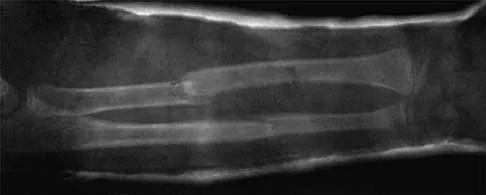
Standard weight-bearing radiographs of the foot—including anteroposterior (AP), lateral, and oblique views—are essential for preoperative templating. For the Terminal Syme procedure, radiographs are scrutinized for the extent of osteolysis, periosteal reaction, or cortical destruction indicative of osteomyelitis within the distal phalanx. The surgeon must template the exact level of bone resection, ensuring the proximal base and the FDL insertion remain untouched. For combined interphalangeal deformities, radiographs confirm the presence of rigid PIP and DIP flexion and rule out concurrent MTP joint subluxation or dislocation. The surgeon must evaluate the length of the proximal and middle phalanges to estimate the volume of bone resection required to decompress the joints adequately.
Patient Positioning and Anesthesia
The patient is positioned supine on the operating table. A bump may be placed under the ipsilateral hip to internally rotate the lower extremity, bringing the foot into a neutral, easily accessible position. These procedures are typically performed under a digital block using a long-acting local anesthetic, such as 0.5% bupivacaine or 0.75% ropivacaine, mixed with 1% lidocaine for rapid onset. While historical teaching strictly forbade the use of epinephrine in digital blocks, contemporary literature supports its safety in healthy patients; however, in vasculopathic patients, epinephrine should be strictly avoided. Monitored anesthesia care (MAC) or a regional ankle block may be utilized for patient comfort.
Equipment, Instrumentation, and Hemostasis
A bloodless surgical field is absolutely critical, particularly during the Terminal Syme procedure, to accurately identify and completely excise the pearly-white tissue of the germinal matrix. Following exsanguination of the digit via elevation or an Esmarch bandage, a digital tourniquet—such as a sterile Penrose drain or a specialized silicone digital ring—is applied at the base of the toe. Instrumentation should include fine microsurgical gear: Beaver blades for precise matrix excision, small periosteal elevators (e.g., Freer), double-action bone rongeurs, a microsagittal saw for precise phalangeal osteotomies, fine bone rasps, and 0.045-inch or 0.062-inch Kirschner wires (K-wires) for arthroplasty stabilization.
Step-by-Step Surgical Approach and Fixation Technique
The Terminal Syme Procedure
The execution of the Terminal Syme procedure requires meticulous soft-tissue handling and precise osseous resection to create a functional, pain-free terminal stump.
1. The Incision: An elliptical incision is meticulously planned to entirely encircle the nail complex. The incision must include 2 to 3 mm of the normal skin of the lateral nail folds and the distal hyponychium. Crucially, the incision must extend 3 to 4 mm proximally to the eponychium. This proximal extension is the most vital step to ensure the complete capture and removal of the germinal nail matrix, including its deep lateral horns.
2. Dissection and Bone Exposure: The incision is carried sharply down to the periosteum of the distal phalanx proximally. The surgeon carefully skirts the lateral margins and the distal tip of the ungual tuft. Using a fine periosteal elevator or a scalpel, the plantar pulp tissue is freed from the plantar aspect of the distal half of the distal phalanx. Surgical Warning: Dissection must not proceed too proximally on the plantar aspect. The insertion of the flexor digitorum longus tendon at the broad plantar base of the distal phalanx must remain completely undisturbed to prevent an iatrogenic extension deformity.
3. Bone Resection: With the distal half of the phalanx exposed, a microsagittal saw or a sharp double-action rongeur is used to resect the ungual tuberosity (the tuft). A bone rasp is then employed to aggressively smooth the remaining distal bony stump. Any residual sharp bony prominences will cause point-pressure necrosis or chronic pain beneath the advanced plantar pulp flap.
4. Matrix Inspection and Ablation: The proximal margin of the wound is carefully inspected. The proximal skin edge is everted using fine skin hooks to ensure absolutely no germinal matrix tissue remains. The matrix presents as distinct, pearly-white, striated tissue. If any residual matrix is identified, particularly in the deep lateral gutters, it must be sharply excised or aggressively curetted down to bone.
5. Flap Advancement and Closure: The digital tourniquet is released prior to closure to achieve meticulous hemostasis using bipolar electrocautery. Hematoma formation beneath the flap is a primary cause of excessive tension, subsequent infection, and catastrophic flap necrosis. The plantar pulp flap is advanced dorsally over the smoothed bony stump and sutured to the proximal dorsal skin margin using interrupted 4-0 nonabsorbable sutures (e.g., Nylon or Prolene). The closure must be completely tension-free; if tension is noted, additional bone must be resected from the distal phalanx.
Combined PIP and DIP Resection Arthroplasty
Addressing a rigid combined hammer and mallet toe requires aggressive decompression of both interphalangeal joints.
1. Incision and Exposure: Two separate transverse elliptical incisions are typically made directly over the PIP and DIP joints. This approach allows for the simultaneous excision of the hyperkeratotic double corns and direct exposure of the underlying joints. Alternatively, a single dorsal longitudinal incision can be utilized, though this requires separate, meticulous sharp excision of the corns. The extensor tendon is either tenotomized or split longitudinally to expose the dorsal articular surfaces.
2. Bone Resection (The Critical Step): At the PIP joint, the collateral ligaments are sharply released, and the head and neck of the proximal phalanx are delivered dorsally into the wound. A microsagittal saw is used to resect the head of the proximal phalanx just proximal to the condyles. Similarly, at the DIP joint, the collateral ligaments are sectioned, and the head of the middle phalanx is exposed and resected.
3. The 10% Recurrence Rule: Orthopedic literature dictates an approximately 10% recurrence rate of deformity following combined DIP and PIP resection arthroplasties. This unacceptably high failure rate is almost exclusively attributed to inadequate bone resection. The surgeon must ensure that a sufficient volume of bone is removed to allow the toe to lie perfectly flat without any soft-tissue tension or resistance. If the toe springs back into flexion upon release, more bone must be aggressively resected from the phalangeal shafts. The toe should feel somewhat "flail" prior to fixation.
4. Deformity Correction and Fixation: Once adequate bone is resected, the toe is placed in a rectus alignment. Temporary stabilization is achieved using a smooth 0.045-inch or 0.062-inch K-wire. The wire is driven antegrade through the remaining middle and distal phalanges, exiting the tip of the toe. It is then driven retrograde across the DIP and PIP joints, embedding securely into the medullary canal of the proximal phalanx. The extensor tendon is left to scar in an elongated position, and the skin is closed with 4-0 nonabsorbable sutures.
Complications, Incidence Rates, and Salvage Management
Despite meticulous surgical technique, complications following distal forefoot reconstruction can occur, often necessitating complex salvage procedures. The surgeon must be intimately aware of the specific pitfalls associated with both the Terminal Syme procedure and combined resection arthroplasties.
For the Terminal Syme procedure, the most frequent and frustrating complication is the regrowth of painful nail spicules, occurring in up to 5-10% of cases depending on the surgeon's experience. This is a direct result of incomplete ablation of the germinal matrix, specifically the deep lateral horns. When a spicule regrows, it often pierces the advanced plantar flap, leading to chronic pain, recurrent infection, and the need for revision surgery. Ischemic necrosis of the plantar pulp flap is a catastrophic complication, usually stemming from unrecognized preoperative vascular insufficiency, excessive tension on the closure, or an undrained postoperative hematoma.
In the context of combined resection arthroplasties, the "10% Recurrence Rule" highlights the most common pitfall: recurrence of the flexion deformity due to under-resection of bone. Conversely, over-resection of bone, combined with a failure to utilize K-wire stabilization, can result in a flail toe—a functionally useless, floppy digit that causes significant impingement in footwear. Pin tract infections are a recognized risk when utilizing percutaneous K-wires, requiring vigilant postoperative monitoring and oral antibiotic therapy if erythema develops. Vascular embarrassment post-straightening is a rare but critical emergency; if a chronically contracted toe is suddenly straightened and pinned, the digital arteries may go into spasm or be anatomically tethered, leading to a white, ischemic digit. If releasing the pin does not restore perfusion, the toe must be slightly flexed to relieve arterial tension.
| Complication | Incidence Rate | Primary Etiology | Salvage Management |
|---|---|---|---|
| Nail Spicule Regrowth | 5% - 10% | Retained lateral horns of the germinal matrix. | Revision sharp matrixectomy or aggressive phenolization of the spicule tract. |
| Flap Necrosis (Syme) | 1% - 3% | Vascular insufficiency, closure under tension, hematoma. | Debridement, local wound care, healing by secondary intention, or proximal amputation if gangrenous. |
| Deformity Recurrence | ~10% | Inadequate bone resection at PIP/DIP joints. | Revision arthroplasty with further bone resection, or conversion to a PIP/DIP arthrodesis. |
| Flail Toe | 2% - 5% | Excessive bone resection without adequate soft-tissue scarring/fixation. | Syndactylization (buddy-taping) to adjacent digit, or bone grafting with arthrodesis (rare). |
| Vascular Embarrassment | < 1% | Arterial spasm or tethering upon straightening a chronic contracture. | Immediate removal of K-wire, warm compresses, slight flexion of the digit. If irreversible, amputation. |
Phased Post-Operative Rehabilitation Protocols
Phase I: Acute Healing and Protection (Weeks 0-2)
The immediate postoperative goal is the protection of the surgical site, mitigation of edema, and ensuring the viability of the soft-tissue envelope. Following a Terminal Syme procedure or combined arthroplasty, a non-adherent dressing (e.g., Adaptic or Xeroform) is applied, followed by sterile gauze and a mildly compressive cohesive wrap. The patient is placed in a rigid-soled postoperative shoe to protect the distal toe from incidental trauma and to prevent bending forces across the surgical sites. Weight-bearing is permitted strictly on the heel or flat-footed as tolerated. Elevation of the extremity above the level of the heart is absolutely critical during the first 72 hours to minimize edema and prevent hematoma formation. The initial dressing is typically left intact until the first postoperative visit at 10 to 14 days, at which point sutures are removed.
Phase II: Subacute Stabilization and Alignment (Weeks 2-6)
During the subacute phase, the focus shifts to maintaining alignment and monitoring for delayed complications. For the Terminal Syme procedure, once sutures are removed, the patient may transition to a wide-toe-box athletic shoe, and local wound care is continued if any superficial delayed healing is present. No splinting is necessary for the Terminal Syme, as the preserved proximal base of the distal phalanx and the intact FDL insertion maintain biomechanical stability.
For combined resection arthroplasties, this phase requires meticulous management of the percutaneous K-wires. Patients are instructed on daily pin site care using alcohol or betadine solutions. The toe must remain rigidly protected in the postoperative shoe. If K-wires were not utilized, the toe must be securely buddy-taped to the adjacent normal toe 24 hours a day to dictate the axis of scar tissue formation and maintain rectus alignment.
Phase III: Long-Term Maintenance and Return to Function (Weeks 6+)
At 4 to 6 weeks postoperatively, K-wires are removed in the clinic setting. This is a simple, relatively painless procedure that does not require local anesthesia. Following pin removal, the patient is encouraged to perform gentle active and passive range-of-motion exercises of the MTP joint to prevent stiffness, while the PIP and DIP joints are allowed to fibrose in their new, corrected positions. Desensitization massage of the Terminal Syme stump is encouraged to prevent hypersensitivity. Patients are counseled on permanent footwear modifications, emphasizing shoes with a deep and wide toe box to prevent the recurrence of friction and the potential reformation of hyperkeratotic lesions. In diabetic or neuropathic patients, lifelong monitoring and the use of custom accommodative orthoses are mandatory to prevent contralateral or adjacent digit ulcerations.
Summary of Landmark Literature and Clinical Guidelines
The evolution of distal forefoot reconstruction is deeply rooted in historical surgical innovation and refined by modern biomechanical studies. The Terminal Syme procedure owes its conceptual origin to Sir James Syme, who in 1842 described a transmalleolar ankle amputation. The principles of complete ablation of the offending pathology while utilizing a robust, weight-bearing plantar flap were later elegantly adapted to the terminal digits. Landmark anatomical studies by Zook et al. in the late 20th century revolutionized our understanding of the nail matrix microanatomy. Zook’s definitive mapping of the germinal matrix, particularly the proximal and plantar extent of the lateral horns, established the modern surgical mandate for aggressive proximal resection during terminal amputations, directly reducing the historical incidence of nail spicule regrowth.
In the realm of lesser toe deformities, the seminal work by Coughlin and Mann remains the cornerstone of operative decision-making. Their extensive long-term follow-up studies on resection arthroplasties versus arthrodesis for hammer and mallet toes established the notorious "10% Recurrence Rule." Coughlin’s literature definitively demonstrated that failure to resect adequate bone at the interphalangeal joints leads to soft-tissue tension and inevitable recurrence of the flexion contracture. This literature forms the basis of the current clinical guideline that a toe must be rendered completely flaccid prior to K-wire fixation during a resection arthroplasty.
Current consensus guidelines from the American Academy of Orthopaedic Surgeons (AAOS) and the American Orthopaedic Foot & Ankle Society (AOFAS) emphasize the critical nature of preoperative vascular assessment. Guidelines strictly recommend against elective forefoot reconstruction in patients with an absolute toe pressure of less than 30 mm Hg or a TcPO2 of less than 30 mm Hg, due to the unacceptably high risk of flap necrosis and subsequent limb loss. Furthermore, in the diabetic population, current literature advocates for a multidisciplinary approach, combining aggressive surgical offloading (via arthroplasty or amputation) with stringent glycemic control (HbA1c < 8.0%) to optimize wound healing and prevent the recurrence of osteomyelitis. By adhering to these evidence-based guidelines and respecting the intricate anatomy of the
