- Shoulder 2. ### Osteology
- Scapula
- Clavicle
- Spans second through seventh ribs and serves as an attachment for 17 muscles
- Anteverted on chest wall approximately 30 degrees relative to the body
- Glenoid retroverted approximately 5 degrees relative to scapular body
- Os acromiale: incomplete fusion of secondary ossification centers, most commonly between mesoacromion and metaacromion
- Coracoid: attachments to coracoid include coracoacromial ligament, coracoclavicular ligaments (conoid [medial] and trapezoid [lateral]), conjoined tendon (coracobrachialis and short head of biceps), and pectoralis minor.
-
Suprascapular notch: suprascapular artery passes superior to superior transverse scapular ligament and suprascapular nerve passes inferior to ligament through notch (mnemonic: “Army over Navy” for artery over nerve).
- Spinoglenoid notch: both the artery and nerve inferior to the inferior transverse scapular ligament
-
Coracoacromial ligament contributes to anterosuperior stability in rotator cuff deficiency and should be preserved with irreparable cuff tears to prevent anterosuperior escape.
- Acromial branch of the thoracoacromial artery runs on medial aspect of the coracoacromial ligament.
- First bone in the body to ossify (at 5 weeks’ gestation) and last to fuse (medial epiphysis at 25 years of age)
- Fracture of clavicle is the most common musculoskeletal birth injury.
- Proximal humerus
- Humeral head retroverted 30 degrees relative to transepicondylar axis of humerus
- Head height approximately 5.6 cm above superior border of pectoralis major tendon (important for arthroplasty)
- Anatomic neck (directly below humeral head) attachment for the shoulder capsule
- Surgical neck more distal and more often involved in fractures
- Transverse humeral ligament important stabilizer of the biceps tendon
-
Arthrology: one major articulation (glenohumeral joint) and two minor articulations (sternoclavicular, acromioclavicular)
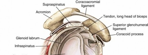
- Glenohumeral joint ( Fig. 2.1): ball and socket; greatest joint range of motion in body; motion is at the expense of stability, which is provided by static and dynamic restraints
- Static restraints: articular anatomy, glenoid labrum, glenohumeral ligaments, capsule, and negative intraarticular pressure
- Dynamic stabilizers: rotator cuff and biceps tendon; scapulothoracic mechanics contribute to stability
- Important glenohumeral stabilizers summarized in Table 2.1
- Fibrocartilaginous glenoid labrum deepens socket 50% and provides bumper to translation.
- Labral anatomic variants include sublabral foramen (anterosuperior) and Buford complex (absence of anterosuperior labrum and cordlike middle glenohumeral ligament).
-
Use caution repairing anterosuperior labral variant—may cause loss of external rotation.
- Sternoclavicular joint ( Fig. 2.2)
- Double gliding with an articular disc
-
Only true joint connecting upper extremity with axial skeleton

 FIG. 2.1 Glenohumeral joint.
From Miller MD et al:
Orthopaedic surgical approaches,
Philadelphia, 2008, Saunders, Figure SA-4.
FIG. 2.1 Glenohumeral joint.
From Miller MD et al:
Orthopaedic surgical approaches,
Philadelphia, 2008, Saunders, Figure SA-4.
- Posterior sternoclavicular ligament strongest and primary restraint to anteroposterior instability
- Rotates 30 degrees with shoulder motion
- Acromioclavicular (AC) joint
- Plane/gliding joint with a fibrocartilaginous disc
- Ligaments (see Fig. 2.2):
- AC ligaments: prevent anteroposterior displacement
- Coracoclavicular ligaments: prevent superior displacement of distal clavicle
- Trapezoid (anterolateral): approximately 25 mm from AC joint
- Conoid (posteromedial and stronger): approximately 45 mm from AC joint
- When the arm is maximally elevated, about 5 to 8 degrees of rotation are possible at the AC joint, although the clavicle rotates approximately 40 to 50 degrees.
Table 2.1 Glenohumeral Stabilizers Structure Function --- Glenoid labrum | Increases surface area, deepens socket, static stabilizer Coracohumeral ligament | Restrains inferior translation and external rotation of adducted arm Superior glenohumeral ligament | Restrains external rotation and inferior translation of adducted or slightly abducted arm Middle glenohumeral ligament (absent in up to 30% of shoulders) | Restrains anterior translation with arm abducted to 45 degrees Inferior glenohumeral ligament, anterior band | Restrains anterior and inferior translation with arm externally rotated and abducted to 90 degrees (position of apprehension) Inferior glenohumeral ligament, posterior band | Restrains posterior and inferior translation with arm internally rotated and abducted to 90 degrees
- Scapulothoracic joint
- Though not a true joint, this attachment allows scapular movement against the posterior rib cage and contributes to glenohumeral joint positioning and mechanics.
- Fixed primarily by the scapular muscular attachments
- Positions the glenoid for glenohumeral motion. Glenohumeral motion in comparison with scapulothoracic motion is in a 2:1 ratio.
- Muscles of the shoulder girdle are shown in Fig. 2.3 and outlined in Table 2.2. Rotator cuff: supraspinatus, infraspinatus, teres minor, subscapularis
- Function: depress and stabilize the humeral head against the glenoid; force-couple larger shoulder muscles to maintain humeral head center of rotation
- Shoulder internal rotators (pectoralis major, latissimus dorsi, teres major, and subscapularis) are stronger than external rotators (teres minor and infraspinatus), which is why posterior shoulder dislocations may occur with electric shock and seizures.
- Three important (and testable) spaces formed by muscles around the posteromedial shoulder: quadrangular space, triangular space, and triangular interval ( Fig. 2.4 , Table 2.3)
- Triangular interval is inferior to quadrangular space.
- Mnemonic: triangular interv al is dist al

 FIG. 2.2 Joints and ligaments of the clavicle (anterior view).
From Drake RL et al, editors:
Gray’s atlas of anatomy,
ed 2, Philadelphia, 2015, Churchill Livingstone.
FIG. 2.2 Joints and ligaments of the clavicle (anterior view).
From Drake RL et al, editors:
Gray’s atlas of anatomy,
ed 2, Philadelphia, 2015, Churchill Livingstone.


 FIG. 2.3
Muscle attachments of the shoulder and arm (anterior and posterior views). From Drake RL et al, editors:
Gray’s atlas of anatomy,
ed 2, Philadelphia, 2015, Churchill Livingstone.
FIG. 2.3
Muscle attachments of the shoulder and arm (anterior and posterior views). From Drake RL et al, editors:
Gray’s atlas of anatomy,
ed 2, Philadelphia, 2015, Churchill Livingstone.
Table 2.2
Muscles of the Shoulder Girdle Muscle | Origin | Insertion | Action | Innervation | ---|---|---|---|---| Trapezius | SP C7–T12
| Clavicle,
scapula
| Rotating scapula
| Cranial nerve XI
| |
(acromion, SP)
| |
| ---|---|---|---|---|
Latissimus dorsi
| SP T6–S5, ilium
| Humerus
(ITG)
| Extending, adducting, internally rotating humerus
| Thoracodorsal nerve
Rhomboid major
| SP T2–T5
| Scapula
(medial border)
| Adducting scapula
| Dorsal scapular nerve
Rhomboid minor
| SP C7–T1
| Scapula
(medial spine)
| Adducting scapula
| Dorsal scapular nerve
Levator scapulae
| Transverse
process C1–4
| Scapula
(superior medial)
| Elevating, rotating scapula
| C3, C4 nerves
Pectoralis major
| Sternum, ribs, clavicle
| Humerus
(lateral ITG)
| Adducting, internally rotating arm
| Medial and lateral pectoral nerves
Pectoralis minor
| Ribs 3–5
| Scapula
(coracoid)
| Protracting scapula
| Medial pectoral nerve
Subclavius
| Rib 1
| Inferior
clavicle
| Depressing clavicle
| Upper trunk nerves
Serratus anterior
| Ribs 1–9
| Scapula
(ventral medial)
| Preventing winging
| Long thoracic nerve
Deltoid
| Lateral clavicle, scapula
| Humerus
(deltoid tuberosity)
| Abducting arm
| Axillary nerve
Teres major
| Inferior scapula
| Humerus
(medial ITG)
| Adducting, internally rotating, extending arm
| Lower
subscapular nerve
Subscapularis
| Ventral scapula
| Humerus
(lesser tuberosity)
| Internally rotating arm, providing anterior stability
| Upper and lower subscapular nerves
Supraspinatus
| Superior
scapula
| Humerus
(GT)
| Abducting and externally rotating arm, providing
| Suprascapular nerve
| |
| stability
| ---|---|---|---|---
Infraspinatus
| Dorsal scapula
| Humerus
(GT)
| Providing stability, externally rotating arm
| Suprascapular nerve
Teres minor
| Scapula
(dorsolateral)
| Humerus
(GT)
| Providing stability, externally rotating arm
| Axillary nerve
GT, Greater tuberosity; ITG, intertubercular groove; SP, spinous process.


 FIG. 2.4 Borders of the key spaces and intervals around the shoulder, including the suprascapular nerve course as well as the quadrangular space and triangular space/interval. ### Table 2.3
Shoulder Spaces Space Borders Nerve Artery --- Quadrangular (quadrilateral) space | Superior: lower border of teres minor
Inferior: upper border of teres major
Medial: long head of triceps Lateral: surgical neck of humerus
| Axillary
| Posterior
humeral circumflex
Triangular space
| Superior: lower border of teres minor
Inferior: upper border of teres major
Lateral: long head of triceps
| |
Circumflex
scapular
Triangular interval
| Superior: lower border of teres major
Medial: long head of triceps Lateral: shaft of humerus
| Radial
| Profunda
brachii
