Introduction & Epidemiology
Sciatic nerve palsy (SNP) following total hip replacement (THR) is a serious and potentially debilitating complication, representing a significant challenge in orthopedic surgery. While relatively rare, its impact on patient morbidity, functional outcome, and quality of life is profound. The term "hidden danger" aptly describes this complication, as its onset can be immediate or delayed, its symptoms varied, and its diagnosis sometimes obscured by post-operative pain or sedation. Understanding the precise mechanisms of injury, meticulous surgical technique, and prompt, accurate diagnosis are paramount to minimizing its incidence and optimizing recovery.
The reported incidence of sciatic nerve palsy after primary THR ranges from 0.1% to 2.8%. However, this rate can be significantly higher in revision THR (up to 7.6%) or in cases involving complex anatomies such as developmental dysplasia of the hip (DDH) requiring substantial limb lengthening (reported up to 11%). Factors contributing to this variability include the surgical approach, patient comorbidities, magnitude of limb lengthening, and the definition of nerve injury itself (transient neuropraxia vs. complete transection). Given the global volume of THR procedures performed annually, even a low incidence translates to a substantial number of affected individuals, underscoring the importance of this topic for every orthopedic surgeon.
Surgical Anatomy & Biomechanics
A thorough understanding of the sciatic nerve's intricate anatomical course and its relationship to the hip joint and surrounding musculature is fundamental to preventing iatrogenic injury during THR.
The sciatic nerve, the largest nerve in the body, originates from the sacral plexus (L4-S3 nerve roots). It exits the pelvis through the greater sciatic foramen, typically inferior to the piriformis muscle, though anatomical variations exist where it may pierce or pass superior to the piriformis. Its course is posterior to the hip joint capsule and deep to the gluteus maximus. It then descends between the greater trochanter and the ischial tuberosity, superficial to the short external rotator muscles (superior gemellus, obturator internus, inferior gemellus, and quadratus femoris) before continuing distally into the thigh.
Crucially, the sciatic nerve lies in close proximity to the posterior aspect of the acetabulum and proximal femur. During a posterior approach to the hip, the nerve is exposed after dissection of the gluteus maximus and release of the short external rotators. Retractors, particularly those used to expose the acetabulum or to mobilize the femur, can exert direct pressure on the nerve within the greater sciatic notch or along the posterior column.
Biomechanical mechanisms of sciatic nerve injury during THR include:
- Traction (Stretch Injury): This is widely considered the most common mechanism, particularly with limb lengthening exceeding 3-4 cm. Significant elongation can stretch the nerve beyond its physiological elasticity limits, leading to demyelination, axonal damage, and impaired blood supply. This risk is amplified in patients with pre-existing neuropathies, diabetes, or those undergoing revision surgery where the nerve may be scarred or tethered.
-
Direct Compression:
- Retractor Placement: Misplaced or excessively large retractors, especially in the greater sciatic notch, can compress the nerve.
- Hematoma: Post-operative bleeding can lead to an expanding hematoma that exerts compressive force on the nerve, particularly in the confined space of the greater sciatic notch.
- Cement Extrusion: In cemented THR, excessive cement migration posteriorly can directly impinge on the nerve, often exacerbated by the exothermic reaction of polymerization.
- Implant Impingement: Malpositioned acetabular components (e.g., retroverted, excessively prominent posterior rim) or screws perforating the posterior column can cause direct mechanical irritation or compression.
- Direct Laceration or Transection: Although rare, direct injury from osteotomes, reamers, drills, or sharp retractors is possible, especially during revision surgery in a scarred field or with challenging osteophyte removal.
- Thermal Injury: The exothermic reaction during cement polymerization can potentially cause thermal damage to the sciatic nerve if cement extrudes in close proximity.
- Ischemia: Prolonged tourniquet use, vessel damage, or compressive dressings can compromise the nerve's blood supply.
- Patient Positioning: Inadequate padding or prolonged positioning on the operating table can cause nerve compression, though typically this would involve the peroneal division at the fibular head or the femoral nerve in the groin. However, extreme hip flexion or internal rotation can increase tension on the sciatic nerve.
Anatomical variations, such as the sciatic nerve piercing the piriformis muscle, can predispose it to greater tension or compression with hip movements or in the presence of an inflammatory response. Awareness of these possibilities during pre-operative planning and intraoperative assessment is crucial.
Indications & Contraindications
While "indications and contraindications" for sciatic nerve palsy itself are not strictly defined as for a surgical procedure, this section focuses on identifying patients and surgical scenarios that indicate a heightened risk for sciatic nerve injury during THR and therefore necessitate meticulous preventative measures. Conversely, certain patient conditions may contraindicate aggressive limb lengthening strategies.
Indications for Heightened Vigilance and Preventative Strategies (Risk Factors for SNP):
- Pre-existing Neuropathy: Patients with diabetes mellitus, peripheral vascular disease, previous spinal surgery with radiculopathy, or other neurological conditions are inherently more susceptible to nerve injury from stretch or compression. Baseline neurological assessment is crucial.
- Significant Pre-operative Leg Length Discrepancy (LLD): Patients with substantial LLD, particularly those with long-standing DDH, post-traumatic arthritis, or protrusio acetabuli, where significant limb lengthening (typically >3-4 cm) is planned to restore leg length and hip biomechanics. The acute lengthening can stretch the chronically shortened sciatic nerve beyond its elastic limits.
- Revision Total Hip Arthroplasty: The presence of extensive scarring, distorted anatomy, retained cement, or heterotopic ossification from previous surgeries increases the difficulty of tissue dissection and the risk of nerve entrapment or direct injury.
- Congenital Hip Dysplasia (DDH): Often associated with proximal femoral migration, severe anteversion, and LLD, requiring complex reconstruction and potentially significant lengthening.
- Ankylosing Spondylitis / Spinal Fusion: Patients with stiff spines may transmit more stress to the hip joint, and their fixed spinal posture can alter nerve mechanics.
- Extremes of Body Mass Index (BMI): Both very low and very high BMI can present challenges in exposure and retraction, potentially increasing nerve risk.
- Prolonged Operative Time: Extended periods of exposure, retraction, or patient positioning increase overall risk.
- Specific Surgical Approaches: The posterior approach, while versatile, inherently places the sciatic nerve at greater risk due to its anatomical proximity and the need to mobilize the short external rotators.
Contraindications for Aggressive Limb Lengthening (Relative):
In patients with known significant risk factors for SNP, particularly pre-existing neuropathy or a history of prior nerve injury, aggressive limb lengthening should be carefully considered and potentially modified.
- Established Severe Neuropathy: In cases where pre-operative neurological deficits are severe and documented, the threshold for acceptable limb lengthening should be lowered.
-
Unavoidable Excessive Lengthening:
If pre-operative templating indicates that restoring hip biomechanics will necessitate lengthening beyond 4-5 cm, alternative strategies should be explored. These might include:
- Accepting a residual LLD.
- Performing a concomitant femoral shortening osteotomy to mitigate nerve stretch.
- Selecting a different component design (e.g., short-stem femoral components, constrained liners) that minimizes soft tissue tension if feasible.
Table 1: Decision-Making in High-Risk Patients / Post-Operative Management of Sciatic Nerve Palsy
| Category | Operative Intervention / Strategy (Preventative/Acute Management) | Non-Operative Management / Observation |
|---|---|---|
| Pre-operative High-Risk | Meticulous templating to limit limb lengthening. Consider femoral shortening osteotomy for >4-5 cm predicted lengthening. Intraoperative neuromonitoring (SSEP/EMG). Careful choice of approach (e.g., less posterior retraction). Patient education on nerve injury risk. | Careful patient selection. Pre-operative neurological assessment and documentation. Optimize co-morbidities (e.g., diabetes control). Counseling on realistic LLD correction expectations. |
| Acute Post-op Palsy (Complete) | Immediate investigation (CT/MRI for hematoma/component impingement). Surgical exploration for suspected correctable causes (e.g., large hematoma, gross cement extrusion, malpositioned component/screw) within days to weeks. Neurolysis if entrapment is identified. | Immediate conservative management: strict bed rest, nerve observation, pain control. Neurological exam monitoring (serial). Consider EMG/NCS at 3-4 weeks. Ankle-foot orthosis (AFO) for foot drop. |
| Delayed/Partial Palsy | Consider surgical exploration if no improvement or worsening after 3-6 months, or if imaging reveals a clear anatomical impingement that is surgically correctable. Removal of offending agent, neurolysis. | Aggressive physical therapy, occupational therapy. Neuropathic pain management. AFO. Regular neurological assessment. Serial EMG/NCS studies to track recovery. Patient education and psychological support. |
| Chronic Non-Recovery | Tendon transfers (e.g., posterior tibial tendon transfer for foot drop) for functional improvement. Arthrodesis or ankle fusion in rare, severe, and persistent cases with instability. | Long-term AFO use. Continued physical therapy for strength and gait training. Consideration for adaptive equipment. |
Pre-Operative Planning & Patient Positioning
Meticulous pre-operative planning and careful intraoperative patient positioning are critical steps in minimizing the risk of sciatic nerve palsy.
Pre-Operative Planning
-
Comprehensive History and Physical Examination:
- Neurological Baseline: Document any pre-existing neurological deficits, including sensory loss, motor weakness, or neuropathic pain. Specific attention to the sciatic nerve distribution (foot drop, lateral calf numbness) is essential.
- Comorbidities: Identify risk factors such as diabetes, peripheral vascular disease, previous lumbar spine surgery, or prior nerve injuries.
- Previous Hip Surgery: Assess for scarring, heterotopic ossification, or retained hardware.
- Leg Length Discrepancy (LLD): Clinically measure apparent and true LLD. Assess compensatory mechanisms (pelvic tilt, scoliosis).
- Range of Motion (ROM): Document hip ROM, specifically internal and external rotation, which can influence soft tissue tension.
-
Radiographic Assessment and Templating:
- High-Quality Imaging: Obtain standardized AP pelvis, lateral hip, and potentially standing full-length leg radiographs to accurately assess LLD, femoral offset, and hip center of rotation.
- Digital Templating: Use digital templating software to meticulously plan component sizing, position, and predicted limb length restoration. This allows the surgeon to visualize the anticipated lengthening and determine if it exceeds acceptable limits. Aim to restore anatomical leg length while being cognizant of the sciatic nerve's elasticity. Generally, lengthening >3-4 cm significantly increases SNP risk.
- Identify High-Risk Anatomy: Recognize severe DDH with proximal femoral migration, protrusio acetabuli, or significant osteophytes that may tether the nerve or complicate exposure.
-
Anesthesia Consultation: Discuss the use of nerve blocks (e.g., spinal vs. general anesthesia, use of post-operative blocks) and their potential to mask early signs of nerve injury. Intraoperative wake-up tests for leg motion are rarely practical but have been described in extreme cases.
-
Neuromonitoring: In high-risk cases (e.g., severe LLD, revision surgery, pre-existing neuropathy), intraoperative neuromonitoring (IONM) with somatosensory evoked potentials (SSEPs) and electromyography (EMG) can be considered. While controversial and not universally adopted due to cost and interpretation challenges, a significant drop in SSEP amplitude or increased spontaneous EMG activity could indicate nerve irritation or stretch, prompting intraoperative adjustments. However, it requires an experienced neurophysiology team and real-time feedback.
-
Patient Education: Inform the patient about the potential risk of nerve injury, its symptoms, and the typical course of recovery. Manage expectations regarding LLD correction, particularly in cases where full correction would entail unacceptable nerve risk.
Patient Positioning
The choice of surgical approach (posterior, anterior, anterolateral) dictates specific positioning, but fundamental principles apply to all to protect the sciatic nerve.
-
Lateral Decubitus Position (Common for Posterior/Anterolateral Approaches):
- Stable Positioning: Ensure the patient is securely positioned to prevent inadvertent movements during surgery. Use beanbags, kidney rests, and tape as necessary.
-
Adequate Padding:
Crucial for all pressure points.
- Peroneal Nerve: Pad the fibular head on the dependent leg to prevent compression of the common peroneal nerve, which is a division of the sciatic nerve and prone to injury at this site.
- Axilla: Pad the axilla on the dependent side to prevent brachial plexus compression.
- Contralateral Leg: Ensure the contralateral leg is not excessively flexed, abducted, or internally rotated, as these positions can strain the sciatic nerve or common peroneal nerve.
- Hip Flexion/Extension: Avoid extreme hip flexion or extension that can tension the sciatic nerve.
- Torso Alignment: Maintain neutral spine alignment to prevent undue tension on the nerve roots.
-
Supine Position (Common for Anterior Approach):
- Padding: Ensure all bony prominences are adequately padded.
- Lower Extremity Positioning: Avoid excessive external rotation or hyperflexion of the operative leg, which can tension the sciatic nerve. The non-operative leg should be positioned neutrally.
- Foot Drop Prevention: Ensure the feet are in a neutral position, avoiding prolonged plantarflexion that could compromise the peroneal nerve.
General Principles for All Positions:
- Avoid Excessive Traction: Traction on the operative limb during dislocation or reduction should be controlled and brief.
- Monitor Anesthetic Depth: Deep anesthesia can mask reflexive muscle contractions that might indicate nerve irritation.
- Communication with Anesthesiologist: Report any concerns regarding patient positioning or limb manipulation.
Detailed Surgical Approach / Technique
This section focuses on intraoperative strategies, particularly within the context of the posterior approach, to meticulously protect the sciatic nerve during THR. While the general steps of THR are standard, emphasis here is placed on the nuances that mitigate nerve injury.
Posterior Approach
The posterior approach provides excellent exposure to the acetabulum and femur, but necessitates identification and protection of the sciatic nerve due to its direct proximity.
-
Incision and Initial Dissection:
- Standard curvilinear incision centered over the greater trochanter.
- Careful dissection through subcutaneous tissue to the fascia of the gluteus maximus.
-
Gluteus Maximus Splitting:
- The gluteus maximus is split in line with its fibers, typically 3-5 cm posterior to the greater trochanter.
- Avoid excessive medial retraction of the gluteus maximus muscle, as this can directly impinge on the sciatic nerve lying deep to it.
-
Identification and Release of Short External Rotators:
- The short external rotators (piriformis, superior gemellus, obturator internus, inferior gemellus, quadratus femoris) are identified. These muscles lie superficial to the posterior hip capsule and deep to the gluteus maximus.
- The sciatic nerve typically lies superficial to the obturator internus and gemelli, and deep to the gluteus maximus.
- The short external rotators are sharply released from their insertions on the posterior femur, with careful attention to dissecting along their tendinous attachments to avoid inadvertent nerve injury. The piriformis tendon is often the first and most superior rotator encountered; its release provides initial exposure to the nerve.
- Crucial Step: Sciatic Nerve Identification and Protection: Before any deep retraction or capsular incision, the sciatic nerve must be directly visualized. It is a large, whitish cord, typically lying just inferior to the piriformis and superior to the quadratus femoris, as it crosses the posterior aspect of the hip joint.
- Once identified, the nerve should be gently mobilized and protected with a broad, blunt retractor (e.g., Darrach or Hohmann retractor) placed around the nerve, ensuring no direct pressure or traction is applied. The retractor should always be placed deep to the nerve, on the bone (posterior column), never directly on the nerve or compressing it into the soft tissues.
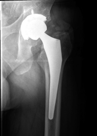
Figure 1: Illustration of the sciatic nerve's anatomical course in relation to the posterior aspect of the hip, showing its proximity to the short external rotators and the posterior acetabulum. Meticulous identification and protection are paramount during posterior approach THR. -
Capsulotomy and Hip Dislocation:
- A posterior capsulotomy is performed.
- The hip is dislocated, typically by internally rotating, adducting, and flexing the femur. Controlled dislocation minimizes undue soft tissue tension.
-
Acetabular Preparation:
- Retractor Placement: Acetabular retractors should be placed carefully. A posterior wall retractor should be placed subperiosteally on the posterior column, ensuring it does not impinge on the sciatic nerve in the greater sciatic notch.
- Reaming and Component Insertion: Reaming should be performed within the confines of the bony acetabulum. Avoid over-reaming posteriorly. When inserting the acetabular component, ensure it is flush with the posterior rim, avoiding posterior overhang, which can directly compress the nerve. Screw placement for acetabular fixation must avoid penetrating the posterior column or violating the greater sciatic notch.
-
Femoral Preparation:
- Femoral Head Resection: Resect the femoral head based on pre-operative templating, aiming for restoration of femoral offset and limb length without excessive lengthening.
- Broaching and Reaming: Maintain gentle traction on the limb to facilitate femoral canal access, but avoid forceful rotation or excessive adduction that could tension the nerve.
- Cemented Stems: If using a cemented stem, ensure careful cement pressurization to prevent posterior extrusion. The exothermic reaction of cement polymerization can also contribute to nerve injury if it is in direct contact with extruded cement.
-
Limb Lengthening Assessment and Trial Reduction:
- Intraoperative Measurement: Prior to definitive component implantation, measure limb length. This can be done by comparing the distance from the anterior superior iliac spine (ASIS) to the medial malleolus, or by assessing the relative position of the greater trochanter to the ASIS. Comparing the amount of femoral head resected to the thickness of the trial femoral head is also useful.
- Soft Tissue Tension: During trial reduction, assess the tension in the soft tissues. If the hip feels excessively tight, or if there is significant resistance to reduction, this may indicate excessive limb lengthening or impingement. Consider using a shorter femoral neck or reducing the overall leg length correction.
- Range of Motion: Test stability and ROM with trial components.
-
Component Implantation and Final Reduction:
- Implant definitive components, ensuring proper orientation and fixation.
- Carefully reduce the hip.
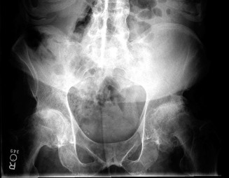
Figure 2: Intraoperative view showing a well-placed acetabular component. Careful attention to posterior component placement and avoidance of overhang or prominent screws is crucial to prevent sciatic nerve impingement. -
Closure:
- Hemostasis: Achieve meticulous hemostasis to prevent post-operative hematoma formation, which can compress the nerve.
- Repair Short External Rotators: Reattach the short external rotators (e.g., with transosseous sutures through the greater trochanter). This can help restore posterior hip stability but ensure the repair does not inadvertently entrap the sciatic nerve.
- Wound Closure: Standard fascial, subcutaneous, and skin closure.
Anterior Approach Considerations
While the anterior approach is generally considered to have a lower risk of sciatic nerve injury due to the nerve's posterior location, it is not immune:
- Excessive Lengthening: Any approach that results in significant limb lengthening carries a risk of stretch injury to the sciatic nerve.
- Retractor Placement: Ill-placed or excessively deep retractors, particularly during posterior acetabular reaming or component insertion, could theoretically impinge on the nerve, although this is rare.
- Femoral Nerve Risk: The anterior approach has a higher risk of femoral nerve injury, which must be differentiated from sciatic nerve palsy.
Regardless of the approach, the fundamental principle remains: constant awareness of the sciatic nerve's anatomy and meticulous surgical technique to avoid direct trauma, compression, or excessive stretch.
Complications & Management
Sciatic nerve palsy is a serious complication, but it is one of several potential adverse events following THR. Prompt identification and appropriate management are crucial for optimizing patient outcomes.
Clinical Presentation of Sciatic Nerve Palsy
- Motor Deficits: The most common presentation is foot drop, indicating weakness of ankle dorsiflexion and eversion (peroneal division involvement). Weakness in knee flexion (hamstrings) and toe flexion/ankle plantarflexion (tibial division) may also be present, depending on the extent of injury. A complete sciatic palsy would affect all these muscle groups.
- Sensory Deficits: Numbness, paresthesia, or dysesthesia in the foot (dorsal, plantar, lateral) and lateral calf (peroneal distribution) or posterior calf (tibial distribution).
- Pain: Neuropathic pain, described as burning, shooting, or electric-like, may be a prominent symptom.
- Timing: Symptoms can be immediate post-operatively or have a delayed onset, often due to hematoma formation.
Differential Diagnosis
It is essential to differentiate sciatic nerve palsy from other conditions:
*
Lumbar Radiculopathy:
Pre-existing or new onset, can mimic sciatic symptoms. Imaging of the spine may be warranted.
*
Femoral Nerve Palsy:
Weakness of hip flexion and knee extension (quadriceps), sensory loss over anterior thigh/medial calf. More common with anterior approaches.
*
Acute Compartment Syndrome:
Rare but critical. Severe pain, tense compartment, pain with passive stretch, paresthesia.
*
Deep Venous Thrombosis (DVT) / Popliteal Artery Injury:
Distal edema, pain, warmth, diminished pulses.
Investigation
- Clinical Examination: Detailed motor, sensory, and reflex examination immediately post-operatively and serially.
-
Electromyography (EMG) and Nerve Conduction Studies (NCS):
- Gold Standard: For diagnosis, localization of the lesion, assessment of severity (demyelinating vs. axonal), and prognosis.
- Timing: Typically performed 3-4 weeks post-operatively when denervation changes become evident. Early studies may be normal.
-
Imaging:
- Plain Radiographs: Assess component position, identify gross malposition, or obvious hardware impingement.
- CT Scan: Superior for evaluating component position, identifying cement extrusion, bony impingement, or heterotopic ossification near the nerve.
- MRI Scan: Useful for detecting soft tissue hematoma, nerve edema, or scarring around the nerve.
Management
-
Conservative Management:
- Observation: Many sciatic nerve palsies, particularly stretch-induced neuropraxias, improve spontaneously over weeks to months.
- Physical Therapy: Early mobilization, range-of-motion exercises, and strengthening of unaffected muscles.
- Ankle-Foot Orthosis (AFO): Prescribed for foot drop to prevent equinus deformity and facilitate safe ambulation.
- Pain Management: Neuropathic pain may require specific medications (e.g., gabapentin, pregabalin, tricyclic antidepressants).
- Patient Education: Reassurance, realistic expectations for recovery, and instructions on skin care for insensate areas.
-
Surgical Exploration:
-
Indications:
- Complete palsy with no signs of recovery: Typically, surgical exploration is considered if there is no evidence of clinical or electrophysiological recovery after 3-6 months.
- Acute, severe palsy with identifiable correctable lesion: If imaging (CT/MRI) reveals a clear compressive lesion (e.g., large hematoma, gross cement extrusion, malpositioned component or screw) that is amenable to surgical intervention, early exploration (within days to weeks) may be warranted.
- Progressive neurological deficit: Worsening symptoms mandate urgent investigation and potential exploration.
- Timing: Early exploration within weeks for acute, compressive lesions. Delayed exploration (3-6 months) for non-recovering palsies to perform neurolysis or remove chronic impingements.
-
Technique:
- Re-exposure of the sciatic nerve via the posterior approach.
- Careful removal of any offending agent (hematoma evacuation, cement removal, revision of impinging components/screws).
- Neurolysis: Gentle release of the nerve from scar tissue (external neurolysis) to relieve compression.
- If the nerve is found to be transected (rare), primary repair or nerve grafting may be considered, although functional outcomes are often poor.
-
Indications:
Table 2: Common Complications of Total Hip Replacement and Management Strategies
| Complication | Incidence (approximate) | Salvage Strategies |
|---|---|---|
| Sciatic Nerve Palsy | 0.1-2.8% (Primary) | Conservative (observation, PT, AFO, pain meds). Surgical exploration (hematoma evacuation, component revision, neurolysis) for complete or non-recovering palsies. Tendon transfer for chronic foot drop. |
| Dislocation | 1-5% (Primary) | Closed reduction (acute). Hip precautions. Bracing. If recurrent: revision of component position (acetabular/femoral), constrained liner, larger femoral head, soft tissue repair/advancement, trochanteric advancement. |
| Periprosthetic Joint Infection (PJI) | 0.5-2% | Debridement, antibiotics, implant retention (DAIR) for acute early infections. One-stage or two-stage revision arthroplasty (excision arthroplasty, temporary spacer, intravenous antibiotics, reimplantation). Chronic suppressive antibiotics in select cases. |
| Periprosthetic Fracture | 0.1-1% (Intraoperative) | Intraoperative: Cerclage wiring, cables, plates, revision to larger stem, longer stem. Postoperative: Open reduction internal fixation (ORIF), revision arthroplasty (stem, cementless vs. cemented, augments). |
| Deep Venous Thrombosis (DVT) | 1-10% (clinical) | Anticoagulation (oral/injectable). Compression stockings. Early mobilization. IVC filter for recurrent DVT/PE with failed anticoagulation or contraindications. |
| Pulmonary Embolism (PE) | 0.1-1% | Therapeutic anticoagulation. Oxygen support. IVC filter. Surgical embolectomy (rare). |
| Leg Length Discrepancy | Varies | Shoe lift (small discrepancy). Physiotherapy. If functional issues or significant cosmetic concern: component revision (femoral stem, head length) to adjust leg length (challenging, higher risk for nerve injury with further lengthening). |
| Aseptic Loosening | <1% per year | Revision arthroplasty (component exchange, stem revision, bone grafting). |
| Heterotopic Ossification | 5-15% (mild) | Non-steroidal anti-inflammatory drugs (NSAIDs) or radiation prophylaxis (high-risk patients). Excision if symptomatic (impingement, decreased ROM). |
Post-Operative Rehabilitation Protocols
Rehabilitation for a patient who has sustained a sciatic nerve palsy post-THR requires a specialized and multidisciplinary approach, focusing on maximizing nerve recovery, preventing secondary complications, and restoring functional independence.
Initial Phase (Days to Weeks Post-Op)
-
Protection and Education:
- Ankle-Foot Orthosis (AFO): Immediately fitted for patients with foot drop to prevent equinus deformity, protect the insensate foot from pressure sores, and facilitate a safer gait pattern.
- Skin Care: Meticulous skin care instructions for areas of sensory loss to prevent breakdown, burns, or other injuries.
- Patient Education: Reassurance regarding the potential for recovery (especially for neuropraxias). Education on avoiding prolonged pressure on the affected limb and proper body mechanics.
-
Early Mobilization and Joint Protection:
- Weight-Bearing: As per THR protocol, usually protected weight-bearing initially, progressing to full weight-bearing as tolerated.
- Hip Precautions: Strict adherence to post-THR hip precautions (if applicable to the surgical approach) to prevent dislocation.
- Range of Motion (ROM): Passive and active-assisted ROM exercises for the hip and knee within pain-free limits. Gentle, passive stretching of the affected musculature to prevent contractures, but avoid aggressive stretching that could further tension the nerve.
-
Pain Management:
- Neuropathic pain is common. Pharmacological management with gabapentinoids (gabapentin, pregabalin) or tricyclic antidepressants may be initiated.
- Referral to a pain specialist if pain is severe or refractory.
Intermediate Phase (Weeks to Months Post-Op)
-
Strengthening and Motor Re-education:
- Targeted Exercises: Focus on strengthening muscles innervated by the sciatic nerve, particularly those showing early signs of reinnervation. This includes hamstrings, and if foot drop is present, ankle dorsiflexors (tibialis anterior), evertors (peroneals), and toe extensors.
- Biofeedback: May be used to help patients identify and activate weak muscles.
- Functional Electrical Stimulation (FES): Can be used to stimulate dorsiflexors during gait, providing a more natural walking pattern and potentially aiding muscle re-education.
-
Gait Training:
- Progressive gait training with appropriate assistive devices (walker, crutches, cane) and the AFO.
- Focus on improving balance, coordination, and proprioception.
-
Sensory Re-education:
- For patients with significant sensory loss, specific exercises to improve tactile discrimination and proprioception can be beneficial.
- Protection of the insensate limb remains critical.
-
Electrodiagnostic Monitoring:
- Follow-up EMG/NCS studies (e.g., at 3, 6, 9, 12 months) to monitor for signs of reinnervation and to gauge the extent and pattern of recovery. This helps guide prognosis and further intervention decisions.
Advanced Phase (Months to Years Post-Op)
- AFO Weaning: If significant motor recovery occurs, gradual weaning from the AFO can be attempted under the guidance of a physical therapist.
- Return to Activity: Progressive return to desired activities and sports, adapted as necessary.
-
Long-term Management for Non-Recovery:
- Persistent Foot Drop: If significant foot drop persists after 12-18 months with no further signs of recovery, tendon transfer procedures (e.g., posterior tibial tendon transfer to the dorsum of the foot) may be considered to restore active dorsiflexion and improve gait.
- Chronic Pain: Ongoing management of neuropathic pain may be required.
- Psychological Support: The long-term impact of nerve injury can be significant, and psychological counseling or support groups can be valuable.
Throughout all phases, a collaborative approach involving the orthopedic surgeon, physical therapist, occupational therapist, neurologist, and pain specialist is paramount to ensure comprehensive care and optimize functional outcomes for patients affected by sciatic nerve palsy post-THR.
Summary of Key Literature / Guidelines
The body of literature concerning sciatic nerve palsy following total hip arthroplasty underscores its multifactorial etiology and the importance of preventive strategies. Several key themes emerge from current research and clinical guidelines:
-
Incidence and Risk Factors:
- Studies consistently report a higher incidence of SNP in revision THR, in cases of significant limb lengthening (particularly >3-4 cm), and in patients with pre-existing neurological conditions such as diabetes or lumbar radiculopathy. Rates vary from 0.1% to 11% depending on the population and definition of palsy.
- The posterior approach has traditionally been associated with a higher direct risk to the sciatic nerve due to anatomical exposure, although modern techniques emphasizing direct nerve visualization and careful retraction aim to mitigate this. Anterior approaches have a lower sciatic nerve risk but a higher femoral nerve risk.
- Anatomical variations, such as the sciatic nerve piercing the piriformis muscle, are recognized as predisposing factors to stretch injury.
-
Mechanism of Injury:
- Stretch injury due to limb lengthening is considered the predominant mechanism. The critical threshold for lengthening is debated but generally accepted to be around 3-4 cm. Beyond this, the risk increases substantially.
- Direct compression from retractors, hematoma, cement extrusion, or malpositioned components/screws are also well-documented causes. Thermal injury from exothermic cement polymerization is a less common but recognized mechanism.
-
Prevention Strategies:
- Meticulous Pre-operative Planning: Accurate templating to predict and manage limb length discrepancy is crucial. Accepting a small residual LLD may be preferable to significant nerve stretch.
- Intraoperative Vigilance: Direct visualization and protection of the sciatic nerve, especially during the posterior approach, is emphasized. Careful retractor placement, avoiding direct nerve compression, and meticulous hemostasis are critical.
- Controlled Limb Lengthening: Intraoperative assessment of limb length and soft tissue tension during trial reduction is vital. Strategies like controlled femoral shortening osteotomy may be considered in cases of severe LLD requiring extensive lengthening.
- Neuromonitoring: While not universally adopted, intraoperative neuromonitoring (SSEPs, EMG) is increasingly considered in high-risk cases, though its efficacy in preventing permanent injury and cost-effectiveness remain subjects of ongoing debate.
-
Diagnosis and Prognosis:
- Clinical examination is the initial diagnostic tool, followed by EMG/NCS at 3-4 weeks post-operatively to confirm diagnosis, localize the lesion, and assess severity.
- Imaging (CT, MRI) helps identify compressive lesions such as hematomas, cement extrusion, or implant impingement.
- Most sciatic nerve palsies are neuropraxias and demonstrate spontaneous recovery, often partial. Complete recovery is less common. Proximal lesions or axonal injuries carry a poorer prognosis.
-
Management Principles:
- Conservative management with observation, physical therapy, and AFO use is the mainstay for most cases, especially neuropraxias.
- Surgical exploration is indicated for acute, complete palsies with an identifiable, surgically correctable cause (e.g., large hematoma, gross cement extrusion, malpositioned hardware) that can be relieved. It is also considered for non-recovering palsies after 3-6 months.
- Tendon transfers are a recognized option for chronic, persistent foot drop to improve function.
Current guidelines emphasize a proactive, preventive approach through thorough pre-operative assessment, meticulous surgical technique, and judicious intraoperative decision-making regarding limb length restoration. Early recognition and a structured management pathway are essential for optimizing outcomes in the unfortunate event of sciatic nerve palsy. Continued research focuses on refining risk prediction models, improving neuromonitoring efficacy, and developing enhanced surgical techniques to further minimize this challenging complication.
