Comprehensive Introduction and Patho-Epidemiology
The lesser rays of the foot function as a highly complex, dynamic biomechanical unit explicitly designed to distribute weight-bearing forces during the stance phase of gait, facilitate forward propulsion, and provide sensory feedback for postural equilibrium. Pathologies of the lesser toes—encompassing metatarsophalangeal (MTP) joint instability, hammer toes, mallet toes, intractable plantar keratosis (IPK), bunionettes, and Freiberg infraction—arise from a disruption in the delicate, highly evolved balance between intrinsic and extrinsic musculature, capsuloligamentous structures, and osseous alignment. These deformities represent a spectrum of progressive mechanical failure rather than isolated anatomic anomalies.
Epidemiologically, lesser toe deformities are exceedingly common, disproportionately affecting the female population with a female-to-male ratio approaching 4:1 to 5:1 in various epidemiological cohorts. The peak incidence occurs in the fifth to seventh decades of life. The etiology is undeniably multifactorial. Extrinsic factors, most notably the prolonged use of constricting, narrow-toebox, and high-heeled footwear, serve as a significant mechanical catalyst. These shoes force the lesser digits into a non-physiologic position of MTP joint extension and proximal interphalangeal (PIP) joint flexion, chronically stretching the plantar plate and shortening the extensor apparatus. Intrinsic factors include underlying biomechanical abnormalities such as a hypermobile first ray, severe hallux valgus (which physically crowds the second digit, initiating the "crossover toe" cascade), gastrocnemius-soleus equinus contracture, and systemic inflammatory arthropathies like rheumatoid arthritis, which enzymatically degrade the capsuloligamentous restraints.
The pathoanatomy of these deformities is characterized by a predictable cascade of structural failure. In the second MTP joint, for instance, repetitive microtrauma and chronic hyperextension lead to the progressive attenuation and eventual rupture of the plantar plate, typically initiating at its lateral and distal insertion on the proximal phalanx. This loss of primary static plantar restraint allows the proximal phalanx to subluxate dorsally. Subsequently, the lateral collateral ligament complex fails, allowing the medial intrinsic musculature to overpower the lateral structures, culminating in the classic medial and dorsal subluxation known as crossover toe deformity. Understanding this precise sequence of pathoanatomic deterioration is absolute paramount for the orthopedic surgeon, as surgical intervention must meticulously reconstruct or compensate for each specific point of failure to achieve durable, long-term correction.
Detailed Surgical Anatomy and Biomechanics
A profound mastery of forefoot surgical anatomy and kinematics is the absolute prerequisite for executing reconstructive procedures of the lesser rays. The osseous architecture of the forefoot is defined by the metatarsal parabola, typically described by the Maestro criteria, wherein the second metatarsal is the longest, followed sequentially by the third, fourth, first, and fifth. This parabolic alignment ensures an even distribution of plantar pressures during the terminal stance phase of gait. Any iatrogenic or pathologic alteration of this cascade—such as excessive shortening of a metatarsal during osteotomy—will inevitably lead to transfer metatarsalgia, shifting the mechanical burden to the adjacent, unprepared metatarsal head.
The primary static stabilizer of the lesser MTP joints is the plantar plate, a robust, rectangular, fibrocartilaginous structure composed predominantly of Type I collagen. It originates from the plantar aspect of the metatarsal neck via a relatively thin, compliant synovial reflection, which allows for physiologic joint excursion. Conversely, its distal insertion onto the plantar base of the proximal phalanx is thick, rigid, and acts as the primary restraint against dorsal translation of the phalanx. The plantar plate is intimately contiguous with the deep transverse metatarsal ligament (DTML), which tethers the adjacent metatarsal heads, and the collateral ligament complex. The collateral complex consists of the proper collateral ligament (restraining varus/valgus stress) and the accessory collateral ligament (inserting directly into the plantar plate to suspend it beneath the metatarsal head).
Dynamically, the lesser toes are governed by a complex interplay of extrinsic and intrinsic musculature. The extrinsic extensors (extensor digitorum longus [EDL] and extensor digitorum brevis [EDB]) insert via the extensor hood mechanism. The extrinsic flexors (flexor digitorum longus [FDL] and flexor digitorum brevis [FDB]) insert on the distal and middle phalanges, respectively. The intrinsic musculature, comprising the lumbricals and interossei, is crucial for stabilizing the MTP joint in a neutral position during weight-bearing. The lumbricals, originating from the FDL tendons, pass plantar to the DTML to insert on the medial aspect of the extensor expansion, acting as primary flexors of the MTP joint and extensors of the interphalangeal joints. When the MTP joint hyperextends due to plantar plate failure, the intrinsic muscles displace dorsal to the joint's center of rotation. In this pathologic position, they paradoxically become MTP joint extensors, exacerbating the dorsal subluxation and creating a self-perpetuating cycle of deformity.
Exhaustive Indications and Contraindications
Surgical intervention for lesser toe deformities is strictly indicated only after the exhaustion of a comprehensive trial of conservative management. Conservative modalities—including wide-toe-box shoe modifications, custom orthoses with metatarsal offloading pads, budin splints, taping techniques (e.g., plantarflexion taping), and judicious use of intra-articular corticosteroid injections—must be documented to have failed to alleviate pain or halt progressive deformity. Surgery is never indicated for purely cosmetic reasons due to the inherent risks of stiffness, neurovascular compromise, and altered foot biomechanics.
The decision-making process requires a meticulous assessment of the patient's physiologic age, vascular status, neurologic function, and functional demands. The presence of a rigid deformity dictates osseous correction (arthrodesis or osteotomy), whereas flexible deformities may be amenable to soft-tissue rebalancing procedures (tendon transfers, capsulorrhaphies). Below is an exhaustive breakdown of the indications and contraindications guiding surgical decision-making.
| Pathology / Procedure | Primary Surgical Indications | Absolute Contraindications | Relative Contraindications |
|---|---|---|---|
| MTP Joint Instability / Plantar Plate Repair | Painful, progressive dorsal/medial subluxation; positive Lachman test >50%; failed taping/orthoses; flexible or semi-rigid deformity. | Active local or systemic infection; severe peripheral arterial disease (ABI < 0.5); Charcot neuroarthropathy. | Uncontrolled diabetes mellitus (HbA1c > 8.0%); heavy tobacco use; severe osteopenia compromising hardware fixation. |
| Hammer Toe / PIP Arthrodesis | Rigid PIP flexion contracture; recurrent dorsal PIP ulceration/corns; failure of conservative shoe modifications. | Active osteomyelitis of the phalanx; critical limb ischemia; non-ambulatory patient status. | Flexible deformities (better suited for tenotomy/transfer); history of keloid formation; non-compliant patient. |
| IPK / Weil Metatarsal Osteotomy | Recalcitrant plantar keratosis beneath a specific metatarsal head; elongated metatarsal relative to the parabola; associated MTP subluxation. | Inadequate bone stock; active plantar ulceration directly over the surgical approach site; complex regional pain syndrome (CRPS). | Pre-existing transfer metatarsalgia in adjacent rays; significant midfoot arthritis; high-demand athletic status (risk of floating toe). |
| Bunionette / 5th Metatarsal Osteotomy | Painful lateral eminence; Type II (lateral bowing) or Type III (IMA > 8 degrees) deformity; failed wide footwear. | Severe vascular insufficiency; active lateral ulceration with exposed bone. | Mild Type I deformity (better suited for simple condylectomy); severe peripheral neuropathy. |
| Freiberg Infraction / Gauthier Osteotomy | Smillie Stage II-IV with viable plantar cartilage; persistent pain despite 3-6 months of offloading/boot immobilization. | Smillie Stage V (end-stage arthrosis - requires arthroplasty or resection); active infection. | Advanced age with low functional demand; severe osteoporosis precluding stable wedge closure. |
Pre-Operative Planning, Templating, and Patient Positioning
Thorough preoperative clinical and radiographic evaluation is the cornerstone of successful forefoot reconstruction. The clinical examination must systematically evaluate the entire lower extremity, beginning with an assessment of the Achilles tendon and gastrocnemius-soleus complex. A positive Silfverskiöld test indicates isolated gastrocnemius tightness, which may necessitate a concurrent gastrocnemius recession to decrease forefoot loading pressures. The lesser toes are evaluated for flexibility using the Kelikian push-up test; upward pressure on the plantar aspect of the metatarsal heads will reduce a flexible deformity but will not alter a rigid, structural contracture. The MTP joints are subjected to the "drawer test" (Lachman test of the MTP). The examiner stabilizes the metatarsal head and applies a dorsal translation force to the proximal phalanx. Translation greater than 50% of the joint surface, or a lack of a firm endpoint, indicates high-grade plantar plate incompetence.
Standard weight-bearing radiographs are mandatory, including anteroposterior (AP), lateral, and oblique views of the foot. On the AP view, the surgeon must meticulously evaluate the metatarsal cascade. The first metatarsophalangeal joint must be scrutinized; a severe hallux valgus deformity will physically impede the correction of a crossover second toe. If hallux valgus is present, it must be addressed concurrently to prevent immediate recurrence of the lesser toe deformity. The intermetatarsal angles, particularly the 4-5 IMA for bunionette evaluation, are measured. For Freiberg infraction, the AP and oblique views are utilized to stage the disease according to the Smillie classification, assessing the degree of subchondral collapse and joint space narrowing.
Preoperative digital templating is particularly critical for the Weil osteotomy. The surgeon must calculate the exact amount of proximal translation required to decompress the MTP joint and restore the parabolic curve, typically ranging from 2 to 5 millimeters. Excessive shortening must be avoided.
Patient positioning is standardized for the vast majority of these procedures. The patient is placed in the supine position on the operating table. A small bump may be placed under the ipsilateral hip to correct natural external rotation of the lower extremity, ensuring the foot rests strictly neutral to the ceiling. A well-padded pneumatic tourniquet is applied to either the calf or the thigh, depending on surgeon preference and whether concomitant hindfoot or proximal soft-tissue procedures are planned. The foot is prepped and draped in a standard sterile fashion, ensuring the toes are completely exposed. The fluoroscopy unit (C-arm) is positioned either perpendicular to the table or entering from the contralateral side, allowing for unobstructed AP, lateral, and oblique intraoperative imaging.
Step-by-Step Surgical Approach and Fixation Technique
Surgical execution in the forefoot demands meticulous soft-tissue handling and precise osseous cuts. The following protocols detail the gold-standard approaches for the most common lesser ray pathologies.
Management of Metatarsophalangeal Joint Instability
The modern standard of care for MTP joint instability and crossover toe deformity involves a direct anatomic repair of the plantar plate combined with a joint-decompressive Weil osteotomy.
- Incision and Dissection: A dorsal longitudinal incision is made centered over the affected MTP joint, typically the second ray. Dissection is carried down through the subcutaneous tissue, meticulously preserving the dorsal venous network and the dorsal cutaneous nerves.
- Soft Tissue Release: The extensor apparatus is identified. The EDL tendon is lengthened via a Z-plasty or step-cut technique, and the EDB tendon is tenotomized. A dorsal capsulotomy is performed. The collateral ligaments are sharply released from the metatarsal head to allow for complete plantarflexion of the proximal phalanx, exposing the articular surface.
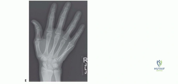
- Weil Osteotomy: Using a microsaw, an intra-articular osteotomy is initiated at the dorsal margin of the articular cartilage of the metatarsal head. The saw blade must be oriented strictly parallel to the weight-bearing surface of the foot, NOT parallel to the metatarsal shaft. This parallel orientation is critical; if the cut is made parallel to the shaft, proximal translation will result in plantar depression of the metatarsal head, causing severe postoperative IPK. The capital fragment is translated proximally (typically 2-4 mm) to decompress the joint. Fixation is achieved with a single dorsal-to-plantar 2.0 mm twist-off screw or threaded K-wire.
- Plantar Plate Repair: With the metatarsal shortened, excellent visualization of the plantar plate is achieved. The tear is typically avulsive at the phalangeal insertion. The proximal phalanx base is decorticated to create a bleeding bone bed. Using a specialized suture-passing device (e.g., Viper or Mini-Scorpion), two strands of #0 non-absorbable suture are passed through the healthy proximal stump of the plantar plate. Drill holes are made from plantar-to-dorsal through the base of the proximal phalanx. The sutures are passed through these osseous tunnels and tied securely over the dorsal bony bridge while the toe is held in 10-15 degrees of plantarflexion.
Correction of Hammer and Mallet Toe Deformities
For rigid hammer toe deformities, a PIP joint arthrodesis is the most reliable procedure to restore alignment and prevent recurrence.
- Incision: A dorsal longitudinal or transverse elliptical incision is made directly over the PIP joint. If a symptomatic dorsal hyperkeratotic lesion (corn) is present, it is excised elliptically with the skin.
- Exposure: The extensor tendon is split longitudinally or transected. The collateral ligaments of the PIP joint are excised, completely destabilizing the joint and allowing the middle phalanx to be hyper-plantarflexed, exposing the condyles of the proximal phalanx.
- Bone Resection: Using a microsaw or bone rongeur, the head and neck of the proximal phalanx are resected at the metaphyseal-diaphyseal junction. The articular cartilage at the base of the middle phalanx is denuded using a curette or a small drill bit to expose subchondral bleeding bone.
- Fixation: Traditional fixation utilizes a 0.045-inch or 0.062-inch smooth K-wire. The wire is driven antegrade through the central axis of the middle and distal phalanges, exiting the tip of the toe just plantar to the nail bed. The toe is then reduced, and the wire is driven retrograde into the medullary canal of the proximal phalanx. Modern alternatives include intramedullary shape-memory nitinol implants or two-piece threaded implants, which eliminate the need for percutaneous wires and reduce infection risk, though they significantly complicate revision surgery if nonunion occurs.
- Mallet Toe Variation: For an isolated mallet toe, the procedure is identical but localized to the DIP joint. A concurrent percutaneous FDL tenotomy at the plantar crease of the DIP joint is often required to release the dynamic deforming force.
Surgical Strategies for Intractable Plantar Keratosis
When conservative offloading of an IPK fails, surgical elevation or shortening of the offending metatarsal is mandated.
- Weil Osteotomy for IPK: As described above, the Weil osteotomy is highly effective for shortening a long metatarsal without significantly elevating it. It is the procedure of choice when the IPK is secondary to an elongated metatarsal.
- Helal (Distal Oblique) Osteotomy: If elevation is primarily required, a Helal osteotomy may be considered. A dorsal approach exposes the distal metatarsal shaft. A simple oblique osteotomy is made from dorsal-proximal to plantar-distal at a 45-degree angle. The distal fragment is allowed to slide proximally and dorsally. Historically, this was left unfixed to allow the metatarsal to "seek its own level," but due to unacceptable rates of nonunion and transfer metatarsalgia, contemporary practice mandates rigid internal fixation with a screw once the desired 1-2 mm of elevation is achieved.
Bunionette Deformity Correction
Surgical correction of a bunionette is dictated by the radiographic classification.
- Type I (Enlarged Lateral Condyle): A simple lateral condylectomy is performed via a dorsolateral approach. The lateral one-third of the metatarsal head is resected flush with the metatarsal shaft.
- Type II and Mild Type III (Distal Chevron Osteotomy): A V-shaped osteotomy with the apex pointing distally is created in the metatarsal neck. The capital fragment is translated medially by up to 50% of the shaft width. Fixation is secured with a single K-wire or a 2.0 mm low-profile screw. The prominent lateral edge of the proximal shaft is then beveled smooth.
- Severe Type III (Diaphyseal/Basal Osteotomy): For an IMA greater than 10-12 degrees, a distal osteotomy cannot provide sufficient translation. A basal closing wedge osteotomy or an oblique diaphyseal osteotomy (Coughlin procedure) is required. The oblique cut allows for massive medial translation and rotation of the entire fifth ray. Due to the high torsional forces on the lateral column during gait, rigid fixation with a dynamic compression plate or multiple interfragmentary screws is absolutely mandatory.
Freiberg Infraction Joint Preservation
For early to mid-stage Freiberg infraction (Smillie Stages II-IV), joint preservation via a dorsal closing wedge osteotomy (Gauthier procedure) is the gold standard.
- Exposure and Debridement: A dorsal approach to the MTP joint is utilized. A thorough synovectomy is performed, and all loose osteochondral bodies are extracted. The necrotic, collapsed dorsal cartilage is meticulously debrided.
- Osteotomy: A dorsal-based wedge of bone is resected from the metatarsal neck, immediately proximal to the articular surface. The plantar cortex is intentionally left intact to serve as a stabilizing hinge.
- Rotation and Fixation: The metatarsal head is rotated dorsally, closing the bony wedge. This critical maneuver rotates the necrotic dorsal cartilage out of the primary weight-bearing articulation and brings the healthy, viable plantar cartilage into articulation with the proximal phalanx. The osteotomy is secured with crossed K-wires, a dorsal titanium staple, or a low-profile plate.
Complications, Incidence Rates, and Salvage Management
Despite meticulous surgical technique, lesser toe surgery carries a distinct complication profile. The biomechanical complexity of the forefoot means that a minor technical error can lead to profound functional impairment. The surgeon must be adept at recognizing and managing these complications.
| Complication | Estimated Incidence | Pathophysiology & Etiology | Salvage Management & Revision Strategy |
|---|---|---|---|
| Floating Toe Deformity | 15% - 30% (Post-Weil Osteotomy) | Over-shortening of the metatarsal alters the intrinsic muscle axis. Lumbricals and interossei move dorsal to the center of rotation, acting as MTP extensors. Inadequate plantar plate repair. | Non-operative: Plantarflexion taping for 3 months. Operative: Flexor-to-extensor tendon transfer (Girdlestone-Taylor), collateral ligament release, revision plantar plate repair. |
| K-Wire Tract Infection | 5% - 10% | Bacterial migration along the percutaneous pin tract, typically Staphylococcus aureus. Exacerbated by early weight-bearing or poor hygiene. | Immediate removal of the K-wire. Oral culture-directed antibiotics. If deep osteomyelitis is suspected, formal surgical debridement and IV antibiotics are required. |
| Transfer Metatarsalgia | 10% - 20% | Disruption of the metatarsal parabola. Over-elevation or over-shortening of a single metatarsal transfers weight-bearing forces to the adjacent, unprepared metatarsal head. | Initial management with custom orthoses and metatarsal pads. Surgical salvage requires osteotomy of the newly affected metatarsal or revision lengthening/plantarflexion of the index metatarsal. |
| Nonunion / Malunion | 2% - 5% | Inadequate fixation, thermal necrosis from the saw blade, patient non-compliance with weight-bearing restrictions, or poor vascularity (especially in basal 5th metatarsal osteotomies). | Revision open reduction and internal fixation (ORIF) with rigid plating. Utilization of autologous bone graft or orthobiologics (e.g., BMP, DBM) to stimulate osteogenesis. |
| Flail Toe | 1% - 3% | Over-resection of the proximal phalanx head during PIP arthroplasty, destroying the collateral ligament attachments and leaving a structurally incompetent, floppy digit. | Syndactylization (buddy taping) to the adjacent normal toe. In severe cases, structural bone grafting and revision arthrodesis, or amputation if painful and non-functional. |
| Recurrence of Deformity | 5% - 15% | Failure to address the primary biomechanical driver (e.g., leaving a severe hallux valgus uncorrected, missed equinus contracture, or unrecognized systemic inflammatory disease). | Comprehensive re-evaluation of foot biomechanics. Revision surgery must address the root cause. May require salvage MTP joint arthroplasty or arthrodesis in recalcitrant, end-stage cases. |
Phased Post-Operative Rehabilitation Protocols
The success of forefoot reconstruction is as dependent on rigorous postoperative rehabilitation as it is on the surgical execution. The rehabilitation protocol is phased to protect the osseous fixation and soft-tissue repairs while progressively restoring range of motion and functional strength.
Phase 1: Maximum Protection and Tissue Healing (Weeks 0-2)
Immediately postoperatively, the foot is placed in a bulky, well-padded compressive dressing to control edema, which is the primary enemy of wound healing in the distal extremity. The patient is fitted with a rigid-soled postoperative shoe or a controlled ankle motion (CAM) boot. Weight-bearing is strictly restricted to the heel to completely offload the forefoot. Strict elevation above the level of the heart is mandatory for the first 72 to 96 hours. The patient is instructed to perform active ankle pumps to prevent deep vein thrombosis.
Phase 2: Transition and Early Mobilization (Weeks 2-6)
At the 2-week mark, the initial dressing is removed, and the incisions are inspected. Sutures are typically removed between days 14 and 21. If percutaneous K-wires were utilized for PIP arthrodesis or MTP stabilization, they remain in place and are meticulously cleaned with alcohol or betadine. A critical component of this phase is the initiation of plantarflexion taping. The toes are taped securely into 10-15 degrees of plantarflexion to counteract the tendency for dorsal contracture and to protect the plantar plate repair. Weight-bearing remains restricted to the heel or flat-foot in a rigid shoe. At 4 to 6 weeks, depending on radiographic evidence of early consolidation, the K-wires are pulled in the clinic. Once the wires are removed, passive and active range of motion (ROM) exercises of the MTP joints are initiated to prevent debilitating stiffness.
Phase 3: Strengthening and Return to Function (Weeks 6-12+)
Radiographs are obtained at 6 weeks to confirm osseous union of osteotomies and arthrodeses. Once union is confirmed, the patient is transitioned into a wide-toe-box, stiff-soled athletic shoe. Aggressive physical therapy is instituted. The focus shifts to intrinsic muscle strengthening, utilizing exercises such as "towel scrunches" and marble pick-ups to restore the dynamic stabilizers of the forefoot. Manual therapy, including scar mobilization and aggressive plantar plate stretching, is employed to maximize MTP joint dorsiflexion. Patients are counseled that residual swelling may persist for up to 6 to 9 months postoperatively. Return to high-impact sports and unrestricted footwear is typically permitted between 3 and 4 months, contingent upon the complete resolution of pain and the restoration of normal gait kinematics.
Summary of Landmark Literature and Clinical Guidelines
The evolution of surgical management for lesser toe deformities is deeply rooted in several landmark biomechanical and clinical studies. The foundational understanding of crossover toe deformity was championed by Coughlin et al., who exhaustively detailed the pathoanatomic cascade of plantar plate attenuation and collateral ligament failure, establishing the absolute necessity of addressing the soft-tissue envelope rather than relying solely on osseous correction.
The introduction of the Weil osteotomy revolutionized the management of MTP instability and IPK. Originally popularized by L.S. Weil in the 1990s, the procedure provided a reliable method to decompress the joint without the catastrophic elevation associated with earlier osteotomies. Subsequent biomechanical studies, however, highlighted the risk of the "floating toe" complication, leading to the modern synthesis of combining the Weil osteotomy with direct anatomic plantar plate repair. Recent prospective cohorts have demonstrated that this combined approach yields superior American Orthopaedic Foot and Ankle Society (AOFAS) clinical scores and significantly lower recurrence rates compared to isolated osteotomies or flexor tendon transfers.
For Freiberg infraction, Smillie’s original classification system remains the undisputed gold standard for guiding surgical decision-making. Gauthier’s long-term follow-up studies on the dorsal closing wedge osteotomy demonstrated that rotating viable plantar cartilage into the functional articulation provides durable pain relief and delays the need for joint-destructive procedures for decades.
By adhering strictly to these rigorously established biomechanical principles, respecting the intricate soft-tissue anatomy, and executing precise surgical techniques, orthopedic surgeons can reliably restore forefoot kinematics, alleviate debilitating pain, and achieve excellent, durable functional outcomes in patients afflicted with complex lesser toe deformities.
