Introduction & Epidemiology
Proximal humeral fractures (PHFs) represent a significant orthopaedic burden, particularly in the elderly population. They are the third most common fracture in individuals over 65 years of age, following hip and distal radius fractures, accounting for approximately 5-6% of all fractures. The incidence exhibits a bimodal distribution: high-energy trauma typically causes PHFs in younger, active individuals, while low-energy falls on an outstretched hand are the predominant mechanism in osteoporotic elderly patients. The increasing longevity of the global population and the rising prevalence of osteoporosis contribute to a projected increase in PHHF incidence.
Understanding PHFs necessitates a robust classification system to guide clinical decision-making and facilitate communication. The Neer classification system, based on the displacement of four major segments (humeral head, greater tuberosity, lesser tuberosity, and humeral shaft), remains widely utilized for its clinical utility. A "part" is considered displaced if it demonstrates >1 cm translation or >45 degrees of angulation. The AO/OTA classification, a more comprehensive alphanumeric system, offers a detailed description of fracture morphology, comminution, and articular involvement. While both systems have inter- and intra-observer variability, they are indispensable tools for assessing fracture severity and predicting outcomes.
Optimal treatment of PHFs depends on a multitude of factors beyond mere fracture morphology. These include bone quality (e.g., degree of osteoporosis, comminution), the integrity and blood supply to the humeral head, the preexisting condition of the rotator cuff, and crucially, the patient’s functional demands, comorbidities, and overall medical status. A comprehensive and systematic assessment (e.g., ATLS approach for trauma patients) is paramount to identify and treat life- and limb-threatening conditions, followed by a focused evaluation of the shoulder region, including neurovascular status and a diligent search for associated injuries (scapular, cervical spine, or other upper extremity). Pain, swelling, deformity, and/or an open wound are common presenting signs requiring immediate attention.
Surgical Anatomy & Biomechanics
A thorough understanding of the complex anatomy and biomechanics of the proximal humerus is fundamental for effective treatment of PHFs.
Anatomical Landmarks
The proximal humerus consists of several critical segments:
*
Humeral Head:
Articulates with the glenoid fossa. Its articular surface faces medially, superiorly, and posteriorly (retroversion, typically 20-30 degrees relative to the epicondylar axis).
*
Anatomical Neck:
The demarcation between the articular surface of the head and the tuberosities. Fractures here often disrupt the blood supply to the head.
*
Greater Tuberosity:
Located laterally, it serves as the insertion site for the supraspinatus, infraspinatus, and teres minor tendons (posterior cuff). Displacement can lead to significant functional loss and impingement.
*
Lesser Tuberosity:
Located anteriorly, it is the insertion site for the subscapularis tendon (anterior cuff). Displacement can lead to internal rotation deficits.
*
Bicipital Groove (Intertubercular Sulcus):
Lies between the greater and lesser tuberosities, housing the long head of the biceps tendon.
*
Surgical Neck:
Distal to the tuberosities, this is the most common site for PHFs.
Vascularity of the Humeral Head
The blood supply to the humeral head is critical for its viability and fracture healing. It is predominantly derived from branches of the axillary artery:
*
Anterior Humeral Circumflex Artery (AHCA):
The dominant blood supply, ascending along the bicipital groove to form the arcuate artery, which penetrates the head from an anterosuperior direction. It provides numerous anastomotic branches to the greater tuberosity and metaphysis.
*
Posterior Humeral Circumflex Artery (PHCA):
Contributes a smaller, more variable supply, typically entering the posterior aspect of the greater tuberosity and surgical neck.
Disruption of these vessels, particularly the AHCA, can lead to avascular necrosis (AVN) of the humeral head, a significant complication, especially in Neer 4-part fractures where multiple fragments are devitalized. The integrity of the medial calcar and its periosteal attachments is also crucial for vascularity.
Neural Structures
Several important nerves are in close proximity to the proximal humerus and are vulnerable to injury during trauma or surgical intervention:
*
Axillary Nerve:
Exits the quadrangular space, coursing inferiorly and laterally to wrap around the surgical neck of the humerus, approximately 5-7 cm distal to the acromial edge. It innervates the deltoid and teres minor muscles, and provides sensory innervation to the lateral shoulder. It is at significant risk during lateral plate placement or extensive soft tissue dissection around the surgical neck.
*
Musculocutaneous Nerve:
Located more medially, it innervates the biceps and brachialis muscles.
*
Radial Nerve:
Although more distal, it can be at risk with extensive shaft extension of a proximal fracture.
*
Brachial Plexus:
Vulnerable to traction injuries with high-energy trauma.
Biomechanics and Muscle Forces
The muscles inserting on the proximal humerus exert significant deforming forces on fracture fragments:
*
Rotator Cuff (Supraspinatus, Infraspinatus, Teres Minor, Subscapularis):
These muscles attach to the tuberosities.
* Supraspinatus (superior aspect of greater tuberosity): Tends to superiorly displace the greater tuberosity fragment.
* Infraspinatus and Teres Minor (posterior aspect of greater tuberosity): Contribute to external rotation of the fragment.
* Subscapularis (lesser tuberosity): Tends to internally rotate the lesser tuberosity fragment.
*
Deltoid:
Attaches to the deltoid tuberosity on the humeral shaft. Its pull tends to abduct and superiorly displace the humeral shaft relative to the humeral head, especially in surgical neck fractures.
*
Pectoralis Major, Latissimus Dorsi, Teres Major:
These muscles insert more distally on the shaft and can cause adduction of the shaft.
Fracture patterns are often a result of these unopposed muscle pulls. For instance, in Neer 2-part surgical neck fractures, the humeral head often remains in slight abduction due to capsular attachments and ligamentous support, while the shaft is adducted by the pectoralis major and displaced superiorly by the deltoid. Understanding these deforming forces is critical for achieving and maintaining reduction.
Indications & Contraindications
The decision-making process for managing proximal humeral fractures is complex and multifaceted, requiring careful consideration of patient factors, fracture characteristics, and potential risks versus benefits of operative intervention.
Non-Operative Indications
Non-operative management remains the preferred treatment for the majority of PHFs, particularly those that are minimally displaced or stable.
*
Minimally Displaced or Stable Fractures (Neer 1-part):
This constitutes the largest group, where fracture fragments are well-aligned (angulation <45 degrees, translation <1 cm).
*
Impacted Valgus Fractures:
Fractures where the humeral head is impacted into the shaft in a valgus position are inherently stable and have a good prognosis with non-operative treatment, often demonstrating an intact medial calcar blood supply.
*
Elderly, Low-Demand Patients with Significant Comorbidities:
For patients with severe medical comorbidities that contraindicate surgery, or those with very low functional demands, a non-operative approach can minimize surgical risks, even for moderately displaced fractures. Acceptable alignment in this population might be broader.
*
Fractures with Significant Bone Loss/Osteoporosis where Fixation is Unlikely to Hold:
In rare cases of extreme osteoporosis, the bone quality may be too poor to achieve stable fixation, making non-operative care or arthroplasty (if indicated for other reasons) more suitable.
Operative Indications
Surgical intervention is generally reserved for displaced or unstable fractures in active patients with reasonable bone quality, or when specific criteria for malreduction are met.
*
Significantly Displaced Fractures (Neer 2-part, 3-part, 4-part):
*
Angulation >45 degrees or Translation >1 cm:
These thresholds often indicate mechanical instability and a higher risk of malunion or poor function with non-operative treatment.
*
Articular Step-off or Gap >2mm:
For intra-articular components, significant incongruity can lead to post-traumatic arthritis.
*
Greater Tuberosity Displacement >5mm (particularly in younger, active patients):
Significant displacement of the greater tuberosity impairs rotator cuff function and can lead to impingement. Up to 10mm may be tolerated in the elderly.
*
Lesser Tuberosity Displacement:
Less common but can impact subscapularis function.
*
Irreducible Fracture-Dislocations:
When the humeral head is dislocated and cannot be reduced closed due to interposition of soft tissues (e.g., biceps tendon, capsule) or severe comminution.
*
Open Fractures:
Require urgent surgical debridement and stabilization to prevent infection.
*
Associated Neurovascular Injury Requiring Exploration:
If a significant neurovascular deficit is present and suspected to be caused by direct impingement from fracture fragments, surgical exploration and reduction are indicated.
*
Young, Active Patients:
Younger patients generally have higher functional demands and better bone quality, making them better candidates for operative fixation to restore anatomical alignment and maximize function.
*
Head-Split Fractures:
These are intra-articular fractures involving the articular cartilage, often requiring reduction and internal fixation or arthroplasty.
*
Displaced Anatomical Neck Fractures:
Due to the high risk of AVN, anatomical neck fractures with significant displacement may warrant surgical consideration, though fixation can be challenging.
Relative Contraindications
- Severe Medical Comorbidities: Patients with uncontrolled systemic diseases (e.g., severe cardiac, pulmonary, renal, neurological conditions) that significantly increase anesthetic and surgical risks.
- Active Infection: Absolute contraindication for elective internal fixation or arthroplasty.
- Extensive Soft Tissue Compromise: Severe open wounds, compromised skin, or extensive contusions may delay or contraindicate immediate internal fixation due to high risk of infection or wound complications.
- Patients Unwilling or Unable to Participate in Post-Operative Rehabilitation: Successful outcomes rely heavily on diligent participation in physical therapy.
Summary Table: Operative vs. Non-Operative Indications
| Factor | Non-Operative Management | Operative Management |
|---|---|---|
| Fracture Type | Neer 1-part, impacted valgus fractures | Displaced Neer 2-, 3-, 4-part; Head-split; Fracture-dislocation |
| Displacement | Angulation <45°, Translation <1 cm, GT <5mm | Angulation >45°, Translation >1 cm, GT >5-10mm (age-dependent) |
| Articular Involve. | None or minimally displaced articular step-off (<2mm) | Articular step-off/gap >2mm |
| Patient Age | Elderly, low-demand | Young, active, high-demand |
| Bone Quality | Osteoporotic, poor bone quality (if stable) | Good to moderate bone quality for fixation |
| Comorbidities | Severe comorbidities precluding surgery | Medically fit for surgery |
| Special Cases | Open fractures, neurovascular compromise (requiring exploration) |
Pre-Operative Planning & Patient Positioning
Meticulous pre-operative planning and appropriate patient positioning are critical for successful surgical management of proximal humeral fractures, minimizing intraoperative complications, and optimizing outcomes.
Pre-Operative Assessment & Imaging
- Clinical Assessment: Confirm neurovascular integrity. Assess for skin integrity and soft tissue compromise. Document active and passive range of motion (within pain limits) and baseline function.
-
Radiographic Assessment:
- Standard Trauma Series: True AP, scapular Y, and axillary lateral views are mandatory. A "true AP x-ray" is made with the central ray tangential to the glenoid surface, often requiring the patient to be rotated 30-45 degrees obliquely. The scapular Y view provides an orthogonal projection to the AP, essential for assessing anterior-posterior displacement and dislocation. The axillary lateral view is critical for glenoid congruity and humeral head position relative to the glenoid, but can be challenging to obtain in trauma due to pain or patient positioning limitations.
-
Computed Tomography (CT) Scan:
Indispensable for detailed assessment, especially in complex fractures. 3D reconstructions provide invaluable insights into:
- Degree of comminution (metaphyseal and articular).
- Extent of articular involvement and head-split fragments.
- Precise tuberosity displacement (especially greater tuberosity superior and posterior displacement).
- Identification of impacted fragments or glenoid bone loss in fracture-dislocations.
- Pre-operative templating for plate size, screw length, and arthroplasty component sizing (if applicable).
- MRI: Rarely indicated in acute settings but may be considered if concomitant rotator cuff tear is suspected and will significantly alter treatment strategy.
Patient Optimization
Address any correctable medical comorbidities. Optimize nutritional status, manage diabetes, and advise smoking cessation to enhance wound healing and reduce infection risk. Discuss realistic expectations and the demanding rehabilitation protocol with the patient and family.
Anesthesia
General anesthesia is typically employed. A regional anesthetic block (e.g., interscalene block) can be a valuable adjunct for excellent post-operative pain control. However, careful consideration should be given to its potential masking of intraoperative neurological compromise, particularly the axillary nerve. Some surgeons prefer to avoid it or use a single-shot block after nerve monitoring has been performed.
Patient Positioning
Two primary positions are utilized for proximal humeral fracture surgery:
1.
Beach Chair Position:
*
Setup:
Patient is semi-recumbent (30-70 degrees of back elevation) on a standard operating table with the backrest elevated. The head is secured in a headrest (e.g., Mayfield clamp) to prevent movement. The operative arm is draped free, and the torso is often tilted away from the surgeon (reverse Trendelenburg) to improve exposure.
*
Advantages:
Provides excellent access for the deltopectoral approach, allows dynamic intraoperative assessment of shoulder range of motion, and facilitates posterior access if needed. It minimizes facial swelling compared to supine.
*
Disadvantages:
Risk of cerebral hypoperfusion, especially in hypotensive patients; potential for brachial plexus stretch; challenge with C-arm positioning for perfect axillary views.
2.
Supine Position on a Radiolucent Table:
*
Setup:
Patient lies flat on their back. The ipsilateral side of the chest may be elevated with a bump to allow for greater shoulder extension and a more vertical approach angle. The operative arm is draped free or suspended on a traction table/outrigger.
*
Advantages:
Excellent fluoroscopic control with easy C-arm manipulation for AP, lateral, and axillary views. Better for complex fractures requiring sustained traction and for managing severe multi-trauma patients. Easier for anesthesiologists to manage airway.
*
Disadvantages:
Can be challenging to achieve full shoulder mobility for range of motion checks. Potential for brachial plexus traction injuries with prolonged overhead traction.
Fluoroscopy Setup
Regardless of positioning, the C-arm must be readily available and able to acquire true AP, scapular Y, and axillary lateral views. The image intensifier is typically positioned contralateral to the surgeon. For axillary views in beach chair, the C-arm is often brought from the foot of the bed and angled cephalad. In the supine position, it can be brought in from either side. Confirm adequate views pre-incision and throughout the procedure.
Detailed Surgical Approach / Technique
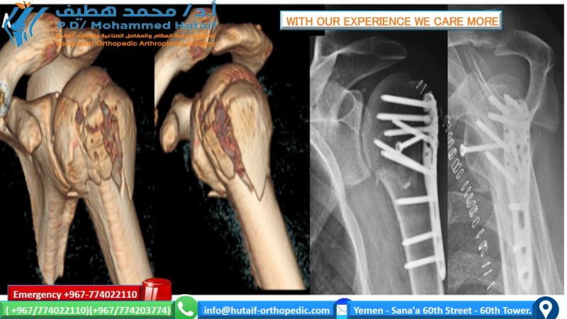
The choice of surgical approach and fixation technique for proximal humeral fractures is dictated by fracture morphology, surgeon preference, patient factors, and bone quality. The most common technique for displaced PHFs amenable to osteosynthesis is open reduction and internal fixation (ORIF) with an anatomical locking plate via the deltopectoral approach.
Choice of Approach
- Deltopectoral Approach: This is the workhorse approach for most PHFs requiring ORIF or arthroplasty. It provides extensive exposure of the anterior aspect of the proximal humerus and glenohumeral joint, allowing for direct reduction and stable plate placement.
- Anterolateral (Deltoid Split) Approach: Less invasive, primarily used for minimally displaced tuberosity fractures or for minimally invasive plating techniques. Limited exposure and risk of axillary nerve injury with splits greater than 5 cm from the acromion.
- Posterior Approach: Rarely used for PHFs, primarily for posterior dislocations or specific posterior tuberosity fractures.
Deltopectoral Approach (ORIF with Locking Plate)
- Incision: A curvilinear incision is made from the coracoid process, extending distally along the deltopectoral groove for 8-12 cm, parallel to the anticipated plate length.
- Interval Identification: The deltopectoral interval is identified by locating the cephalic vein, which lies within this groove. The vein is carefully mobilized and typically retracted laterally with the deltoid, but can be ligated if necessary. The lateral border of the pectoralis major muscle is retracted medially.
-
Deep Dissection:
- The clavipectoral fascia is incised longitudinally to expose the coracoid process and the conjoined tendon (short head of biceps, coracobrachialis).
- The subdeltoid space is entered. The deltoid muscle is carefully retracted laterally.
- Protect the axillary nerve : It courses around the surgical neck, approximately 5-7 cm distal to the acromial edge. It is vital to stay superficial to the nerve and protect it throughout the procedure, especially during distal plate application and screw insertion.
- Hematoma Evacuation & Visualization: The fracture hematoma is evacuated to improve visualization of the fracture fragments, especially the humeral head and tuberosities.
-
Reduction:
This is the most challenging and critical step. The goal is anatomical restoration of the humeral head articular surface, proper height, version, and reduction of tuberosities.
- Indirect Reduction: Ligamentotaxis (traction on the arm) can help restore length and align major fragments.
-
Direct Reduction:
- Humeral Head: Often internally rotated and adducted. Using a blunt Hohmann retractor or a K-wire joystick directly into the head can facilitate manipulation.
- Tuberosities: Reduced anatomically to the head and shaft. The greater tuberosity is often displaced superiorly and posteriorly, while the lesser tuberosity may be internally rotated. Rotator cuff sutures (non-absorbable) can be passed through the rotator cuff tendons and tuberosities pre-emptively to aid in reduction and later for fixation augmentation.
- Shaft: Reduced relative to the humeral head. Restore proper length, alignment, and version. The medial calcar should be reduced anatomically; medial comminution can lead to varus collapse.
- Temporary Fixation: K-wires are invaluable for holding reduction while preparing for definitive fixation.
- Bone Grafting: For significant metaphyseal defects or comminution, particularly in osteoporotic bone, autologous cancellous bone graft or allograft can be packed into the void to provide structural support and promote healing, reducing the risk of varus collapse.
-
Fixation with Locking Plate:
- Plate Selection: Anatomical locking plates designed specifically for the proximal humerus (e.g., PHILOS, Stryker VariAx) are chosen.
- Plate Placement: The plate is carefully positioned on the lateral aspect of the humerus, typically 5-8 mm lateral to the bicipital groove and inferior to the greater tuberosity. Proper placement avoids impingement with the acromion or rotator cuff, minimizes risk to the biceps tendon, and ensures optimal screw trajectory into the humeral head. Ensure the plate is not too distal, as this risks axillary nerve injury.
- Distal Screws: At least 2-3 bicortical locking screws are inserted into the humeral shaft distally.
- Proximal Screws (Humeral Head): Multiple locking screws (typically 4-6) are directed into the humeral head, aiming for optimal purchase in the densest bone (subchondral bone) to create a "calcar screw" or "raft of screws" construct. Avoid joint penetration; fluoroscopy is essential here. The medial calcar screw is particularly important for preventing varus collapse.
- Suture Augmentation: Non-absorbable sutures passed through the rotator cuff tendons (supraspinatus, infraspinatus, subscapularis) and secured through plate holes (tension band technique) can enhance tuberosity reduction and provide additional rotational stability, especially for osteoporotic bone or comminuted tuberosity fragments.
-
Final Assessment:
- Confirm reduction and fixation stability under fluoroscopy in multiple planes (AP, Y, axillary).
- Check for hardware prominence and impingement by performing gentle range of motion (internal/external rotation, abduction/adduction).
- Confirm integrity of neurovascular structures.
- Wound Closure: Layered closure, ensuring secure soft tissue coverage over the implant.
Hemiarthroplasty (HA) / Reverse Total Shoulder Arthroplasty (RTSA)
- Indications: Irreparable 4-part PHFs, head-split fractures not amenable to fixation, severe comminution with poor bone quality, or in elderly patients with pre-existing rotator cuff arthropathy (RTSA).
- Technique Overview (HA): Resection of the comminuted humeral head. Preparation of the intramedullary canal. Trial prosthesis to determine appropriate head size, height, and version. Final implantation of the humeral stem and head. Crucially, anatomical repair and secure fixation of the tuberosities around the prosthetic head are paramount for achieving good function and preventing pain, though often challenging.
- Technique Overview (RTSA): Resection of humeral head. Glenoid reaming and placement of a baseplate and glenosphere. Preparation of the humeral intramedullary canal and implantation of the humeral stem and polyethylene cup. Tuberosity reattachment is less critical for function in RTSA, as the deltoid becomes the primary elevator of the arm, but can still improve pain and rotational stability. RTSA provides more predictable pain relief and active elevation compared to HA in selected elderly patients with unreconstructable 4-part fractures or poor tuberosity repair.
Complications & Management
Despite meticulous surgical technique, complications following proximal humeral fracture management can occur, ranging from minor to devastating. Proactive recognition and appropriate management are crucial for salvage and optimizing patient outcomes.
General Surgical Complications
-
Infection:
- Incidence: 1-5% for ORIF, potentially higher in open fractures or with prolonged surgery.
- Management: Superficial infections may respond to oral antibiotics. Deep infections require aggressive surgical debridement, intravenous antibiotics, and potentially hardware removal (if fracture is healed) or a two-stage revision (debridement, antibiotic spacer, delayed revision arthroplasty).
-
Bleeding/Hematoma:
- Incidence: Variable.
- Management: Meticulous hemostasis during surgery. Post-operative drains may be used. Large hematomas can cause nerve compression or increase infection risk, potentially requiring evacuation.
-
Neurovascular Injury:
- Incidence: Axillary nerve injury is the most common (up to 20% transient neuropraxia, 1-5% permanent). Brachial plexus or vascular injuries are rare but severe.
- Management: Intraoperative protection is key. Post-operative deficit requires immediate evaluation (clinical exam, EMG/NCS after 3 weeks). Neuropraxia often resolves spontaneously. Persistent deficit may warrant neurolysis or nerve grafting.
Specific Complications of Proximal Humeral Fracture Surgery
| Complication | Incidence (Approximate) | Salvage Strategies / Management |
|---|---|---|
| Avascular Necrosis (AVN) of Humeral Head | 10-30% (higher in 4-part, anatomical neck fractures) | If symptomatic: Arthroplasty (Hemiarthroplasty or Reverse Total Shoulder Arthroplasty). Non-operative management with pain control for mild, asymptomatic cases. |
| Nonunion / Malunion | 5-20% (higher with poor bone quality, comminution, inadequate fixation) | Nonunion: Revision ORIF with bone grafting (autograft/allograft), stronger fixation, or conversion to arthroplasty (HA/RTSA). Malunion: If symptomatic (pain, stiffness, impingement): Corrective osteotomy (rare), hardware removal (if prominent), arthroplasty (HA/RTSA) for significant deformity. |
| Screw Cutout / Perforation | 5-15% | Occurs typically with varus collapse or inadequate screw length/placement in osteoporotic bone. If symptomatic (pain, loss of reduction): Revision ORIF with shorter screws, re-reduction, or conversion to arthroplasty. |
| Loss of Reduction | 5-15% | Typically due to inadequate fixation, poor bone quality, or early aggressive rehabilitation. If symptomatic: Revision surgery (re-reduction, stronger fixation, bone graft) or conversion to arthroplasty. |
| Subacromial Impingement | 5-10% | Can result from prominent hardware (plate positioned too superiorly) or malunion (greater tuberosity nonunion/malunion, superior humeral head migration). If symptomatic and refractory to conservative management: Hardware removal (if fracture healed), acromioplasty, or revision for tuberosity malunion. |
| Stiffness / Adhesive Capsulitis | Very common (up to 20-30% significant limitation) | Aggressive physical therapy, pain management, activity modification. If persistent and severe after 6-9 months: Manipulation under anesthesia (MUA), arthroscopic capsular release. |
| Rotator Cuff Pathology | 5-10% (new or exacerbated tear) | Can be a pre-existing condition, exacerbated by trauma or surgery, or due to impingement. If symptomatic: Arthroscopic repair (if amenable) or conversion to RTSA for irreparable tears with pseudoparalysis. |
| Hardware Prominence / Pain | 10-20% | If symptomatic and fracture is healed (typically >1 year post-op): Hardware removal. |
| Deltoid Dysfunction | Rare, typically due to axillary nerve injury | Management as per neurovascular injury. May lead to pseudoparalysis or severe weakness. |
| Biceps Tendon Pathology | Variable | Bicipital groove irritation from plate, subluxation, or rupture. If symptomatic: Tenotomy or tenodesis. |
Post-Operative Rehabilitation Protocols
Post-operative rehabilitation is a critical determinant of functional outcome following surgical management of proximal humeral fractures. A structured, phased approach is essential, balancing protection of the surgical repair with gradual restoration of motion and strength. Protocols must be individualized based on fracture stability, fixation quality, patient age, bone quality, and concomitant injuries. Close communication between the surgeon, physical therapist, and patient is paramount.
Phase 1: Immobilization & Protection (0-6 Weeks Post-Op)
The primary goal is to protect the surgical repair, control pain and swelling, and initiate gentle passive range of motion.
*
Immobilization:
The arm is typically immobilized in a sling (e.g., UltraSling) continuously, except for hygiene and prescribed exercises. A body-brace sling may be used for greater stability in unstable fractures.
*
Pain & Swelling Management:
Cryotherapy, NSAIDs, and analgesics as needed.
*
Passive Range of Motion (PROM):
*
Pendulum Exercises:
Initiate 2-3 days post-op. Patient leans forward, allowing the arm to hang freely and gently swing in small circles, side-to-side, and forward-backward. These are gravity-assisted and do not involve active muscle contraction.
*
Supine Passive Flexion:
Therapist or patient (with non-operative hand) passively elevates the operative arm in the sagittal plane, typically limited to 90 degrees initially.
*
Supine Passive External Rotation:
Therapist or patient passively rotates the arm externally, keeping the elbow at the side. Often limited to 0-30 degrees initially, depending on tuberosity repair.
*
Scapular Stabilization Exercises:
Gentle retraction and protraction, elevation, and depression exercises to maintain scapular mobility and neuromuscular control.
*
Precautions:
*
NO active motion
of the shoulder.
*
NO lifting
, pushing, or pulling with the operative arm.
*
NO weight-bearing
on the operative arm.
* Avoid sudden, uncontrolled movements.
* External rotation may be restricted, especially after greater tuberosity repair.
* Internal rotation and extension may also be limited depending on lesser tuberosity repair.
Phase 2: Early Active Motion & Light Strengthening (6-12 Weeks Post-Op)
The goal is to gradually increase range of motion and initiate controlled active muscle contraction as fracture healing progresses.
*
Discontinuation of Sling:
As tolerated and as per surgeon's discretion, typically around 6 weeks.
*
Active-Assisted Range of Motion (AAROM):
* Begin with therapist assistance or using the non-operative hand, pulleys, or a wand.
* Progress to gentle
Active Range of Motion (AROM)
as pain allows and bone healing is confirmed radiographically.
* Continue working towards full flexion, abduction, and rotation within pain limits.
*
Isometric Strengthening:
* Initiate gentle isometric exercises for the rotator cuff and deltoid, with the arm in neutral positions (e.g., pushing against a wall for internal/external rotation, flexion, abduction).
* Low-load, high-repetition exercises.
*
Scapular Strengthening:
Continue exercises to improve scapular control and strength.
*
Precautions:
* Continue to avoid heavy lifting or sudden movements.
* Avoid positions of extreme abduction with external rotation until sufficient healing and strength are achieved.
Phase 3: Progressive Strengthening (12 Weeks to 6 Months Post-Op)
The focus shifts to restoring full strength, endurance, and advanced range of motion.
*
Progressive Resistive Exercises:
* Begin with light resistance bands, light weights, or bodyweight exercises for all planes of shoulder motion.
* Gradually increase resistance and repetitions.
* Focus on rotator cuff strengthening (internal/external rotation, scaption), deltoid strengthening, and periscapular muscle strengthening.
*
Endurance Training:
Incorporate activities to improve muscle endurance.
*
Proprioceptive Training:
Exercises to improve joint position sense and neuromuscular control.
*
Sport-Specific Training:
For athletes or individuals with high functional demands, begin incorporating movements specific to their activities.
*
Precautions:
* Avoid pain-provoking activities.
* Progress incrementally.
Phase 4: Return to Activity (6+ Months Post-Op)
The final phase focuses on returning to full, unrestricted activities, including sports or heavy manual labor, with continued maintenance.
*
Gradual Return to Full Activities:
Based on strength, range of motion, and pain levels.
*
Maintenance Program:
Encourage continuation of strengthening and stretching exercises to prevent recurrence of stiffness or weakness.
*
Long-Term Monitoring:
Periodic follow-up to assess long-term function and address any evolving issues.
Key Considerations
- Pain Management: Effective pain control throughout rehabilitation is crucial to allow patient participation.
- Radiographic Healing: Progression through phases should ideally correlate with radiological signs of fracture healing.
- Individualized Progression: Patients progress at different rates. The protocol serves as a guideline, but adjustments must be made based on individual response and surgeon guidance.
- Surgeon-Therapist Communication: Regular communication ensures the rehabilitation program aligns with the surgical goals and fracture stability.
Summary of Key Literature / Guidelines
The landscape of proximal humeral fracture management is dynamic, with ongoing research refining treatment algorithms. Several pivotal studies and clinical guidelines inform current practice.
Operative vs. Non-Operative Management
- Minimally Displaced Fractures: Strong consensus supports non-operative management for Neer 1-part fractures, with studies consistently showing equivalent or superior outcomes compared to surgical intervention, avoiding surgical risks.
-
Displaced 2- and 3-Part Fractures:
The optimal treatment for these fractures, particularly in the elderly, remains a subject of debate.
- The PROFIT trial (Proximal Fracture of the Humerus: an International Trial) , a large multicenter randomized controlled trial published in The Lancet (2015), compared locking plate fixation with non-operative treatment for displaced PHFs. It found no significant difference in functional outcome (Oxford Shoulder Score) at 2 years between the operative and non-operative groups, but reported a higher rate of complications in the operative group. This trial has significantly influenced practice, leading to increased consideration of non-operative management for many displaced fractures, particularly in older patients.
- Subsequent meta-analyses and systematic reviews have largely corroborated these findings, suggesting that for many elderly patients with displaced 2- and 3-part fractures, non-operative management yields comparable functional outcomes to ORIF, often with fewer early complications. However, patient selection based on functional demands, bone quality, and fracture pattern remains critical.
Role of Locking Plate Fixation (ORIF)
- Locking plate technology has revolutionized ORIF for PHFs, offering superior angular stability compared to conventional screw fixation, particularly in osteoporotic bone.
- Studies demonstrate that locking plates can provide stable fixation, enabling early rehabilitation. However, their use is associated with a specific complication profile, including screw cutout, AVN, and hardware-related impingement. Proper surgical technique, including avoiding joint penetration, achieving anatomical reduction, and medial calcar support, is crucial to mitigate these risks.
- The effectiveness of locking plates versus other fixation methods (e.g., intramedullary nails, tension band wiring) often depends on the specific fracture pattern and bone quality, with no universally superior method across all scenarios.
Arthroplasty for Complex Fractures
- Hemiarthroplasty (HA): Traditionally considered the gold standard for unreconstructable 4-part fractures and head-split fractures, especially in older patients. Outcomes are highly dependent on the secure and anatomical reattachment of the tuberosities, which is often challenging and unpredictable. Poor tuberosity healing is a major cause of unsatisfactory results and revision.
-
Reverse Total Shoulder Arthroplasty (RTSA):
Has gained significant traction for the treatment of complex PHFs, particularly in elderly patients with poor bone quality, pre-existing rotator cuff dysfunction, or those with highly comminuted 3- and 4-part fractures where tuberosity healing is unlikely.
- RTSA has demonstrated more predictable pain relief and improved active elevation compared to HA in these challenging scenarios, as it bypasses the need for a functional rotator cuff and reliable tuberosity healing for elevation.
- Several studies support RTSA over HA in selected elderly populations, showing better functional scores and fewer revisions for primary fracture treatment. However, RTSA has its own complication profile, including infection, instability, and scapular notching.
General Guidelines & Consensus
- Shared Decision-Making: Current trends emphasize shared decision-making, where patient preferences, functional goals, and comorbidities are carefully weighed alongside fracture characteristics.
- Pre-Operative Planning: Comprehensive imaging (including CT scans) and meticulous pre-operative planning are universally advocated to optimize surgical strategy.
- Rehabilitation: Adherence to structured, progressive rehabilitation protocols is critical for maximizing post-operative function, regardless of treatment modality.
- Evidence-Based Practice: Clinicians are encouraged to critically appraise the evolving literature and integrate the best available evidence into their practice, acknowledging the nuances of individual patient care.
In conclusion, mastering proximal humeral fractures requires a deep understanding of anatomy, biomechanics, comprehensive patient assessment, judicious application of classification systems, meticulous surgical planning and execution when indicated, and a commitment to structured rehabilitation. The landscape continues to evolve, with ongoing research refining our understanding and treatment paradigms.
