Comprehensive Introduction and Patho-Epidemiology
The operative management of forefoot fractures requires an exceptionally nuanced understanding of foot biomechanics, particularly regarding the dynamic weight-bearing distribution across the first ray during the gait cycle. While the surgical treatment of phalangeal fractures of the lesser toes is relatively uncommon—given that the vast majority can be treated successfully and definitively by conservative measures—injuries to the hallucal sesamoid complex present a distinct, highly morbid, and formidable clinical challenge. The disparity in clinical significance between these two entities cannot be overstated; a missed or mismanaged lesser toe fracture may result in a mild cosmetic deformity or manageable symptomatic malunion, whereas a mismanaged sesamoid fracture can precipitate catastrophic functional decline, chronic debilitating pain, and the end of an athletic career.
The epidemiology of sesamoid fractures is heavily skewed toward active populations, particularly dancers, gymnasts, runners, and athletes participating in field sports on artificial turf. The advent of lighter, more flexible athletic footwear combined with harder playing surfaces has led to a documented increase in the incidence of "turf toe" variants and acute sesamoid fractures. Furthermore, the orthopedic surgeon is frequently confronted with the diagnostic dilemma of differentiating an acute sesamoid fracture from a symptomatic bipartite sesamoid. Approximately 10% to 30% of the general population possesses a bipartite sesamoid—most commonly the tibial (medial) sesamoid—which is bilateral in up to 85% of these cases. Acute fractures typically present with sharp, irregular, and uncorticated edges on radiography, whereas bipartite variants exhibit smooth, sclerotic, and well-rounded margins. However, a stress fracture through a bipartite synchondrosis blurs these radiographic lines, necessitating advanced imaging and astute clinical correlation.
Pathophysiologically, sesamoid injuries exist on a spectrum from acute, high-energy traumatic fractures to insidious, repetitive-stress injuries culminating in microtrabecular failure and overt fracture. Acute fractures typically result from a direct axial load (e.g., landing from a significant height) or a sudden, forceful hyper-dorsiflexion of the first metatarsophalangeal (MTP) joint. This hyper-dorsiflexion can cause catastrophic disruption of the flexor hallucis brevis (FHB) musculotendinous unit, leading to wide proximal displacement of the sesamoid fragments. Stress fractures, conversely, are the sequelae of chronic, repetitive mechanical overloading that outpaces the bone's physiological remodeling capacity. The tenuous vascular supply to the sesamoids makes them uniquely susceptible to delayed union, nonunion, and avascular necrosis (AVN) following both acute trauma and chronic stress.
In contrast, phalangeal fractures of the lesser toes typically result from direct blunt trauma, such as a crush injury from a falling object, or an indirect axial load mechanism, such as the classic "bedpost" injury (an abduction-extension force applied to the fifth toe). Epidemiologically, these are among the most common fractures of the foot. While inherently stable due to the robust surrounding soft tissue envelope and the splinting effect of adjacent digits, the orthopedic surgeon must remain vigilant for the rare, high-risk phalangeal fractures. Intra-articular fractures with significant step-off, grossly unstable spiral fractures leading to rotational malalignment, and open fractures require definitive operative intervention to prevent chronic MTP or interphalangeal joint arthrosis, intractable keratoses, and overlapping toe deformities.
Detailed Surgical Anatomy and Biomechanics
To master the operative interventions of the forefoot, the orthopedic surgeon must first appreciate the intricate, highly specialized anatomy of the first MTP joint and the hallucal sesamoid complex. The hallucal sesamoids (tibial/medial and fibular/lateral) are two distinct ossicles embedded within the thick plantar plate and the two distinct tendinous slips of the FHB. The tibial sesamoid is generally larger, more elongated, and bears significantly more weight than the smaller, more circular fibular sesamoid. They articulate dorsally with the plantar facets of the first metatarsal head, separated by the intersesamoidal crista.
The ligamentous and tendinous attachments to the sesamoids form a highly complex, interdependent stabilizing network. The FHB muscle originates from the cuboid and lateral cuneiform, bifurcating into a medial and lateral head. The medial head, along with the abductor hallucis tendon, inserts into the tibial sesamoid and the medial base of the proximal phalanx. The lateral head, conjoined with the adductor hallucis tendon, inserts into the fibular sesamoid and the lateral base of the proximal phalanx. The sesamoids are tethered to each other by the robust intersesamoidal ligament, and to the metatarsal head via the metatarsosesamoid suspensory ligaments. This entire complex is continuous with the plantar plate, forming a dynamic hammock that stabilizes the first MTP joint.
Biomechanically, the sesamoids serve three primary, indispensable functions. First, they provide lever arm augmentation. The tibial sesamoid, in particular, significantly increases the mechanical advantage of the FHB muscle during the push-off phase of the gait cycle, amplifying the plantarflexion force essential for normal ambulation. Second, they serve a critical protective role. By elevating the first metatarsal head, the sesamoids create a protective anatomical groove that shields the flexor hallucis longus (FHL) tendon from direct, crushing compressive forces against the ground during weight-bearing. Third, they function as vital load distributors. During terminal stance, the sesamoids absorb up to 300% of the body's weight, dispersing this massive load across the plantar aspect of the first ray. Excision of the sesamoids drastically alters these biomechanics, decreasing push-off power and increasing focal contact pressures beneath the metatarsal head.
The vascular anatomy of the sesamoids is perhaps the most critical anatomical consideration for the operating surgeon, as it directly dictates the propensity for nonunion and avascular necrosis. The blood supply is highly variable but generally derives from the medial plantar artery and the first plantar metatarsal artery. These vessels form a pre-sesamoid arterial network that penetrates the ossicles predominantly from their proximal and plantar aspects. The distal poles of the sesamoids represent a vascular watershed area. Consequently, transverse fractures—the most common fracture pattern—frequently disrupt the intraosseous blood supply to the distal fragment, rendering it highly susceptible to avascular necrosis. Surgical approaches and dissection must be meticulously executed to preserve the remaining extraosseous vascular contributions from the plantar plate and FHB capsule.
Exhaustive Indications and Contraindications
The decision-making process regarding the operative versus non-operative management of forefoot fractures requires a careful synthesis of patient demographics, functional demands, fracture morphology, and the temporal presentation of the injury. The overarching philosophy in sesamoid management is the preservation of the osteoarticular complex whenever clinically feasible, given the profound biomechanical consequences of sesamoidectomy. However, persistent pain, functional limitation, and specific acute fracture patterns mandate decisive surgical intervention.
For hallucal sesamoid fractures, an exhaustive trial of conservative management—typically spanning 3 to 6 months—is the absolute prerequisite for surgical consideration in the setting of stress fractures or minimally displaced acute fractures. Operative intervention becomes indicated when patients experience recalcitrant pain and an inability to return to their baseline level of activity despite strict immobilization, prolonged non-weight-bearing, and customized orthotic offloading. In the acute trauma setting, absolute indications for immediate surgical intervention include gross disruption of the FHB musculotendinous unit with wide displacement of the sesamoid fragments (typically >5 mm), as this represents a profound loss of the plantar tension band mechanism. Symptomatic nonunions and painful bipartite sesamoids that fail orthotic management are also prime candidates for operative intervention, utilizing either osteosynthesis or partial excision.
In the context of lesser toe phalangeal fractures, the threshold for operative intervention is significantly higher. The vast majority of these injuries are managed successfully with buddy taping and rigid-soled footwear. However, surgical fixation is absolutely indicated for open fractures requiring formal debridement, grossly unstable or irreducible fractures that compromise the soft tissue envelope, and intra-articular fractures with a significant articular step-off (greater than 2 mm) affecting either the MTP or interphalangeal joints. Furthermore, spiral or oblique fractures resulting in severe rotational deformity must be reduced and pinned, as malrotation will lead to overlapping toes, intractable painful keratoses, and profound difficulty with shoe wear.
Contraindications to operative intervention in the forefoot must be strictly respected, as the complication profile in compromised hosts is exceptionally high. Active local or systemic infection is an absolute contraindication to internal fixation. Severe peripheral vascular disease (PVD) precludes surgical intervention due to the unacceptably high risk of catastrophic wound breakdown, infection, and subsequent amputation; optimization of vascular status via surgical or endovascular bypass is a prerequisite in these patients. Profound peripheral neuropathy, particularly in the setting of active Charcot neuroarthropathy or poorly controlled diabetes mellitus, represents a strong relative contraindication, as these patients are at extreme risk for hardware failure, nonunion, and postoperative Charcot exacerbation. Finally, non-ambulatory patients or those with severe medical comorbidities precluding safe anesthesia should be managed non-operatively.
| Clinical Condition | Operative Indications | Absolute/Relative Contraindications |
|---|---|---|
| Hallucal Sesamoid Fractures | - Acute displacement > 5 mm - Disruption of FHB complex - Symptomatic nonunion (>6 months) - Painful bipartite sesamoid refractory to orthotics - Avascular necrosis with collapse |
- Active local/systemic infection - Severe peripheral vascular disease - Active Charcot neuroarthropathy - Uncontrolled diabetes (HbA1c > 8.0%) - Non-ambulatory patient status |
| Phalangeal Fractures (Lesser Toes) | - Open fractures requiring I&D - Irreducible dislocations/fractures - Intra-articular step-off > 2 mm - Severe rotational malalignment - "Floating toe" variants |
- Minimally displaced/stable fractures - Patient non-compliance with NWB - Compromised dorsal soft tissue envelope - Severe peripheral neuropathy (relative) |
Pre-Operative Planning, Templating, and Patient Positioning
Thorough pre-operative planning is the cornerstone of successful forefoot surgery. The clinical evaluation must be supplemented with high-quality, weight-bearing radiographs of the foot, including anteroposterior (AP), lateral, and specialized axial sesamoid views. The axial sesamoid view is paramount, as it allows the surgeon to assess the articulation between the sesamoids and the plantar facets of the first metatarsal head, evaluating for arthrosis, subluxation, or subtle osteochondral defects. In cases of delayed union, nonunion, or suspected avascular necrosis, advanced imaging is mandatory. Magnetic Resonance Imaging (MRI) is highly sensitive for detecting bone marrow edema, assessing the viability of the sesamoid fragments, and evaluating the integrity of the plantar plate and FHB tendons. Computed Tomography (CT) is invaluable for precisely defining fracture morphology, assessing the degree of displacement, and confirming the presence of osseous bridging in suspected nonunions.
Templating and meticulous implant selection are critical, particularly given the diminutive size of the osseous structures involved. For phalangeal fractures, the surgeon must ensure the availability of appropriately sized Kirschner wires (typically 0.045-inch or 0.062-inch) for percutaneous pinning, as well as low-profile mini-fragment plates (1.2 mm to 1.5 mm systems) for complex intra-articular reconstructions. For sesamoid open reduction and internal fixation (ORIF), the surgical inventory must include 1.5-mm and 2.0-mm headless compression screws, cortical mini-fragment screws, and high-tensile strength non-absorbable sutures (e.g., #2 FiberWire or Orthocord) for figure-of-eight tension band wiring. Furthermore, the surgeon must be prepared for autologous bone grafting; specialized curettes, high-speed burrs, and bone graft harvest systems (for the calcaneus or distal tibia) must be readily available on the sterile field.
Patient positioning is standardized but requires meticulous attention to detail to optimize surgical exposure and fluoroscopic access. The patient is placed supine on a radiolucent operating table. A bump is placed under the ipsilateral hip to internally rotate the leg, neutralizing the natural external rotation of the lower extremity and bringing the medial border of the foot perfectly perpendicular to the floor. This is particularly crucial for the medial approach to the tibial sesamoid. A well-padded thigh or calf tourniquet is applied to ensure a bloodless surgical field, which is absolutely essential for identifying the delicate neurovascular structures of the plantar forefoot. The foot is prepped and draped in a standard sterile fashion, ensuring that the toes are freely mobile to allow for intraoperative manipulation and assessment of tendon tension.
Anesthetic considerations typically involve a combination of monitored anesthesia care (MAC) or general anesthesia, supplemented with a regional block for optimal intraoperative hemodynamics and prolonged postoperative analgesia. A popliteal sciatic nerve block, combined with a saphenous nerve block, provides excellent, comprehensive anesthesia to the entire forefoot. Alternatively, a highly specific ankle block or Mayo block can be utilized. Pre-operative prophylactic antibiotics, typically a first-generation cephalosporin (e.g., Cefazolin), are administered intravenously within one hour prior to tourniquet inflation. Meticulous skin preparation, utilizing chlorhexidine-alcohol or povidone-iodine solutions, is performed, paying special attention to the interdigital web spaces to minimize the risk of surgical site infection.
Step-by-Step Surgical Approach and Fixation Technique
Phalangeal Fracture Fixation (CRPP and ORIF)
When operative intervention is mandated for lesser toe phalangeal fractures, Closed Reduction and Percutaneous Pinning (CRPP) is the workhorse technique. The toe is manipulated under fluoroscopic guidance using longitudinal traction and targeted digital pressure to correct angular and rotational deformities. Once reduced, a 0.045-inch or 0.062-inch Kirschner wire is driven axially. The wire is typically introduced at the tip of the distal phalanx, just plantar to the nail bed to avoid nail matrix injury, and advanced from distal to proximal across the distal interphalangeal (DIP) joint, the proximal interphalangeal (PIP) joint, and the fracture site, terminating in the subchondral bone of the proximal phalanx base. The pin is left protruding through the skin and bent to prevent proximal migration. For complex, displaced intra-articular fractures, an open approach via a dorsal longitudinal or lazy-S incision is required. The extensor mechanism is split or retracted, the fracture is anatomically reduced under direct visualization, and fixation is achieved using multiple 1.0-mm or 1.2-mm mini-fragment screws or a low-profile condylar plate.
Sesamoid Open Reduction and Internal Fixation (ORIF)
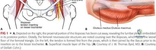
If the sesamoid fragments are of roughly equal size, viable, and the displacement is significant (> 5 mm), ORIF is the procedure of choice to restore the functional integrity of the FHB and preserve the biomechanical advantages of the sesamoid. A medial longitudinal incision is utilized for the tibial sesamoid. The incision, approximately 4 to 5 cm in length, is centered over the palpable medial eminence of the first MTP joint, placed just dorsal to the plantar-medial border to avoid creating a painful plantar scar. Meticulous, superficial dissection is absolutely critical to identify and protect the plantar medial cutaneous nerve of the hallux, which courses directly over or immediately adjacent to the medial capsule and sesamoid. Retraction of this nerve must be gentle to prevent neuropraxia or subsequent neuroma formation.
The medial capsule of the MTP joint is incised longitudinally, and the abductor hallucis is reflected plantarly. The fracture site or nonunion is identified within the substance of the FHB tendon. In the setting of a nonunion, the fibrous pseudarthrosis tissue interposed between the fragments is meticulously debrided using a fine curette or a #15 blade. The sclerotic bone ends must be decorticated using a 1.5-mm drill bit or a high-speed burr until punctate bleeding, healthy cancellous bone is exposed (the "paprika sign"). Autologous cancellous bone graft, harvested from the ipsilateral calcaneal tuberosity or the distal tibial metaphysis, is densely packed into the prepared defect to stimulate osteogenesis.
Fixation is highly dependent on fragment size. For robust fragments, one or two 1.5-mm or 2.0-mm headless compression screws are placed from proximal to distal. The guide wires are advanced under strict fluoroscopic guidance to ensure they do not violate the dorsal articular surface of the sesamoid. Alternatively, and highly effectively in cases of smaller or comminuted fragments, a figure-of-eight tension band wiring technique is employed. A heavy, non-absorbable suture (e.g., #2 FiberWire) is passed through drill holes in the proximal and distal poles, or woven directly through the robust FHB tendinous insertions. The suture is tied in a figure-of-eight configuration over the plantar aspect of the sesamoid, converting the tensile forces of the FHB into dynamic compression across the fracture site during hallux dorsiflexion.
Partial and Complete Sesamoidectomy
Modern orthopedic practice heavily favors partial sesamoid excision over complete excision for painful bipartite sesamoids or recalcitrant nonunions where one fragment is small and avascular. Through the same medial approach, the capsule is opened. The smaller, typically distal, fragment is carefully shelled out of the plantar plate and FHB tendon. It is absolutely critical to stay strictly subperiosteal during this enucleation; straying beyond the periosteal sleeve risks catastrophic iatrogenic transection of the FHL tendon or disruption of the intersesamoidal ligament. Once the fragment is excised, repairing the resulting defect in the flexor hallucis brevis is the most critical step of the procedure. The remaining tendon must be securely imbricated and sutured to the preserved sesamoid fragment or the robust plantar plate using heavy non-absorbable sutures. Failure to meticulously repair this defect will inevitably lead to weakness in hallux plantarflexion and potential dorsal subluxation of the MTP joint.
Complete sesamoid excision (sesamoidectomy) is reserved strictly as a salvage procedure for severely comminuted fractures, end-stage avascular necrosis with collapse, or failed partial excisions. The entire sesamoid is carefully enucleated from its tendinous envelope. Excision of the tibial (medial) sesamoid fundamentally weakens the medial head of the FHB. If the resulting tendinous defect is not tightly imbricated and repaired, the unopposed dynamic pull of the adductor hallucis (which attaches to the intact fibular sesamoid) will invariably lead to an iatrogenic hallux valgus deformity. Conversely, excision of the fibular (lateral) sesamoid via a plantar or dorsal-web approach can lead to an iatrogenic hallux varus deformity due to the unopposed pull of the abductor hallucis. Bilateral excision of both sesamoids is strongly discouraged and almost universally condemned, as it completely destroys the intrinsic plantarflexion power of the MTP joint, inevitably resulting in a severe, debilitating "cock-up" deformity (hyperextension of the MTP and flexion of the IP joint).
Complications, Incidence Rates, and Salvage Management
The orthopedic surgeon must be acutely vigilant regarding the complications associated with sesamoid surgery, as the delicate soft tissue envelope, complex biomechanics, and tenuous vascularity create a uniquely unforgiving surgical environment. Complications in this anatomical region are not merely radiographic curiosities; they frequently result in profound functional impairment and chronic pain that can be more debilitating than the index injury.
Iatrogenic deformity is perhaps the most feared complication following sesamoid excision. Hallux valgus following tibial sesamoidectomy and hallux varus following fibular sesamoidectomy occur due to the disruption of the delicate balance between the abductor and adductor hallucis muscles. The incidence of these deformities is directly inversely proportional to the meticulousness of the FHB and plantar plate repair at the time of excision. Neurological complications are also highly prevalent. The plantar medial cutaneous nerve is exquisitely vulnerable during the medial approach to the tibial sesamoid. Inadvertent transection, aggressive retraction, or entrapment in scar tissue can lead to a painful neuroma or debilitating numbness along the medial aspect of the hallux.
Nonunion and delayed union are particularly common following ORIF of sesamoid fractures, occurring in up to 15-20% of cases. This is primarily driven by the tenuous, retrograde blood supply to the distal pole, which is frequently disrupted by the fracture itself or the surgical dissection. Arthrofibrosis and stiffness of the first MTP joint are nearly universal following any surgical intervention in this area. The prolonged immobilization required to protect the bony fixation and soft tissue repair inevitably leads to capsular contracture. Aggressive, structured postoperative physical therapy is mandatory once the repair is deemed clinically and radiographically stable.
| Complication | Estimated Incidence | Pathophysiology / Risk Factors | Salvage Management Strategies |
|---|---|---|---|
| Iatrogenic Hallux Valgus | 10% - 15% (Post-Tibial Excision) | Unopposed adductor hallucis pull; Failure to imbricate medial FHB defect. | Medial capsulorrhaphy; Extensor hallucis brevis (EHB) transfer; First MTP Arthrodesis. |
| Iatrogenic Hallux Varus | 5% - 10% (Post-Fibular Excision) | Unopposed abductor hallucis pull; Over-tightening of medial structures. | Abductor hallucis release; Extensor hallucis longus (EHL) transfer; First MTP Arthrodesis. |
| Symptomatic Neuroma | 5% - 12% | Injury to the plantar medial cutaneous nerve during surgical approach. | Conservative (gabapentin, steroid injections); Surgical excision and proximal burying of the nerve stump. |
| Nonunion / AVN | 15% - 20% (Post-ORIF) | Tenuous pre-sesamoid vascular supply; Inadequate decortication/bone grafting. | Revision ORIF with structural bone graft; Conversion to partial or complete sesamoidectomy. |
| Severe Arthrofibrosis | 25% - 40% | Prolonged immobilization; Extensive capsular dissection and scarring. | Aggressive physical therapy; Intra-articular corticosteroid injections; Open or arthroscopic capsular release. |
Phased Post-Operative Rehabilitation Protocols
The postoperative rehabilitation protocol following forefoot surgery is not a mere afterthought; it is a highly structured, critical component of the treatment algorithm. The protocol is strictly dictated by the specific procedure performed, requiring a delicate balance between protecting the tenuous soft tissue repair or bony fixation and initiating early motion to prevent debilitating arthrofibrosis.
Phase 1: Maximum Protection (Weeks 0-2)
Immediately postoperatively, the patient is placed in a bulky, well-padded compressive dressing and a rigid posterior splint to immobilize the ankle and the entire forefoot. Strict, absolute non-weight-bearing (NWB) status is maintained using crutches or a knee scooter. The primary goals during this phase are wound healing, hematoma prevention, and edema control. The patient is instructed to maintain strict elevation of the operative extremity above the level of the heart and to utilize cryotherapy aggressively.
Phase 2: Controlled Mobilization and Tissue Healing (Weeks 2-6)
At the 10-to-14-day mark, the bulky dressing is removed, the wound is inspected, and sutures or K-wires (if applicable and ready) are removed. The patient is transitioned to a rigid fracture boot equipped with a custom toe-spica extension. This extension is critical as it rigidly prevents any dorsiflexion of the MTP joint, thereby neutralizing the tensile pull of the FHB on the repair site. For patients who underwent ORIF and bone grafting, strict NWB is continued until week 4, followed by a very gradual progression to heel-touch weight-bearing. For patients who underwent partial or complete excisions, protected flat-foot weight-bearing within the boot is permitted as tolerated by pain. During this phase, gentle, passive plantarflexion exercises of the MTP joint are initiated by a physical therapist, but active or passive dorsiflexion is strictly prohibited to protect the FHB repair and the fracture fixation.
Phase 3: Strengthening, Proprioception, and Return to Function (Weeks 6-12+)
At 6 weeks postoperatively, new weight-bearing radiographs are obtained to confirm osseous union (in the case of ORIF) or the maintenance of concentric joint alignment (in the case of excisions). Once clinical and radiographic stability is confirmed, the patient transitions from the fracture boot to a stiff-soled athletic shoe. This shoe must be fitted with a custom, full-length rigid carbon fiber plate and a custom orthotic incorporating a dancer's pad. The dancer's pad (a U-shaped felt or dense foam pad) is strategically placed immediately proximal to the first metatarsal head to effectively offload the sesamoid complex during the stance phase of gait. Active range of motion exercises for the MTP joint are aggressively initiated, pushing into dorsiflexion to combat capsular contracture. Progressive resistance exercises utilizing resistance bands are introduced to strengthen the intrinsic foot musculature, the FHB, and the FHL. Proprioceptive training and gait retraining are essential. Return to high-impact sports, running, or dancing is typically delayed until 3 to 4 months postoperatively, and is strictly contingent upon complete radiographic healing, the absence of pain with provocative testing, and the restoration of symmetric push-off strength compared to the contralateral limb.
Summary of Landmark Literature and Clinical Guidelines
The evolution of operative management for sesamoid fractures has been significantly shaped by a deeper understanding of forefoot biomechanics and vascular anatomy. Historically, complete sesamoid excision was the standard of care for failed conservative treatment of sesamoiditis or fractures.
