Peripheral Nerve Injury Classification and Operative Management
Key Takeaway
Peripheral nerve injuries present complex reconstructive challenges. Understanding the Seddon and Sunderland classification systems is paramount for accurate prognostication and surgical decision-making. This comprehensive guide details the histopathology of nerve trauma—from transient neurapraxia to complete neurotmesis—and outlines evidence-based protocols for clinical evaluation, advanced neurodiagnostic imaging, and microsurgical reconstruction. Mastery of these principles ensures optimal functional recovery following debilitating peripheral nerve trauma.
INTRODUCTION TO PERIPHERAL NERVE TRAUMA
Peripheral nerve injuries represent a formidable challenge in operative orthopaedics, often resulting in profound functional impairment, chronic pain, and severe socioeconomic consequences for the patient. The accurate diagnosis, prognostication, and surgical management of these lesions rely entirely on a foundational understanding of nerve microanatomy and the pathophysiological response to injury.
Historically, the classification of nerve injuries has evolved to provide surgeons with a reliable framework for predicting spontaneous recovery versus the absolute necessity for microsurgical intervention. The systems proposed by Seddon (1943) and Sunderland (1951), later modified by Mackinnon, remain the gold standards in peripheral nerve surgery. This masterclass delineates these classification systems, correlates them with histopathological and clinical findings, and provides an exhaustive guide to the operative management of peripheral nerve trauma.
MICROANATOMY AND BIOMECHANICS OF PERIPHERAL NERVES
To fully grasp the classification systems, the orthopedic surgeon must first understand the intricate microarchitecture of a peripheral nerve. A peripheral nerve is not a simple biological wire; it is a complex, highly organized composite structure designed to withstand physiological tension and compression while maintaining uninterrupted axoplasmic flow and electrical conduction.
- The Axon and Myelin Sheath: The fundamental functional unit is the axon, an extension of the neuronal soma located in the anterior horn of the spinal cord (motor) or dorsal root ganglion (sensory). Large myelinated fibers are enveloped by Schwann cells, which form the myelin sheath, separated by Nodes of Ranvier to facilitate rapid saltatory conduction.
- Endoneurium: A delicate layer of loose connective tissue surrounding individual axons and their associated Schwann cells. It forms the endoneurial tube, a critical biological conduit that guides regenerating axonal sprouts following injury.
- Perineurium: A dense, metabolically active layer of connective tissue that bundles groups of axons into fascicles. The perineurium provides the primary biomechanical tensile strength of the nerve and acts as the blood-nerve barrier.
- Epineurium: The outermost layer, divided into the internal epineurium (cushioning fascicles within the nerve) and the external epineurium (defining the outer boundary of the nerve trunk). It protects against compressive forces.
Clinical Pearl: The biomechanical resilience of a nerve is highly dependent on its connective tissue-to-fascicle ratio. Nerves with abundant epineurial connective tissue (e.g., the sciatic nerve in the gluteal region) are more resistant to compression but may be more susceptible to traction injuries compared to nerves with tightly packed fascicles.
SEDDON’S CLASSIFICATION OF NERVE INJURIES (1943)
Sir Herbert Seddon introduced a tripartite classification system based on the macroscopic and microscopic severity of the injury. While generally accepted, its broad categories lack the granular detail required for complex surgical decision-making in modern microsurgery.
1. Neurapraxia
Neurapraxia designates a localized, transient conduction block secondary to minor contusion, compression, or ischemia.
* Pathology: The axis-cylinder (axon) remains intact. There may be localized edema or segmental demyelination, but the structural continuity of the nerve is preserved. Crucially, there is no Wallerian degeneration distal to the injury.
* Clinical Presentation: Transmission of action potentials is physiologically interrupted. Motor function is typically more profoundly affected than sensory function. Sympathetic fibers are highly resistant to neurapraxia; thus, autonomic function (e.g., sweating) is often preserved.
* Prognosis: Spontaneous, complete recovery is expected within days to a maximum of 12 weeks as remyelination occurs.
2. Axonotmesis
Axonotmesis represents a more significant injury, typically secondary to severe crush or traction.
* Pathology: The axon is structurally disrupted, leading to distal Wallerian degeneration. However, the supporting connective tissue framework—specifically the Schwann cell basal lamina and endoneurial tubes—remains completely intact.
* Clinical Presentation: Complete motor, sensory, and sympathetic paralysis distal to the lesion. Because the endoneurial tubes are intact, regenerating axons are perfectly guided back to their original target organs.
* Prognosis: Spontaneous regeneration occurs at a rate of approximately 1 mm/day (1 inch/month). Functional recovery is generally excellent, provided the distance to the target motor endplate is not so vast that irreversible muscle atrophy occurs before reinnervation.
3. Neurotmesis
Neurotmesis is the most severe form of nerve injury, denoting complete anatomical severance or catastrophic crush/avulsion.
* Pathology: The axon, Schwann cells, endoneurial tubes, perineurium, and epineurium are completely disrupted.
* Clinical Presentation: Complete neurological deficit. Without an intact connective tissue scaffold, regenerating axonal sprouts wander blindly into surrounding tissues, forming a painful, disorganized terminal neuroma.
* Prognosis: Zero potential for spontaneous functional recovery. Surgical intervention (repair, graft, or transfer) is absolutely mandatory.
SUNDERLAND’S CLASSIFICATION SYSTEM (1951)
Sir Sydney Sunderland expanded Seddon’s concepts into a five-degree classification system. This system is vastly more applicable clinically, as each ascending degree represents a progressive anatomical disruption of the nerve's microarchitecture, directly correlating with prognosis and surgical strategy.
First-Degree Injury (Corresponds to Neurapraxia)
- Anatomical Disruption: Myelin sheath only. Axon and all connective tissues are intact.
- Pathophysiology: Localized conduction block without Wallerian degeneration.
- Clinical Features: Motor loss predominates. Sensory loss follows a specific hierarchy: proprioception > touch > temperature > pain. Sympathetic function is usually preserved.
- Tinel Sign: Absent (no axonal regeneration is occurring).
- Recovery: Simultaneous return of motor function in proximal and distal musculature. Complete restoration within weeks.
Second-Degree Injury (Corresponds to Axonotmesis)
- Anatomical Disruption: Axon is severed. Endoneurium, perineurium, and epineurium remain intact.
- Pathophysiology: Wallerian degeneration occurs distally. The intact endoneurial tube provides a flawless anatomical conduit for regeneration.
- Clinical Features: Complete motor, sensory, and sympathetic loss.
- Tinel Sign: Present and advancing distally at ~1 inch per month, tracing the progression of the regenerating growth cone.
- Recovery: Progressive "motor march" from proximal to distal. Excellent functional return is expected without surgery.
Third-Degree Injury
- Anatomical Disruption: Axon and Endoneurium are disrupted. Perineurium and epineurium remain intact.
- Pathophysiology: Wallerian degeneration occurs. Because the endoneurial tubes are destroyed, intrafascicular scarring occurs. Regenerating axonal sprouts may be blocked by scar tissue or misdirected into incorrect endoneurial tubes (aberrant regeneration).
- Clinical Features: Complete neurological loss.
- Tinel Sign: Present and advancing, but clinical recovery lags behind the advancing Tinel sign due to axonal misdirection and scarring.
- Recovery: Incomplete and unpredictable. Varying degrees of permanent motor or sensory deficit will remain. Surgical neurolysis or grafting may be required if recovery plateaus.
Fourth-Degree Injury
- Anatomical Disruption: Axon, endoneurium, and perineurium are disrupted. Only the epineurium remains intact.
- Pathophysiology: The fascicular architecture is completely destroyed. Nerve continuity is maintained solely by disorganized scar tissue. Axonal sprouts exit through perineurial defects, creating a dense neuroma-in-continuity.
- Clinical Features: Complete neurological deficit.
- Tinel Sign: Present at the site of injury but does not advance distally, as axons cannot penetrate the dense scar block.
- Recovery: Prognosis for spontaneous return of useful function is uniformly poor. Surgical resection of the neuroma and nerve grafting are required.
Fifth-Degree Injury (Corresponds to Neurotmesis)
- Anatomical Disruption: Complete transection of the entire nerve trunk (Axon, endoneurium, perineurium, and epineurium).
- Pathophysiology: Complete anatomical separation. Retraction of nerve ends occurs due to inherent elasticity.
- Clinical Features: Complete neurological deficit. Often identified during early surgical exploration of open wounds.
- Recovery: No spontaneous recovery. Requires operative repair or reconstruction.
Sixth-Degree Injury (Mackinnon’s Modification)
Introduced by Susan Mackinnon, the sixth-degree injury describes a mixed nerve injury. In a single nerve trunk, some fascicles may sustain a first-degree injury, while others suffer third-, fourth-, or fifth-degree injuries.
* Clinical Challenge: A neuroma-in-continuity develops, but the recovery pattern is mixed. The surgical dilemma lies in resecting the severely damaged fascicles (requiring grafting) without sacrificing the intact, recovering fascicles. This requires meticulous intraoperative internal neurolysis and nerve stimulation.
Surgical Warning: Unnecessary exploration of first- and second-degree injuries can cause iatrogenic damage and disrupt spontaneous recovery. Conversely, delaying surgery for fourth- and fifth-degree injuries leads to irreversible motor endplate degradation. Accurate clinical and electrodiagnostic staging is paramount.
CLINICAL AND ELECTRODIAGNOSTIC EVALUATION
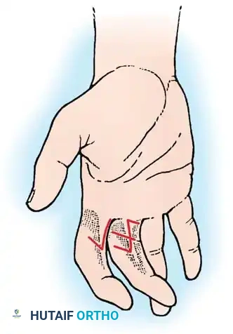
The Advancing Tinel Sign
The Hoffmann-Tinel sign is elicited by gently tapping along the course of the nerve from distal to proximal. A positive sign is the sensation of tingling or "electric shocks" radiating into the nerve's sensory distribution.
* An advancing Tinel sign indicates regenerating axons (seen in 2nd and 3rd-degree injuries).
* A static Tinel sign localized only at the injury site indicates a neuroma-in-continuity or complete transection (4th or 5th-degree injury) where axons are trapped in scar tissue.
Electromyography and Nerve Conduction Studies (EMG/NCS)
Electrodiagnostic studies are an extension of the physical examination.
* Timing is Critical: Wallerian degeneration takes 10 to 21 days to complete. EMG/NCS performed before 3 weeks may yield falsely reassuring results because the distal nerve segment may still conduct action potentials.
* Fibrillation Potentials: The presence of fibrillation potentials and positive sharp waves at 3-4 weeks confirms axonal loss (denervation) and rules out a simple neurapraxia.
* Nascent Motor Unit Potentials (MUPs): The appearance of nascent MUPs on serial EMGs precedes clinical muscle contraction and confirms successful reinnervation, guiding the surgeon to continue observation rather than intervene.
SURGICAL INDICATIONS AND TIMING
The timing of surgical intervention is dictated by the mechanism of injury and the Sunderland classification:
- Clean, Sharp Transections (e.g., glass laceration, knife wound): Indicate immediate or early (within 72 hours) primary epineurial repair. The zone of injury is minimal, allowing for direct, tension-free coaptation.
- Blunt Trauma, Crush, or High-Velocity Gunshot Wounds: These mechanisms create a wide, unpredictable zone of injury. Immediate repair is contraindicated as the extent of intraneural damage is not yet demarcated. These injuries are managed with delayed exploration at 3 months. If clinical and EMG evidence of recovery (advancing Tinel, nascent MUPs) is absent at 3 months, surgical exploration is mandated.
- Closed Traction Injuries (e.g., Brachial Plexus injuries): Managed expectantly for 3 to 6 months. Serial clinical exams and EMGs dictate the need for intervention.
OPERATIVE MANAGEMENT: STEP-BY-STEP PROTOCOLS
When surgical intervention is indicated, meticulous microsurgical technique is required to optimize outcomes.
1. Preoperative Preparation and Positioning
- Anesthesia: General anesthesia is preferred. Crucial: The anesthesiologist must be instructed not to use long-acting neuromuscular blocking agents (paralytics), as intraoperative nerve stimulation is essential for mapping and decision-making.
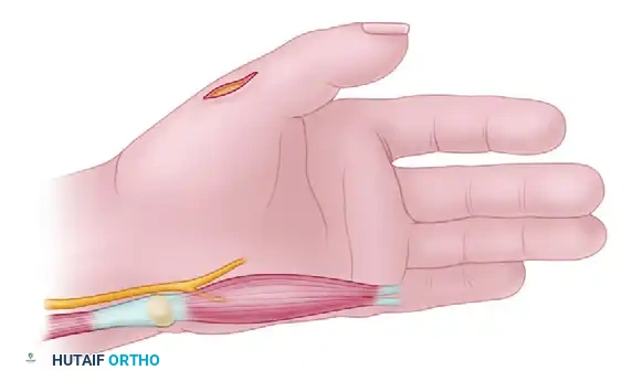
- Positioning: Varies by the nerve involved. Ensure all potential donor sites for nerve autografts (e.g., sural nerve, medial antebrachial cutaneous nerve) are prepped and draped in the sterile field.
- Tourniquet: A pneumatic tourniquet is utilized for hemostasis. However, exsanguination with an Esmarch bandage should be avoided if the surgeon relies on visualizing intraneural microvasculature to assess nerve viability.
2. Surgical Approach and Exposure
- Extensile Incisions: Always utilize generous, extensile incisions.
- Normal to Abnormal: The golden rule of nerve surgery is to identify the nerve in pristine, uninjured tissue both proximally and distally, and then trace it toward the zone of injury (the neuroma or scar bed).
- Vascular Preservation: Preserve the segmental mesoneurial blood supply. Excessive mobilization strips the nerve of its extrinsic vascularity, leading to ischemia and subsequent intraneural fibrosis.
3. Intraoperative Assessment of Neuroma-in-Continuity
When encountering a Sunderland 3rd, 4th, or 6th-degree injury, the surgeon faces a neuroma-in-continuity. The decision to perform neurolysis versus resection and grafting is the most critical intraoperative juncture.
* Intraoperative Nerve Stimulation (IONS): A sterile stimulating probe is applied proximal to the neuroma, and the target muscles are observed for contraction.
* Nerve Action Potential (NAP) Recording: If no visible contraction occurs, NAP monitoring is employed. If a NAP is conducted across the neuroma, it indicates functional regenerating axons (Sunderland 3rd degree); the treatment is external neurolysis (freeing the nerve from surrounding scar) and observation.
* Resection: If no NAP is conducted across the lesion, the injury is a Sunderland 4th degree. The neuroma must be resected back to healthy, bleeding fascicles ("bread-loafing" the nerve ends) until normal pouting fascicles are visualized under the operating microscope.
4. Techniques of Nerve Reconstruction
Primary Direct Repair
Indicated for sharp transections where the nerve ends can be brought together without tension.
* Biomechanics of Repair: Tension across a nerve repair causes ischemia and catastrophic failure of regeneration. If an 8-0 nylon epineurial suture cannot hold the nerve ends together without breaking, the repair is under too much tension, and a graft is required.
* Technique: Under microscopic magnification, the nerve ends are aligned using surface vascular landmarks. Interrupted 8-0 or 9-0 non-absorbable monofilament sutures are placed in the epineurium. Fibrin glue may be used to augment the repair.
Nerve Autografting
Indicated when resection of a neuroma or retraction of nerve ends creates a gap that cannot be closed without tension.
* Donor Nerves: The reversed sural nerve is the workhorse autograft. Reversing the graft prevents regenerating axons from escaping through severed sensory branches.
* Cable Grafting: Because the sural nerve is smaller in diameter than major motor nerves (e.g., sciatic or radial), multiple strands (cables) are used to bridge the gap, matching the cross-sectional area of the injured nerve.
* Coaptation: The grafts are sutured using 9-0 or 10-0 microsutures, ensuring precise fascicular alignment.
Nerve Transfers (Neurotization)
In cases of very proximal injuries (e.g., high brachial plexus avulsions) or delayed presentations (>12 months) where the target muscle is at risk of irreversible motor endplate fibrosis, nerve transfers are indicated.
* Principle: A redundant or expendable motor nerve branch (donor) is transected and coapted directly to the injured nerve (recipient) as close to the target muscle as possible.
* Example: Transferring a fascicle of the ulnar nerve to the musculocutaneous nerve (Oberlin transfer) to restore elbow flexion in upper trunk brachial plexus injuries. This drastically reduces the distance regenerating axons must travel.
POSTOPERATIVE PROTOCOLS AND REHABILITATION
The success of peripheral nerve surgery relies heavily on meticulous postoperative care and specialized hand/neuro-rehabilitation.
- Immobilization: Following primary repair or grafting, the extremity is immobilized in a well-padded orthosis for 3 weeks to protect the micro-coaptations from tensile forces. The joints are positioned to minimize tension on the nerve (e.g., elbow flexion for an ulnar nerve repair at the cubital tunnel).
- Mobilization: At 3 weeks, the splint is removed, and a guided, progressive active and passive range of motion protocol is initiated to prevent joint contractures and promote nerve gliding.
- Sensory Re-education: As sensory axons reach the periphery, patients often experience hypersensitivity or altered perception. Cortical sensory re-education programs are vital to help the brain reinterpret these new, often distorted, afferent signals.
- Motor Retraining: Biofeedback and targeted motor exercises are utilized, particularly following nerve transfers, to train the brain to fire the donor nerve to activate the new recipient muscle.
Clinical Pitfall: Failure to maintain passive joint mobility while awaiting nerve regeneration is a catastrophic error. A perfectly regenerated nerve is functionally useless if it innervates a muscle acting across a rigidly contracted, ankylosed joint.
CONCLUSION
The classification of peripheral nerve injuries is not merely an academic exercise; it is the fundamental blueprint that dictates clinical prognostication and surgical strategy. By mastering the nuances of the Seddon and Sunderland classifications, the orthopedic surgeon can accurately interpret clinical signs, optimize the timing of electrodiagnostic studies, and execute precise, evidence-based microsurgical interventions. Whether managing a transient neurapraxia with reassurance or reconstructing a devastating neurotmesis with complex cable grafting, adherence to these foundational principles is paramount to restoring function and quality of life to the trauma patient.
You Might Also Like