العربية
Part of the Master Guide

Shoulder And Elbow: Review | Dr Hutaif Shoulder & Elbow -...

Ortho Shoulder And Elbow Review | Dr Hutaif Shoulder & - ...
23 Apr 2026 46 min read 124 Views
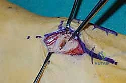
Illustration of response d discussion - Dr. Mohammed Hutaif

Key Takeaway

Your ultimate guide to ORTHO MCQS Shoulder and Elbow 019 starts here. For a radial head fracture with lateral collateral ligament (LCL) avulsion repaired surgically, initial postoperative rehabilitation should include elbow extension exercises with the forearm in pronation. This protects the compromised LCL, crucial for stability. Additional therapeutic insights, including the rationale, are elaborated upon in the response d discussion.

Ortho Shoulder And Elbow Review | Dr Hutaif Shoulder & - ...

Comprehensive 100-Question Exam


00:00

Start Quiz

Question 1

A 25-year-old male undergoes a Latarjet procedure for recurrent anterior shoulder instability with 25% glenoid bone loss. Postoperatively, he presents with profound weakness in elbow flexion and decreased sensation over the lateral aspect of his forearm. Which nerve is most likely injured, and what is the most common mechanism during this procedure?


Explanation

The musculocutaneous nerve is the most frequently injured nerve during a Latarjet procedure. It typically penetrates the coracobrachialis muscle 5 to 8 cm distal to the coracoid process. The primary mechanism of injury is excessive or prolonged medial retraction of the conjoined tendon during the approach and graft preparation. This leads to neuropraxia, presenting with biceps weakness (elbow flexion) and sensory deficits over the lateral forearm (lateral antebrachial cutaneous nerve).

Question 2

A 40-year-old female sustains a 'terrible triad' injury of the elbow following a fall onto an outstretched hand. Open reduction and internal fixation are planned. According to standard evidence-based protocols, which of the following represents the most appropriate surgical sequence to restore elbow stability?


Explanation

The 'terrible triad' of the elbow consists of an elbow dislocation, radial head fracture, and coronoid process fracture. The classic surgical algorithm established by Pugh et al. dictates an 'inside-out' or deep-to-superficial approach. The recommended sequence is: 1) Fixation or repair of the coronoid to restore the anterior buttress; 2) Fixation or arthroplasty of the radial head to restore the lateral column and anterior restraint; 3) Repair of the lateral ulnar collateral ligament (LUCL) to restore posterolateral rotatory stability. The MCL is typically only repaired if the elbow remains unstable after the first three steps and application of a hinged external fixator is not preferred.

Question 3

A 35-year-old bodybuilder undergoes a two-incision technique for repair of a distal biceps tendon rupture. Six months postoperatively, he presents with severely restricted forearm pronation and supination, though elbow flexion and extension are normal. What is the most likely complication he developed from this specific surgical approach?


Explanation

The two-incision technique (modified Boyd-Anderson) for distal biceps repair was developed to reduce the risk of posterior interosseous nerve (PIN) injury associated with the extensile single anterior incision. However, muscle splitting and subperiosteal dissection between the radius and ulna during the posterior approach increases the risk of heterotopic ossification, specifically proximal radioulnar synostosis. This complication leads to profound loss of forearm rotation (pronation/supination). Careful technique to avoid exposing the ulna or breaching the interosseous membrane is critical.

Question 4

A 28-year-old elite volleyball player presents with vague posterior shoulder pain. Magnetic resonance imaging (MRI) reveals a paralabral cyst located strictly in the spinoglenoid notch. Which of the following physical examination findings is most specific to this pathology?


Explanation

A paralabral cyst in the spinoglenoid notch typically compresses the suprascapular nerve after it has innervated the supraspinatus muscle. Therefore, the patient will present with isolated denervation of the infraspinatus muscle, leading to isolated weakness in external rotation. Compression at the suprascapular notch (more proximal) would affect both the supraspinatus and infraspinatus, leading to weakness in both abduction and external rotation. The suprascapular nerve has no significant cutaneous sensory distribution over the lateral shoulder (axillary nerve territory).

Question 5

When performing a reverse total shoulder arthroplasty (RTSA) using the classic Grammont design for a patient with cuff tear arthropathy, how does the prosthesis biomechanically alter the center of rotation (COR) of the glenohumeral joint compared to the native anatomy?


Explanation

The classic Grammont design for a reverse total shoulder arthroplasty (RTSA) biomechanically medializes and distalizes the center of rotation. Medialization decreases the torque on the glenoid component (minimizing the 'rocking horse' effect and risk of baseplate loosening). Distalization tensions the deltoid and significantly increases its moment arm, allowing the deltoid to effectively elevate the arm in the absence of a functional rotator cuff.

Question 6

A 45-year-old male presents to the emergency department after a seizure. Radiographs confirm a locked posterior shoulder dislocation. A CT scan reveals an anteromedial humeral head impaction fracture (reverse Hill-Sachs lesion) involving 30% of the articular surface. The dislocation is successfully reduced, but the shoulder is highly unstable in internal rotation. What is the most appropriate surgical intervention?


Explanation

A reverse Hill-Sachs lesion is an impaction fracture of the anteromedial humeral head resulting from a posterior shoulder dislocation. For defects comprising 20% to 40% of the articular surface, transferring the subscapularis tendon (McLaughlin procedure) or the lesser tuberosity with the attached subscapularis (Modified McLaughlin procedure) into the defect is the treatment of choice. This prevents the defect from engaging the posterior glenoid rim during internal rotation. Defects <20% may be managed non-operatively or with isolated posterior labral repair if non-engaging, while defects >40-50% generally require anatomic head reconstruction via osteochondral allograft or shoulder arthroplasty.

Question 7

In the setting of a Type III acromioclavicular (AC) joint separation, both the AC ligaments and coracoclavicular (CC) ligaments are torn. Which of the following structures acts as the primary restraint to superior translation of the distal clavicle?


Explanation

The coracoclavicular (CC) ligament complex consists of the conoid and trapezoid ligaments. Biomechanical studies have demonstrated that the conoid ligament (which is located posteromedially) is the primary restraint to superior translation of the distal clavicle. The trapezoid ligament (located anterolaterally) serves primarily to resist axial compression of the AC joint. The AC capsular ligaments primarily resist anterior-posterior translation.

Question 8

A 32-year-old female falls onto her outstretched hand and sustains a fracture of the anteromedial facet of the coronoid process of the ulna. Based on this specific fracture pattern, what is the underlying mechanism of injury and the associated ligamentous pathology?


Explanation

Anteromedial facet fractures of the coronoid are the hallmark of Varus Posteromedial Rotatory Instability (VPMRI). This injury pattern is caused by a varus stress applied to the elbow, combined with axial load and posteromedial rotation of the ulna. This forces the anteromedial coronoid facet to impact the trochlea, causing a fracture. The lateral collateral ligament (LCL) complex is classically avulsed or torn, leading to the varus instability. The anterior bundle of the medial collateral ligament (MCL) usually remains intact or is only partially injured.

Question 9

A 22-year-old collegiate baseball pitcher presents with medial elbow pain and decreased pitching velocity. Examination demonstrates pain with the moving valgus stress test. He is diagnosed with an ulnar collateral ligament (UCL) tear. Which distinct portion of the UCL complex serves as the primary restraint to valgus stress between 30 and 120 degrees of elbow flexion?


Explanation

The ulnar collateral ligament (UCL) is composed of three bundles: anterior, posterior, and transverse. The anterior bundle is the primary stabilizer against valgus stress at the elbow throughout the functional range of motion (from roughly 30 to 120 degrees of flexion). The posterior bundle acts as a secondary restraint, specifically functioning in higher degrees of elbow flexion (>90 degrees). The transverse bundle has no significant role in elbow stability, as it originates and inserts on the same bone (ulna).

Question 10

Recent anatomic studies utilizing advanced vascular mapping have challenged traditional orthopedic teaching regarding the principal arterial supply to the proximal humerus. According to current literature (e.g., Hettrich et al.), which artery provides the majority of the blood supply to the humeral head?


Explanation

Historically, the anterolateral branch of the anterior humeral circumflex artery (the arcuate artery) was considered the primary blood supply to the humeral head. However, modern quantitative anatomic studies (such as the landmark 2010 study by Hettrich et al.) demonstrated that the posterior humeral circumflex artery provides approximately 64% of the blood supply to the humeral head, while the anterior humeral circumflex artery supplies only about 36%. This shift in understanding highlights the critical role of the posterior vessels in preventing avascular necrosis following proximal humerus fractures.

Question 11

A 65-year-old male with an isolated, massive, irreparable posterosuperior rotator cuff tear presents with persistent shoulder pain, preserved forward elevation, but a profound external rotation lag sign (positive Hornblower's sign). He is deemed unsuitable for a reverse total shoulder arthroplasty due to lack of glenohumeral arthritis. Which tendon transfer provides the most biomechanically synergistic line of pull to restore active external rotation in this patient?


Explanation

For an irreparable posterosuperior cuff tear with severe external rotation weakness (positive Hornblower's), a lower trapezius transfer is highly favored. The lower trapezius muscle fibers are aligned perfectly with the native infraspinatus, providing a synergistic, in-phase line of pull to restore external rotation. While the latissimus dorsi transfer has historically been used for this indication, it is an internal rotator and adductor out-of-phase with external rotation, requiring extensive cortical re-education and providing a less optimal biomechanical vector.

Question 12

According to the McKee modification of the Bryan and Morrey classification for capitellum fractures, what describes a Type IV injury?


Explanation

The Bryan and Morrey classification divides capitellar fractures into three primary types: Type I (Hahn-Steinthal, large osseous fragment), Type II (Kocher-Lorenz, primarily articular cartilage with a thin layer of bone), and Type III (Broberg-Morrey, comminuted). McKee later modified this classification by adding Type IV, which is a coronal shear fracture that involves not only the capitellum but extends medially to include the majority of the trochlea. Identifying a Type IV fracture is critical, as it requires fixation of both the capitellum and the trochlear fragment to restore elbow biomechanics.

Question 13

A surgeon is performing an open elbow contracture release via an extensile lateral column approach (Kocher). During the release of the anterior capsule to improve elbow extension, which nerve is at the greatest risk of iatrogenic injury and must be meticulously protected?


Explanation

During a lateral approach to the elbow (such as the Kocher or Kaplan intervals) for anterior capsular release, the radial nerve is the most vulnerable neurologic structure. The radial nerve crosses the elbow joint anterior to the radiocapitellar joint and lateral capsule. It can be easily injured if the retractor is placed improperly or if capsulotomy is performed without maintaining a protective layer of the brachialis muscle. The median nerve is situated more medially and is protected by the brachialis, while the ulnar nerve is located posteriorly in the cubital tunnel.

Question 14

A 30-year-old weightlifter feels a 'pop' in his anterior axillary fold during a heavy bench press. MRI confirms a rupture of the pectoralis major tendon at its insertion. In the anatomic footprint of the pectoralis major on the humerus, what is the spatial relationship of the sternal head relative to the clavicular head?


Explanation

The pectoralis major consists of a clavicular head and a sternocostal (sternal) head. As the muscle bellies course laterally toward their insertion on the lateral lip of the bicipital groove of the humerus, the sternal head twists 180 degrees. Consequently, the sternal head tendon passes deep (posterior) to the clavicular head tendon, and its fibers insert more proximally on the humerus. Tears of the pectoralis major most commonly involve isolated rupture of the sternal head, leaving the superficial/distal clavicular head intact.

Question 15

A 13-year-old elite youth baseball pitcher presents with gradually worsening shoulder pain during the late cocking and early acceleration phases of pitching. Radiographs reveal widening and lateral fragmentation of the proximal humeral physis. What is the diagnosis?


Explanation

The presentation of widening, sclerosis, and fragmentation of the proximal humeral physis in a skeletally immature throwing athlete is pathognomonic for 'Little League Shoulder.' The underlying pathology is a stress fracture or epiphysiolysis of the proximal humeral physis caused by repetitive rotational and distraction shear forces during throwing. It is primarily treated with absolute rest from throwing until radiographic resolution and symptom abatement.

Question 16

A 24-year-old male undergoes arthroscopic stabilization for recurrent anterior shoulder instability. Intraoperatively, the surgeon notes an 'engaging' Hill-Sachs lesion and elects to perform a Remplissage procedure in addition to a Bankart repair. The Remplissage procedure specifically involves tenodesis of which of the following structures into the humeral head defect?


Explanation

The Remplissage procedure (French for 'to fill') is performed for anterior shoulder instability accompanied by a large, engaging Hill-Sachs lesion. It involves arthroscopically suturing the posterior capsule and the infraspinatus tendon into the Hill-Sachs defect on the posterolateral humeral head. This essentially makes the Hill-Sachs lesion extra-articular and acts as a dynamic checkrein against excessive external rotation, preventing the humeral head defect from engaging the anterior glenoid rim.

Question 17

A 45-year-old female presents with severe lateral elbow pain exacerbated by lifting objects with the forearm pronated. She is diagnosed with lateral epicondylitis. Histopathologic examination of the affected tissue typically reveals angiofibroblastic hyperplasia rather than acute inflammation. Which tendon is considered the primary site of pathology in this condition?


Explanation

Lateral epicondylitis (tennis elbow) is a tendinopathy (angiofibroblastic tendinosis) primarily involving the origin of the extensor carpi radialis brevis (ECRB) tendon. The ECRB lies deep to the extensor carpi radialis longus (ECRL) and extensor digitorum communis (EDC). The chronic microtrauma at its origin on the lateral epicondyle leads to tissue degeneration rather than active inflammatory cells. The ECRL and EDC can be secondarily involved, but the ECRB is the hallmark site of pathology.

Question 18

A 50-year-old female undergoes a radial head arthroplasty for a comminuted, irreparable radial head fracture (Mason Type III). During the procedure, the surgeon inadvertently implants a prosthesis that is 4 mm too thick. What is the most likely clinical and radiographic consequence of this technical error?


Explanation

Overstuffing the radiocapitellar joint by inserting a radial head prosthesis that is too long causes altered elbow kinematics. It exerts excessive pressure on the capitellum, leading to rapid cartilage wear and subchondral osteolysis. Radiographically, this manifests as asymmetric widening of the ulnohumeral joint (specifically opening of the lateral aspect of the ulnohumeral articulation, creating a 'gap') because the radius is pushing the humerus away from the ulna. It also leads to a severe loss of elbow flexion and extension.

Question 19

A 28-year-old male presents with right shoulder asymmetry. On examination, having the patient perform a wall push-up causes the medial border of the right scapula to become excessively prominent and translate superiorly and medially. Injury to which of the following nerves is responsible for this classic presentation?


Explanation

This is a classic presentation of medial scapular winging, which is caused by paralysis of the serratus anterior muscle due to long thoracic nerve palsy. The serratus anterior normally protracts and upwardly rotates the scapula, keeping the medial border closely applied to the thorax. When it is paralyzed, the medial border lifts off the chest wall (wings medially) and the scapula translates superiorly and medially. This is differentiated from lateral winging (spinal accessory nerve / trapezius palsy), where the scapula translates inferiorly and laterally.

Question 20

A 42-year-old male presents with acute, unprovoked, agonizing pain in his right shoulder that lasted for 2 weeks before gradually subsiding. As the pain improved, he developed profound weakness in shoulder abduction and external rotation. Electromyography (EMG) shows acute denervation in the supraspinatus, infraspinatus, and deltoid. What is the most appropriate initial treatment for this condition?


Explanation

This patient's clinical presentation is pathognomonic for Parsonage-Turner Syndrome (idiopathic brachial neuritis). The classic progression involves a prodromal phase of severe, unrelenting shoulder or arm pain lasting days to weeks, followed by patchy lower motor neuron weakness and muscle atrophy as the pain resolves. Treatment is primarily non-operative, consisting of observation, pain control (gabapentin, NSAIDs), and physical therapy to maintain range of motion. The vast majority of patients recover spontaneously, although full recovery can take 12 to 24 months.

Question 21

A 45-year-old volleyball player presents with painless weakness in shoulder external rotation. Abduction strength is completely normal. Physical examination reveals muscle atrophy localized exclusively to the infraspinatus fossa. Where is the most likely site of nerve compression?


Explanation

Compression at the spinoglenoid notch selectively affects the infraspinatus branch of the suprascapular nerve, causing isolated external rotation weakness. Compression at the suprascapular notch would affect both the supraspinatus and infraspinatus muscles.

Question 22

During an anterolateral approach for internal fixation of a proximal humerus fracture, the axillary nerve is at significant risk of iatrogenic injury. What is the average distance of the axillary nerve from the lateral edge of the acromion?


Explanation

The axillary nerve courses circumferentially from posterior to anterior approximately 7 cm distal to the lateral tip of the acromion. Staying within 5 cm of the acromion during lateral split approaches minimizes the risk of nerve injury.

Question 23

A 65-year-old female undergoes a reverse total shoulder arthroplasty for severe cuff tear arthropathy. Postoperatively, radiographs reveal scapular notching. According to the Sirveaux classification, what defines a Grade 3 notch?


Explanation

In the Sirveaux classification for scapular notching, Grade 1 is confined to the pillar, Grade 2 reaches the inferior screw, Grade 3 extends over the inferior screw, and Grade 4 reaches the central peg. Inferior positioning and eccentric inferior overhang of the glenosphere help prevent this complication.

Question 24

A 30-year-old male presents with elbow pain and a mechanical click during extension and forearm supination following a fall. Examination reveals a positive lateral pivot-shift test. Which ligamentous structure is primarily deficient?


Explanation

Posterolateral rotatory instability (PLRI) of the elbow is caused by insufficiency of the lateral ulnar collateral ligament (LUCL). The lateral pivot-shift test dynamically reproduces the characteristic subluxation and reduction of the radiocapitellar joint.

Question 25

A 55-year-old male undergoes a single-incision anterior repair for an acute distal biceps tendon rupture. Postoperatively, he exhibits an expected neurologic deficit. Which nerve is at greatest risk of stretch injury during the superficial dissection and lateral retractor placement of this approach?


Explanation

The lateral antebrachial cutaneous nerve (LABCN) is at high risk of stretch neurapraxia during the superficial dissection and retraction in a single-incision anterior approach. The posterior interosseous nerve is at higher risk during a two-incision approach or with excessive deep radial retraction.

Question 26

A 28-year-old professional pitcher presents with medial elbow pain during the late cocking phase of throwing. MRI confirms a full-thickness tear of the anterior bundle of the ulnar collateral ligament (UCL). Where is the typical anatomic insertion of this crucial stabilizing bundle on the ulna?


Explanation

The anterior bundle of the medial UCL inserts on the sublime tubercle, located at the anteromedial margin of the coronoid process. This bundle is the primary restraint to valgus stress at the elbow during the throwing motion.

Question 27

A 60-year-old man with a massive, irreparable posterosuperior rotator cuff tear undergoes arthroscopic superior capsule reconstruction (SCR). What is the primary biomechanical objective of this specific procedure?


Explanation

SCR utilizes a dermal allograft or fascia lata autograft attached to the superior glenoid and greater tuberosity to recreate the superior capsular restraint. This depresses the humeral head, prevents superior migration, and improves the mechanical advantage of the deltoid.

Question 28

A 32-year-old powerlifter feels a sharp pop in his anterior chest while bench-pressing heavy weights. Examination reveals loss of the anterior axillary fold and weakness in internal rotation. If surgical repair of the completely avulsed tendon is performed, to which anatomic landmark should the tendon be reattached?


Explanation

The pectoralis major normally inserts onto the lateral lip of the bicipital (intertubercular) groove. Anatomic repair of the sternocostal head to this location is critical to restore maximum adduction and internal rotation strength.

Question 29

A 40-year-old patient falls from a ladder and sustains an Essex-Lopresti injury. Which combination of anatomic lesions defines this highly unstable injury pattern?


Explanation

An Essex-Lopresti lesion is characterized by a longitudinal radioulnar dissociation involving a radial head fracture, tearing of the interosseous membrane, and disruption of the distal radioulnar joint (DRUJ). Radial head excision without replacement in this setting leads to catastrophic proximal radial migration.

Question 30

A 45-year-old poorly controlled diabetic female presents with globally restricted passive and active shoulder range of motion. Radiographs demonstrate no joint space narrowing. What is the characteristic histologic and structural finding in the joint capsule of this specific condition?


Explanation

Adhesive capsulitis is histologically characterized by profound fibroblastic proliferation and type III collagen deposition. Macroscopically, the coracohumeral ligament and the rotator interval capsule become severely thickened and contracted.

Question 31

A 70-year-old female presents with an acute anterior shoulder dislocation. After successful closed reduction, she demonstrates profound inability to actively abduct her shoulder against gravity, despite adequate analgesia. What is the most likely cause of this persistent deficit in this demographic?


Explanation

In patients over the age of 40 (and especially those over 60), anterior shoulder dislocations are highly associated with acute rotator cuff tears. While axillary nerve injuries can occur, an acute rotator cuff tear is the most common structural cause of profound post-reduction weakness.

Question 32

A 16-year-old gymnast presents with chronic elbow pain and mechanical catching. Imaging confirms a large osteochondritis dissecans (OCD) lesion of the capitellum. Which of the following is considered an indication for osteochondral autograft transfer (OATS) rather than arthroscopic microfracture?


Explanation

Arthroscopic debridement and microfracture yield poor functional outcomes for large OCD lesions or those involving the uncontained lateral margin of the capitellum. OATS is specifically indicated for lesions greater than 1 cm or those with lateral margin involvement.

Question 33

A 19-year-old male rugby player sustains a traumatic anterior shoulder dislocation. Advanced imaging reveals a bony Bankart lesion. According to modern biomechanical literature, what is the critical threshold of glenoid bone loss above which isolated arthroscopic soft-tissue repair has an unacceptably high failure rate?


Explanation

Critical glenoid bone loss is widely accepted as 20-25% of the inferior glenoid diameter, though subcritical thresholds (13.5-15%) are now recognized in high-risk athletes. Bone loss exceeding 20% generally necessitates a bony augmentation procedure like a Latarjet.

Question 34

A patient undergoes an ulnar nerve transposition for severe cubital tunnel syndrome. During the approach, the nerve must be carefully mobilized from its native groove. Which fascial structure forms the direct roof of the cubital tunnel and must be released?


Explanation

The roof of the cubital tunnel is formed by Osborne's ligament (the cubital tunnel retinaculum), which spans between the medial epicondyle and the olecranon. Struthers' ligament is located proximally in the arm and is associated with median nerve compression.

Question 35

A 22-year-old boxer complains of prominent medial winging of his right scapula that worsens when doing push-ups against a wall. He sustained a direct blow to his lateral chest wall three months ago. Which nerve was most likely injured, and which muscle is consequently paralyzed?


Explanation

Medial scapular winging is the classic presentation of serratus anterior paralysis, which is innervated by the long thoracic nerve. In contrast, lateral scapular winging is associated with trapezius paralysis from a spinal accessory nerve injury.

Question 36

A 45-year-old male sustains a displaced midshaft clavicle fracture after a bicycle accident. Which of the following represents an absolute, universally accepted indication for acute open reduction and internal fixation?


Explanation

Absolute indications for acute operative intervention of a clavicle fracture include open fractures, impending skin compromise (necrosis), and concomitant neurovascular injury. Shortening, displacement, and standard skin tenting are relative indications based on patient activity and risk profile.

Question 37

A 68-year-old male with a massive, irreparable rotator cuff tear and pseudoparalysis presents for evaluation. Radiographs demonstrate superior migration of the humeral head, acromial acetabularization, but preserved glenohumeral joint space (Hamada grade 3). A Reverse Total Shoulder Arthroplasty (RTSA) is planned. In RTSA, what is the primary biomechanical advantage conferred by the implant design?


Explanation

RTSA medializes and inferiorizes the center of rotation, which increases the lever arm and resting tension of the deltoid. This allows the deltoid to effectively compensate for the deficient rotator cuff.

Question 38

A 45-year-old female with diabetes presents with severe shoulder pain and profound loss of active and passive external rotation for 4 months. She is diagnosed with the 'freezing' stage of adhesive capsulitis. Which cytokine or growth factor is most heavily implicated in the pathogenesis of this condition?


Explanation

TGF-beta is a major driver of fibrosis and plays a central role in the pathogenesis of adhesive capsulitis. It promotes robust fibroblast proliferation and excessive collagen production within the joint capsule.

Question 39

A 32-year-old cyclist falls onto the point of his shoulder. Radiographs reveal a 150% superior displacement of the clavicle relative to the acromion, and the coracoclavicular interval measures 28 mm. According to the Rockwood classification, what is the injury type and optimal management?


Explanation

Displacement of the clavicle between 100% and 300% characterizes a Rockwood Type V injury. This high-grade acromioclavicular joint separation typically requires operative management to restore anatomy and biomechanics.

Question 40

A 14-year-old elite baseball pitcher presents with lateral elbow pain, clicking, and a 15-degree extension deficit. Radiographs reveal a radiolucent lesion of the capitellum with a sclerotic margin and a visible loose body in the joint. What is the most appropriate management?


Explanation

In an adolescent with capitellar osteochondritis dissecans (OCD) presenting with a loose body and mechanical symptoms (indicating an unstable lesion), surgical intervention is required. Arthroscopic loose body removal and marrow stimulation (microfracture) is the standard of care.

Question 41

A 21-year-old collegiate pitcher undergoes a Tommy John surgery (UCL reconstruction). Which specific bundle of the Ulnar Collateral Ligament is the primary restraint to valgus stress at 90 degrees of flexion and is the primary target for this reconstruction?


Explanation

The anterior bundle of the UCL, specifically the anterior band, is the primary restraint to valgus stress between 30 and 120 degrees of elbow flexion. It is the critical structure reconstructed in Tommy John surgery.

Question 42

A 65-year-old female is 3 years post-anatomic total shoulder arthroplasty (TSA). She presents with increasing pain, anterior swelling, and weakness in internal rotation. Radiographs show a well-fixed implant without loosening, but ultrasound confirms a massive subscapularis failure. Which of the following is the most appropriate salvage option if primary repair is impossible?


Explanation

Pectoralis major tendon transfer is the established salvage procedure for an irreparable subscapularis tear following anatomic TSA. Conversely, latissimus dorsi and lower trapezius transfers are utilized for posterosuperior cuff (supraspinatus/infraspinatus) defects.

Question 43

A 72-year-old female with osteoporosis sustains a severely displaced 4-part proximal humerus fracture. She lives independently and is functionally active. Which surgical intervention provides the most predictable functional outcome and pain relief in this demographic?


Explanation

Reverse total shoulder arthroplasty (RTSA) provides more predictable functional outcomes and better pain relief than ORIF or hemiarthroplasty for displaced 4-part proximal humerus fractures in the elderly. This is because RTSA relies on deltoid function rather than tuberosity healing for overhead elevation.

Question 44

A 28-year-old volleyball player presents with insidious onset posterior shoulder pain and profound, isolated weakness in external rotation. MRI reveals a paralabral cyst in the spinoglenoid notch. Which physical examination finding is most likely present?


Explanation

Entrapment of the suprascapular nerve at the spinoglenoid notch affects only the motor branch to the infraspinatus. This results in isolated external rotation weakness and isolated infraspinatus atrophy.

Question 45

A 30-year-old male presents with medial scapular winging and inability to actively elevate his arm past 90 degrees following a heavy traction injury. EMG confirms a complete long thoracic nerve palsy. After 1 year of strict conservative management with no recovery, what is the treatment of choice?


Explanation

Pectoralis major transfer (using fascia lata autograft or direct transfer to the inferior angle of the scapula) is the standard surgical treatment for chronic, irrecoverable serratus anterior palsy causing medial winging.

Question 46

A 35-year-old male develops severe heterotopic ossification (HO) following ORIF of a distal humerus fracture, restricting his elbow arc of motion to 30-60 degrees. When is the optimal time to perform surgical excision of the HO?


Explanation

Excision of HO should be performed when the bone is radiographically mature (distinct trabecular pattern and sharp cortical margins) and the clinical exam demonstrates no further progressive loss of motion. This typically occurs between 6 to 9 months post-injury.

Question 47

A 40-year-old manual laborer undergoes an open subpectoral biceps tenodesis. Postoperatively, he exhibits profound weakness in wrist and finger extension, though elbow extension is intact. Which nerve was most likely injured due to errant retractor placement?


Explanation

The radial nerve runs directly posterior to the humerus and can be compressed or injured with aggressive deep retractor placement along the lateral aspect of the humerus during a subpectoral biceps tenodesis.

Question 48

A 45-year-old golfer fails 6 months of conservative treatment for medial epicondylitis. During open surgical debridement, the surgeon must identify and excise the primary degenerative tissue. Which structure is the primary source of the pathology?


Explanation

Medial epicondylitis (golfer's elbow) primarily involves angiofibroblastic degeneration of the common flexor origin. The most frequently involved structures are the pronator teres and the flexor carpi radialis.

Question 49

A 19-year-old male presents to the trauma bay after a high-speed MVC with shortness of breath, dysphagia, and a prominent depression at the medial end of the clavicle. A posterior sternoclavicular dislocation is suspected. What is the most appropriate imaging modality to confirm the diagnosis and assess associated structures?


Explanation

A CT scan with IV contrast is the gold standard for evaluating posterior sternoclavicular dislocations. It accurately defines the bony displacement and evaluates for potentially life-threatening injuries to the great vessels, trachea, and esophagus.

Question 50

A 22-year-old elite tennis player complains of posterior shoulder pain during the late cocking phase of his serve. Exam shows Glenohumeral Internal Rotation Deficit (GIRD). MRI arthrogram shows a 'peel-back' SLAP lesion and partial articular-sided supraspinatus tear (PASTA). What is the primary underlying pathophysiological mechanism for this internal impingement?


Explanation

Internal impingement in overhead athletes is primarily driven by a contracture of the posterior inferior capsule, which causes GIRD. This contracture shifts the glenohumeral contact point posterosuperiorly during maximum external rotation, trapping the cuff against the labrum.

Question 51

A 34-year-old male sustains a spiral fracture of the distal third of the humeral shaft (Holstein-Lewis fracture) following an arm-wrestling match. He has a complete radial nerve palsy on presentation. What is the most appropriate initial management?


Explanation

Primary radial nerve palsy associated with a closed humeral shaft fracture (including the Holstein-Lewis variant) is initially treated expectantly with splinting or functional bracing. Over 85% of these nerve palsies spontaneously recover without immediate exploration.

Question 52

Scapular notching is a recognized complication of reverse total shoulder arthroplasty (RTSA), most commonly occurring at the inferior scapular neck. Which surgical technical modification reduces the incidence of inferior scapular notching?


Explanation

Inferior overhang of the glenosphere (placing it flush or slightly overhanging the inferior glenoid rim) avoids mechanical impingement of the humeral polyethylene cup against the scapular neck during adduction, thus reducing scapular notching.

Question 53

A 29-year-old weightlifter feels a pop in his anterior axilla while performing a heavy bench press. He presents with extensive bruising, loss of the anterior axillary fold, and weakness in internal rotation. MRI confirms a complete rupture of the pectoralis major. Where is the most common anatomic location for this tear?


Explanation

The most common site of pectoralis major rupture in weightlifters is a direct tendon avulsion from the humeral insertion. This typically involves the sternal head and is optimally managed with early surgical repair.

Question 54

A 42-year-old female sustains a Bryan and Morrey Type I capitellar fracture. During ORIF, headless compression screws are to be placed anterior-to-posterior. Which surgical approach provides the most direct anterior access while minimizing the risk to the lateral ulnar collateral ligament (LUCL)?


Explanation

The Kaplan (lateral) approach utilizes the internervous plane between the ECRB and EDC. It provides more anterior exposure to the capitellum and reduces the risk of iatrogenic injury to the LUCL compared to the more posterior Kocher approach.

Question 55

A 32-year-old volleyball player presents with insidious onset of posterior shoulder pain and weakness in external rotation. Exam reveals isolated infraspinatus atrophy with normal supraspinatus strength. MRI shows a paralabral cyst. Which specific labral tear is most commonly associated with this presentation?


Explanation

A paralabral cyst at the spinoglenoid notch compresses the suprascapular nerve after it innervates the supraspinatus, leading to isolated infraspinatus weakness. This pathology is most commonly associated with posterior or posterosuperior labral tears acting as a one-way valve.

Question 56

A 72-year-old female is 3 years post-operative from a reverse total shoulder arthroplasty (RTSA) utilizing a Grammont-style prosthesis. Radiographs reveal bone loss on the scapular neck extending beyond the inferior screw of the baseplate. According to the Sirveaux classification, what grade is this scapular notching?


Explanation

In the Sirveaux classification for scapular notching in RTSA, Grade 3 describes a notch that extends beyond the inferior screw of the glenoid baseplate. Grade 4 indicates extension up to the central peg.

Question 57

A 65-year-old female sustains a displaced proximal humerus fracture. Which of the following radiographic findings (Hertel's criteria) is the most reliable predictor of subsequent humeral head avascular necrosis (AVN)?


Explanation

Hertel criteria predict ischemia of the humeral head in proximal humerus fractures. The most reliable single predictor is a posteromedial metaphyseal head extension of less than 8 mm, as it indicates loss of the primary blood supply from the posterior humeral circumflex artery.

Question 58

A 45-year-old male sustains an elbow injury. Radiographs show an anteromedial facet fracture of the coronoid. Which specific ligament is most likely injured in this pattern, leading to varus posteromedial rotatory instability?


Explanation

Varus posteromedial rotatory instability (VPMRI) of the elbow occurs due to an anteromedial facet fracture of the coronoid combined with disruption of the lateral ulnar collateral ligament (LUCL). This pattern results from a varus and posteromedial rotational force.

Question 59

A 21-year-old collegiate baseball pitcher presents with vague posterior shoulder pain. Physical examination reveals Glenohumeral Internal Rotation Deficit (GIRD) of 25 degrees compared to the contralateral side, with symmetric total arcs of motion. What is the most appropriate initial management?


Explanation

Glenohumeral Internal Rotation Deficit (GIRD) in overhead athletes is typically caused by posterior capsular contracture. The initial treatment of choice is a targeted stretching program utilizing the "sleeper stretch" to address this tightness.

Question 60

An 80-year-old female with severe rheumatoid arthritis undergoes a semi-constrained total elbow arthroplasty (TEA). Postoperatively, which of the following is an absolute permanent weight-lifting restriction typically recommended for this patient?


Explanation

Patients undergoing total elbow arthroplasty (TEA) are subject to permanent lifetime weight restrictions to prevent catastrophic aseptic loosening or bushing wear. The standard recommendation is a 5-10 pound limit for a single event and 1-2 pounds for repetitive lifting.

Question 61

A 24-year-old male with recurrent anterior shoulder instability undergoes preoperative evaluation. Advanced imaging demonstrates an engaging Hill-Sachs lesion with 10% anterior glenoid bone loss. Which of the following surgical strategies is most appropriate?


Explanation

For recurrent anterior shoulder instability with subcritical glenoid bone loss (< 15-20%) but a large, engaging (off-track) Hill-Sachs lesion, arthroscopic Bankart repair combined with a Remplissage procedure is indicated. This prevents the posterior defect from engaging the anterior glenoid rim.

Question 62

A 28-year-old weightlifter feels a pop in his anterior axilla while bench pressing. Examination reveals an asymmetric axillary fold and weakness in internal rotation. MRI confirms a complete pectoralis major rupture at the sternal head insertion. Which structure provides the most reliable surgical landmark to locate the anatomical insertion site?


Explanation

The insertion of the pectoralis major is located just lateral to the bicipital groove. The long head of the biceps tendon is the most reliable landmark; identifying it and moving laterally allows accurate localization of the pectoralis major footprint for repair.

Question 63

A 30-year-old male sustains a comminuted, unsalvageable radial head fracture and distal radioulnar joint (DRUJ) dislocation. He undergoes radial head excision without arthroplasty. What is the most likely long-term biomechanical complication of this specific management?


Explanation

This patient has an Essex-Lopresti injury (radial head fracture, interosseous membrane disruption, and DRUJ dislocation). Excision of the radial head without replacement in this setting removes the secondary stabilizer, resulting in progressive proximal migration of the radius and severe ulnocarpal impaction.

Question 64

A 45-year-old manual laborer presents with chronic anterior shoulder pain and a positive O'Brien's test. MRI arthrogram reveals a Type II SLAP tear. Given his age and occupation, what is the most appropriate surgical intervention to optimize outcomes and minimize postoperative stiffness?


Explanation

In patients over 40 years old, especially manual laborers, biceps tenodesis is highly preferred over SLAP repair for Type II SLAP lesions. SLAP repairs in this older demographic have higher rates of persistent pain, stiffness, and revision surgery.

Question 65

A 26-year-old female presents with shoulder weakness 4 months after a cervical lymph node biopsy. On exam, her scapula is laterally translated and wings when she attempts to abduct the arm against resistance. The medial border is depressed. Which nerve was most likely injured?


Explanation

Lateral scapular winging with an inability to actively abduct the arm beyond 90 degrees is a hallmark of trapezius paralysis due to spinal accessory nerve injury. Procedures in the posterior cervical triangle, such as lymph node biopsies, carry a high risk for this complication.

Question 66

A 38-year-old female complains of a "clunking" sensation in her elbow when pushing up from a chair. A lateral pivot-shift test is performed to evaluate for posterolateral rotatory instability (PLRI). What is the specific mechanical subluxation that occurs during the provocation phase of this test?


Explanation

Posterolateral rotatory instability (PLRI) is due to lateral ulnar collateral ligament (LUCL) insufficiency. During the pivot-shift test, applying an axial load, valgus stress, and supination causes the radius and ulna to subluxate as a single unit posterolaterally off the humerus.

Question 67

A 32-year-old male experiences a seizure and sustains a locked posterior shoulder dislocation. A CT scan is obtained and demonstrates an anteromedial humeral head impaction fracture (reverse Hill-Sachs lesion) involving 30% of the articular surface. The joint is reduced, but remains unstable in internal rotation. What is the most appropriate surgical management?


Explanation

A reverse Hill-Sachs lesion involving 20 to 40 percent of the articular surface is typically managed with a modified McLaughlin procedure, which involves transferring the lesser tuberosity and subscapularis tendon into the defect. Defects >40% typically require arthroplasty.

Question 68

In planning a Reverse Total Shoulder Arthroplasty (RTSA) for severe rotator cuff tear arthropathy, how does the prosthesis biomechanically alter the shoulder joint to improve active elevation?


Explanation

RTSA medializes and inferiorizes the center of rotation of the glenohumeral joint. This significantly increases the deltoid moment arm and tension, allowing the deltoid to effectively compensate for the deficient rotator cuff.

Question 69

A 45-year-old female undergoes a cervical lymph node biopsy in the posterior triangle of her neck. Postoperatively, she reports a dull ache in her shoulder and difficulty with overhead activities. Examination reveals a laterally displaced and rotated scapula. Which nerve and corresponding muscle were most likely injured?


Explanation

Injury to the spinal accessory nerve during posterior triangle procedures denervates the trapezius muscle. This leads to lateral winging of the scapula, unlike a long thoracic nerve injury which classically causes medial winging.

Question 70

Which ligamentous complex is the primary restraint to posterolateral rotatory instability (PLRI) of the elbow, frequently injured following a fall on an outstretched hand resulting in axial loading, valgus, and supination forces?


Explanation

The lateral ulnar collateral ligament (LUCL) is the primary restraint to posterolateral rotatory instability of the elbow. Reconstruction or repair of this specific structure is critical to restoring stability.

Question 71

A 38-year-old male falls from a ladder and sustains a complex elbow injury. Clinical and radiographic evaluation demonstrates varus posteromedial rotatory instability. Which specific osseous structure is typically fractured in this exact injury pattern?


Explanation

Varus posteromedial rotatory instability of the elbow is classically associated with an anteromedial facet fracture of the coronoid and avulsion or rupture of the lateral collateral ligament complex.

Question 72

A 28-year-old male weightlifter feels a sudden tear in his axilla while performing a heavy bench press. He is diagnosed with a pectoralis major rupture. Which portion of the pectoralis major typically ruptures first in this scenario, and why?


Explanation

The sternal head of the pectoralis major inserts most proximally and inferiorly on the humerus due to its twisted anatomic insertion. During a bench press (extension and external rotation), these fibers are under maximum tension and tear first.

Question 73

According to the Hertel criteria for proximal humerus fractures, which combination of radiographic findings carries the highest positive predictive value for ischemia and subsequent avascular necrosis of the humeral head?


Explanation

Hertel demonstrated that the combination of an anatomic neck fracture, a disrupted posteromedial hinge, and a metaphyseal head extension (calcar length) of less than 8 mm highly predicts humeral head ischemia.

Question 74

A 40-year-old male sustains a severely comminuted radial head fracture and a concomitant distal radioulnar joint (DRUJ) disruption (Essex-Lopresti injury). If the radial head is completely excised without prosthetic replacement, what is the most likely late mechanical complication?


Explanation

In an Essex-Lopresti injury, the interosseous membrane is disrupted. Excision of the radial head removes the proximal longitudinal restraint, leading to proximal radial migration and painful ulnocarpal impingement.

None

Dr. Mohammed Hutaif Clinic
Medically Verified Content by
Prof. Dr. Mohammed Hutaif Clinic
Consultant Orthopedic & Spine Surgeon
Chapter Index
