Operative Management of Frostbite, Chemical Burns, and High-Pressure Injection Injuries of the Hand
Key Takeaway
Frostbite, chemical burns, and high-pressure injection injuries represent severe environmental and industrial threats to the upper extremity. Management requires rapid clinical assessment, targeted medical resuscitation, and often emergent surgical intervention. This guide details the pathophysiology, classification systems, and evidence-based treatment protocols—including triple-phase bone scanning for frostbite, specific neutralization for chemical burns, and urgent radical decompression for high-pressure injection injuries—to optimize tissue salvage and functional outcomes.
Comprehensive Introduction and Patho-Epidemiology
The upper extremity, particularly the hand and digits, serves as the primary interface between the human body and the external environment, rendering it exceptionally susceptible to severe environmental and industrial trauma. Unlike thermal burns or sharp lacerations, which typically present with immediate, visually quantifiable tissue disruption, injuries such as frostbite, chemical burns, and high-pressure injections present unique and insidious pathophysiological challenges. These specific modalities of trauma often exhibit a deceptively benign initial clinical appearance, masking profound underlying tissue ischemia, cellular necrosis, and progressive microvascular thrombosis. For the practicing orthopedic surgeon, hand specialist, or surgical resident, mastering the evidence-based protocols for these injuries is not merely an academic exercise but a critical clinical mandate. Delayed, tentative, or inadequate intervention frequently results in catastrophic functional loss, necessitating radical amputation and resulting in profound patient morbidity.
Frostbite injuries to the hands and feet account for approximately 90% of all reported frostbite cases, with a distinct epidemiological predilection for military personnel, winter sports enthusiasts, and the unhoused population. The pathophysiology of frostbite is fundamentally biphasic. The initial phase involves direct cellular toxicity; as tissue temperatures plummet, extracellular and intracellular ice crystal formation leads to massive osmotic shifts, cellular dehydration, and direct mechanical disruption of the phospholipid bilayer. The secondary, and arguably more destructive, phase is progressive microvascular ischemia. Early cold exposure induces profound, sympathetically mediated vasoconstriction. As tissues freeze and subsequently thaw, severe endothelial damage triggers an overwhelming inflammatory cascade. Seminal research by Heggers and Robson demonstrated that frostbite blister fluid contains highly elevated levels of thromboxane A2 and prostaglandin metabolites. These potent mediators induce intense, sustained vascular constriction followed by microvascular thrombosis, leading to progressive tissue anoxia, demarcation, and ultimate necrosis.
Chemical burns to the upper extremity typically result from industrial spills, splashing, or immersion, presenting a distinct pathophysiological mechanism dependent on the specific agent's pH and chemical composition. Acidic exposures (e.g., sulfuric acid, hydrochloric acid) induce coagulation necrosis. The rapidly denatured tissue proteins form a protective, leathery eschar that paradoxically limits deeper penetration of the chemical, meaning the burn progresses only until the acid is neutralized by the endogenous tissue buffers or by external lavage. Conversely, alkali burns (e.g., lye, sodium hydroxide, wet cement) cause devastating liquefaction necrosis. Alkalis saponify lipids and denature proteins, destroying cellular barriers and allowing the chemical to penetrate deeply and continuously into the fascial planes and tendon sheaths over extended periods. A highly specific and lethal subset includes hydrofluoric acid (HF) burns, which cause severe, continuing vascular constriction and deep tissue necrosis due to the fluoride ion aggressively chelating tissue calcium and magnesium, potentially leading to fatal systemic hypocalcemia and cardiac dysrhythmias if not recognized and treated with calcium gluconate.
High-pressure injection injuries (HPII) represent absolute, limb-threatening surgical emergencies that most commonly occur in industrial or mechanical settings when a worker accidentally wipes the jet opening of a high-pressure paint, grease, or hydraulic fluid gun with their index fingertip. The nozzle pressure in modern industrial equipment can easily exceed 5,000 to 10,000 psi. At these extreme pressures, the fluid effortlessly penetrates the dermis and is forcefully driven proximally along the planes of least resistance—specifically, the neurovascular bundles and the synovial flexor tendon sheaths. This leads to an immediate mechanical compartment syndrome as the sheer volume of injected material balloons the closed fascial spaces, causing profound ischemia. Concurrently, the chemical toxicity of the injectate (especially oil-based paints and solvents) incites a massive, acute inflammatory response. The resulting edema exponentially exacerbates the ischemia, rapidly leading to liquefactive tissue necrosis, systemic fever, and leukocytosis. The entry wound is notoriously deceptive, often appearing as a minuscule, innocuous puncture, leading unwary clinicians to discharge the patient—a catastrophic error that frequently culminates in digital or ray amputation.
Detailed Surgical Anatomy and Biomechanics
A profound understanding of the microvascular network and fascial compartments of the hand is the bedrock upon which successful operative management of these complex injuries is built. The arterial supply to the hand is derived from the superficial and deep palmar arches, which give rise to the common and proper digital arteries. In the context of frostbite, the thermoregulatory function of the hand is paramount. The digits possess a high concentration of glomus bodies—specialized arteriovenous anastomoses located in the reticular dermis, particularly in the nail beds and volar pads. These structures bypass the capillary beds to regulate core body temperature. During extreme cold exposure, these shunts close to preserve core temperature, sacrificing peripheral perfusion and precipitating the profound ischemia characteristic of severe frostbite. The delicate endothelial lining of these digital vessels is highly susceptible to the shear forces of ice crystal formation and the subsequent inflammatory thrombosis, leading to the classic distal-to-proximal pattern of digital necrosis.
The fascial compartments and ligamentous architecture of the digits dictate the trajectory of injected materials in high-pressure injection injuries and the spread of liquefaction necrosis in alkali burns. The digits are compartmentalized by Cleland’s ligaments (dorsal to the neurovascular bundle) and Grayson’s ligaments (volar to the neurovascular bundle). While these structures stabilize the digital skin during pinch and grasp, they also create relatively unyielding longitudinal compartments. When high-pressure fluid breaches the volar pulp, it rapidly fills the closed pulp space, causing immediate microvascular collapse. If the pressure exceeds the bursting strength of the fibrous septa, the fluid tracks proximally. The path of least resistance is invariably the flexor tendon sheath.
The flexor tendon sheath is a complex synovial and retinacular structure designed to provide a frictionless gliding mechanism and mechanical advantage for the flexor digitorum superficialis (FDS) and flexor digitorum profundus (FDP) tendons. The retinacular component consists of five annular (A1-A5) and three cruciform (C1-C3) pulleys. In a high-pressure injection injury, the synovial sheath acts as a conduit, rapidly transporting toxic industrial solvents from the distal phalanx into the deep spaces of the hand. The sheath of the little finger communicates directly with the ulnar bursa, and the sheath of the thumb communicates with the radial bursa. These bursae extend proximally through the carpal tunnel into Parona’s space—the deep fascial space in the distal forearm located between the FDP tendons and the pronator quadratus. Consequently, an injection injury to the thumb or small finger can rapidly track into the forearm, necessitating massive, extensile surgical decompression from the fingertip to the proximal forearm.
The biomechanics of the skin and subcutaneous tissue also play a critical role in the pathophysiology of chemical and thermal injuries. The glabrous skin of the palm and volar digits is thick, highly keratinized, and firmly tethered to the underlying palmar aponeurosis by robust fibrous septa. This structural rigidity, while excellent for gripping, means that even small amounts of edema or injected fluid cause rapid, exponential spikes in interstitial pressure, leading to compartment syndrome. Conversely, the dorsal skin of the hand is thin, pliable, and loosely attached via a highly compliant areolar subcutaneous layer. This allows for massive fluid accumulation (edema) on the dorsum of the hand following frostbite thawing or chemical burns. While this dorsal edema is visually alarming, it is the hidden, high-pressure volar compartments that typically harbor the most severe ischemic threats and require meticulous surgical attention.
Exhaustive Indications and Contraindications
The decision-making matrix for operative intervention in environmental and industrial hand injuries requires a nuanced understanding of the specific injury pathology. The indications and contraindications vary wildly between the three entities discussed, ranging from the absolute necessity of emergent, radical surgery (HPII) to the strict mandate for delayed, conservative observation (Frostbite).
For high-pressure injection injuries, the indication for surgery is absolute and emergent. There is no role for conservative management, regardless of how benign the initial puncture wound appears. The presence of a high-pressure injection injury is an automatic indication for extensile surgical decompression and radical debridement. Delaying surgery beyond 6 hours significantly increases the risk of digital loss, with amputation rates skyrocketing as the interval between injury and decompression lengthens. Contraindications in HPII are primarily related to inappropriate anesthetic techniques; digital blocks are absolutely contraindicated as the injection of additional volume into the already distended, ischemic digit will precipitate irreversible necrosis.
In stark contrast, the surgical management of frostbite is governed by the adage "Frostbite in January, amputate in July." The primary indication for acute surgical intervention in frostbite is the development of wet gangrene, severe uncontrolled infection, or compartment syndrome following the thawing process. Otherwise, early surgical debridement or amputation is strictly contraindicated. The demarcation of necrotic tissue from viable tissue in frostbite can take weeks to months. Premature amputation invariably results in the sacrifice of viable tissue and unnecessary shortening of the digits. Surgical intervention is indicated only when a clear, unequivocal line of demarcation has formed, allowing the surgeon to preserve maximal digit length and functional capacity.
Chemical burns present a middle ground. Superficial chemical burns that are expected to heal within 10 to 14 days are managed non-operatively with rigorous local wound care and topical antimicrobials. However, deep partial-thickness or full-thickness chemical burns represent a clear indication for early tangential excision and split-thickness skin grafting. This approach minimizes the systemic inflammatory response, reduces the risk of hypertrophic scarring, and optimizes functional recovery by preventing severe flexion contractures. A specific contraindication in chemical burn management involves the initial decontamination phase: the use of neutralizing agents (e.g., applying weak acids to alkali burns) is contraindicated, as the resulting exothermic reaction will cause superimposed thermal burns. Copious water lavage is the universal standard, with the notable exception of elemental metal burns (sodium, lithium), where water exposure is contraindicated due to explosive exothermic reactions.
| Clinical Entity | Primary Operative Indications | Absolute & Relative Contraindications | Timing of Intervention |
|---|---|---|---|
| High-Pressure Injection Injury | Confirmed or suspected injection of any substance under high pressure; progressive digital ischemia; severe pain disproportionate to visual injury. | Absolute: Digital blocks (exacerbates ischemia); Primary closure of wounds. Relative: Delaying surgery for imaging if diagnosis is clinically obvious. | Emergent: Within 6 hours of injury to maximize salvage rates. |
| Frostbite (Deep) | Wet gangrene; uncontrollable deep space infection; compartment syndrome post-thawing; definitive demarcation of dry gangrene (late). | Absolute: Early amputation before demarcation (unless infected); early aggressive debridement of dry eschar. | Delayed: Weeks to months for amputation; acute only for infection/compartment syndrome. |
| Chemical Burns | Deep partial-thickness or full-thickness necrosis; circumferential burns causing compartment syndrome; hydrofluoric acid burns unresponsive to topical/injected calcium. | Absolute: Application of chemical neutralizing agents (causes exothermic thermal burn); water lavage for elemental metal burns. | Urgent/Early: Tangential excision and grafting within 3-7 days post-injury. |
Pre-Operative Planning, Templating, and Patient Positioning
Thorough pre-operative planning is critical to navigating the complexities of environmental and industrial hand injuries. For frostbite, the acute phase relies heavily on advanced diagnostic imaging to predict tissue viability and guide long-term surgical planning. The Technetium-99m (Tc-99m) Triple-Phase Bone Scan has emerged as the gold standard for evaluating deep frostbite. This imaging modality should be performed at 48 hours post-injury and repeated at 5 to 7 days if delayed images demonstrate absent flow. The interpretation is highly prognostic: normal blood and bone pool images suggest superficial injury that will survive with observation, whereas absent flow in both pools indicates deep, irreversible microvascular thrombosis and necrosis. This information allows the surgeon to counsel the patient early regarding the likely level of eventual amputation and to begin planning for potential reconstructive options, such as vascularized tissue transfer or local flap coverage.
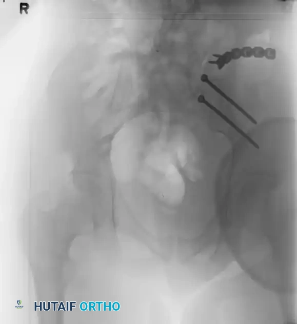
For high-pressure injection injuries, pre-operative planning is a race against time. Plain radiographs of the affected hand and forearm are mandatory to assess for subcutaneous emphysema (indicating the spread of the injectate) and to identify radio-opaque materials such as lead-based paints or metallic debris. However, imaging must never delay the transfer of the patient to the operating room. The surgical team must prepare for an extensile approach. The operating room setup must include loupe magnification, a sterile tourniquet, and copious amounts of normal saline for irrigation. Solvents must never be used to wash out the wound, as they cause profound secondary tissue toxicity.
Anesthesia considerations and patient positioning are critical components of the pre-operative phase. For all three injury types, the patient is positioned supine with the affected upper extremity extended on a radiolucent hand table. Regional anesthesia, specifically supraclavicular or axillary brachial plexus blocks, is highly advantageous. These blocks not only provide excellent intraoperative anesthesia and postoperative analgesia but also induce a profound sympathectomy, maximizing peripheral vasodilation and optimizing blood flow to the compromised digits. As previously emphasized, digital blocks are universally contraindicated in HPII and severe frostbite due to the risk of exacerbating local tissue pressure and ischemia.
Tourniquet management requires specific modifications based on the injury pathology. In high-pressure injection injuries, the limb must absolutely not be exsanguinated with an Esmarch bandage prior to tourniquet inflation. Wrapping the limb tightly can forcefully drive the toxic injectate further proximally into the hand and forearm. Instead, the limb is simply elevated for 3 to 5 minutes to allow for venous drainage by gravity before the pneumatic tourniquet is inflated to 250 mmHg. In cases of chemical burns requiring extensive tangential excision and split-thickness skin grafting, pre-operative planning must also include the preparation of a donor site, typically the ipsilateral or contralateral anterior thigh, ensuring the dermatome and meshing equipment are readily available on the sterile field.
Step-by-Step Surgical Approach and Fixation Technique
The operative techniques for these injuries diverge significantly based on the underlying pathology, requiring the orthopedic surgeon to seamlessly transition between radical decompression, meticulous debridement, and delayed reconstructive salvage.
High-Pressure Injection Injuries: Emergent Decompression
The surgical approach for HPII is defined by aggressive, extensile exposure. Under regional or general anesthesia, and following gravity elevation and tourniquet inflation, the surgeon must trace the entire path of the injected material.
1. Incision: Bold, extensile incisions are mandatory. Mid-axial incisions or volar Bruner (zigzag) incisions are utilized to open the entire involved digit. The incision must extend proximally as far as the injected material has tracked. If the thumb or small finger is involved, the surgeon must be prepared to extend the incision through the carpal tunnel and into the distal forearm to explore Parona's space.
2. Decompression and Exploration: The neurovascular bundles are identified and protected. The flexor tendon sheath is systematically inspected. If injectate is visible within the sheath, it must be opened to relieve pressure and allow for thorough washout. The A1 pulley is frequently released entirely. While efforts should be made to preserve the A2 and A4 pulleys to prevent tendon bowstringing, they must be partially resected or vented if heavily infiltrated with toxic material, prioritizing limb salvage over optimal biomechanics.
3. Radical Debridement: All foreign material, necrotic fat, and devitalized fascia must be meticulously evacuated using curettes and sharp dissection. Copious, low-pressure irrigation with normal saline is performed.
4. Wound Management: The incisions are never closed primarily. They are left entirely open to allow for continued drainage, secondary decompression, and serial washouts. The hand is dressed with a bulky, non-adherent dressing and immobilized in an intrinsic-plus splint.
Deep Frostbite: Delayed Amputation and Reconstruction
Surgical intervention for deep frostbite is typically delayed for 4 to 12 weeks until clear demarcation occurs.
1. Level Selection: The level of amputation is dictated by the line of demarcation. The surgeon must meticulously preserve all viable skin, even if it requires atypical flap designs.
2. Bone Resection and Fixation Technique: The necrotic bone is resected back to healthy, bleeding cortical bone. In cases where multiple digits are involved, the surgeon must prioritize the preservation of the thumb and at least one opposing digit for pinch function. If ray amputations are required, or if local bone stock is compromised requiring arthrodesis for stability (e.g., fusing a flail PIP joint to salvage a functional stump), rigid fixation is achieved using crossed 0.045-inch Kirschner wires or low-profile titanium mini-plates.
3. Soft Tissue Coverage: The bone ends must be adequately covered to prevent painful neuromas and stump breakdown. If local tissue is insufficient, V-Y advancement flaps, cross-finger flaps, or even free tissue transfer (e.g., anterolateral thigh flap) may be indicated to preserve functional length.
Chemical Burns: Tangential Excision and Grafting
For deep partial or full-thickness chemical burns, early excision is paramount.
1. Excision: Using a Weck or Goulian knife, the necrotic eschar is tangentially excised in thin layers. The excision continues sequentially until a bed of healthy, punctate bleeding tissue is encountered, indicating viable dermis or subcutaneous fat.
2. Hemostasis: Meticulous hemostasis is achieved using epinephrine-soaked sponges and pinpoint bipolar electrocautery.
3. Grafting: A split-thickness skin graft (typically 0.012 to 0.015 inches thick) is harvested from the thigh. For the hand, unmeshed sheet grafts are preferred on the dorsum and digits to optimize cosmetic and functional outcomes by reducing secondary contracture. The graft is secured with absorbable sutures or surgical staples and bolstered with a tie-over dressing or negative pressure wound therapy (NPWT) to prevent shear forces and hematoma formation.
Complications, Incidence Rates, and Salvage Management
The complication profile for environmental and industrial hand injuries is profound, often resulting in permanent disability despite optimal surgical management. The systemic and local toxicities associated with these injuries lead to high rates of tissue loss, chronic pain syndromes, and severe biomechanical deficits.
High-pressure injection injuries carry the most devastating prognosis. The overall amputation rate reported in the literature ranges from 16% to 49%. However, this incidence is highly dependent on the nature of the injected material. Oil-based paints and industrial solvents carry an amputation rate approaching 80%, due to their aggressive liquefactive necrosis and intense inflammatory response. Grease and hydraulic fluids, while less acutely toxic, induce chronic, severe granulomatous reactions that lead to profound stiffness, tendon adhesions, and eventual loss of function. If initial decompression fails or if the patient presents late (greater than 24 hours), salvage management invariably involves digital or ray amputation. In cases of massive palmar or forearm necrosis, free tissue transfer or even below-elbow amputation may be required as a life-saving measure to control systemic sepsis.
Frostbite complications are divided into early and late sequelae. Early complications include wet gangrene and secondary bacterial infections, which mandate urgent surgical debridement. Late complications are nearly universal in deep frostbite survivors. Cold intolerance and vasospastic disorders (Raynaud's phenomenon) occur in up to 80% of patients. Complex Regional Pain Syndrome (CRPS) is a frequent and debilitating complication, requiring aggressive multidisciplinary management with sympathetic blocks and neuro-modulating medications. In pediatric patients, frostbite frequently causes premature physeal closure, leading to severe angular deformities and brachydactyly as the child grows. Salvage management for frostbite sequelae includes late reconstructive procedures such as corrective osteotomies, joint arthrodesis for unstable interphalangeal joints, and intrinsic muscle release for ischemic contractures.
Chemical burns frequently result in severe functional impairments due to hypertrophic scarring and contracture formation. The dorsal skin of the hand is particularly susceptible to extensor tendon tethering and the development of severe metacarpophalangeal (MCP) joint extension contractures. Web space syndactyly is a common complication if the web spaces are not adequately separated and grafted during the acute phase. Salvage management for chemical burn contractures involves complex reconstructive surgery, including Z-plasties, release of contracture bands, and the application of full-thickness skin grafts or regional flaps to restore functional joint excursion.
| Complication | Associated Injury Type | Estimated Incidence Rate | Salvage Management & Reconstructive Strategy |
|---|---|---|---|
| Digital/Ray Amputation | High-Pressure Injection (Oil-based paint) | 60% - 80% | Revision amputation, ray resection, toe-to-hand transfer for thumb salvage. |
| Premature Physeal Closure | Frostbite (Pediatric) | 50% - 70% (in deep injuries) | Corrective osteotomies, distraction histogenesis, joint arthrodesis at skeletal maturity. |
| Severe Flexion Contracture | Chemical Burns (Volar) | 30% - 40% | Contracture release, Z-plasty, full-thickness skin grafting, cross-finger flaps. |
| Complex Regional Pain Syndrome | Frostbite / HPII | 15% - 25% | Stellate ganglion blocks, gabapentinoids, intensive desensitization therapy. |
| Tendon Adhesions / Stiffness | HPII (Grease/Hydraulic Fluid) | > 80% | Tenolysis (delayed 6-12 months), aggressive hand therapy, continuous passive motion. |
Phased Post-Operative Rehabilitation Protocols
The surgical intervention in environmental and industrial hand injuries represents only the first step in a protracted journey toward functional recovery. Post-operative rehabilitation must be meticulously phased, highly individualized, and initiated immediately to combat the profound stiffness and edema characteristic of these pathologies. The rehabilitation protocol is generally divided into three distinct phases: Acute/Inflammatory, Proliferative/Early Motion, and Remodeling/Strengthening.
Phase I: Acute/Inflammatory (Days 1-14)
The primary goals during the acute phase are the protection of surgical repairs, management of profound edema, and prevention of intrinsic contractures. Following surgical decompression for HPII or grafting for chemical burns, the hand is immobilized in a bulky, non-compressive dressing and a volar orthosis in the "safe" or intrinsic-plus position: the wrist extended to 30 degrees, the MCP joints flexed to 70-90 degrees, and the interphalangeal (IP) joints in full extension. This position maintains the collateral ligaments of the MCP joints in their elongated state, preventing catastrophic extension contractures. Strict, continuous elevation above the level of the heart is mandatory. For frostbite and open HPII wounds, daily sterile hydrotherapy (whirlpool) is utilized to gently mechanically debride exudate, facilitate dressing changes, and allow for early, gravity-eliminated active range of motion (ROM) of the uninvolved digits.
Phase II: Proliferative/Early Motion (Weeks 2-6)
As the acute inflammation subsides and grafts or wounds begin to stabilize, the focus shifts to preventing tendon adhesions and restoring joint kinematics. This is particularly critical following HPII, where the flexor tendon sheath has been violated and heavily inflamed. Gentle active and active-assisted ROM exercises are initiated. Specific tendon gliding exercises (straight fist, hook fist, composite fist) are meticulously performed to ensure differential glide between the FDS and FDP tendons. Dynamic splinting may be introduced during this phase to provide a low-load, prolonged stretch to developing contractures, particularly in chemical burn patients prone to dorsal skin tightness. In frostbite patients awaiting delayed amputation, this phase focuses on maximizing the ROM of the proximal joints (wrist, elbow, shoulder) to prevent secondary disuse syndromes.
Phase III: Remodeling/Strengthening (Weeks 6 and Beyond)
The final phase focuses on tissue remodeling, scar management, and the return of functional strength. For chemical burn patients, aggressive scar management is instituted, utilizing custom-fitted silicone gel sheeting, elastomer putty, and customized pressure garments to mitigate hypertrophic scarring. Deep friction massage is employed to mobilize scars and prevent tethering of the underlying extensor or flexor tendons. Strengthening exercises are progressively introduced, utilizing therapeutic putty, hand dynamometers, and work-simulation tasks. For patients who have undergone amputations due to frostbite or HPII, this phase involves rigorous stump desensitization protocols (using varied textures and vibration) and training with adaptive equipment or custom prosthetics.
Long-term occupational therapy goals are centered on functional independence and return-to-work assessments. Given the industrial nature of chemical burns and HPII, functional capacity evaluations (FCE) are often required to determine if the patient can safely return to their previous occupation or if vocational retraining is necessary. The orthopedic surgeon must work in close concert with the certified hand therapist (CHT) to continuously monitor progress and identify indications for secondary reconstructive procedures, such as tenolysis or contracture release.
Summary of Landmark Literature and Clinical Guidelines
The operative management of frostbite, chemical burns, and high-pressure injection injuries is heavily guided by a robust body of landmark literature and established clinical guidelines. Mastery of these foundational texts is essential for the academic orthopedic surgeon and serves as the basis for board certification examinations.
In the realm of frostbite, the seminal work by Heggers and Robson in the late 20th century revolutionized the medical management of cold injuries. Their foundational research identified the presence of devastatingly high levels of thromboxane A2 and prostaglandin F2-alpha within the fluid of frostbite blisters. This discovery directly led to the establishment of the modern "Aloe Vera and Ibuprofen" protocol. By demonstrating that topical aloe vera acts as a specific inhibitor of the arachidonic acid cascade, and that systemic ibuprofen blocks the cyclooxygenase pathway (thereby halting thromboxane A2 production), Heggers and Robson provided the first evidence-based medical therapy to combat the progressive microvascular thrombosis that leads to tissue necrosis. This literature firmly established the mandate for aspirating clear blisters (to remove the toxic fluid) while leaving hemorrhagic blisters intact to protect the deep dermal plexus.
The literature surrounding high-pressure injection injuries is anchored by the foundational papers of Stark, Schoo, and later, Hogan and Lee. Schoo's retrospective analyses definitively established the critical prognostic variables in HPII: the type of injectate
