Comprehensive Introduction and Patho-Epidemiology
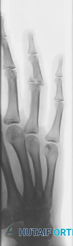
The proximal interphalangeal (PIP) joint is universally recognized as the critical functional hinge of the human digit, responsible for the vast majority of finger flexion arc and overall grip strength. Fracture-dislocations of the PIP joint present a formidable, high-stakes challenge to the orthopedic hand surgeon. These complex injuries typically involve a high-energy axial load combined with forced hyperextension, resulting in a shear-impaction force against the volar lip of the middle phalanx base. This pathomechanical cascade culminates in a volar lip fracture and subsequent dorsal subluxation or frank dislocation of the remaining middle phalanx, severely disrupting the delicate kinematic chain of the digit.
Epidemiologically, these injuries are disproportionately prevalent among young, active individuals, particularly athletes engaged in ball-handling sports such as basketball, volleyball, and baseball, as well as manual laborers exposed to occupational trauma. The incidence of PIP joint fracture-dislocations is estimated at approximately 9 to 12 per 100,000 person-years, though this is likely an underestimation due to frequent misdiagnosis as simple "jammed fingers" in primary care settings. The economic and functional burden of these injuries is substantial, as inadequate initial management almost invariably leads to chronic stiffness, debilitating pain, and early-onset post-traumatic osteoarthritis, severely limiting the patient's vocational and recreational capacities.
Understanding the precise pathophysiology of this injury is paramount for surgical decision-making. The proper collateral ligaments and the robust fibrocartilaginous volar plate insert directly onto the volar base of the middle phalanx. When a fracture involves 50% or more of the articular surface, the collateral ligaments remain anatomically attached to the fractured volar fragment. Consequently, the remaining dorsal portion of the middle phalanx is entirely stripped of its primary static ligamentous stabilizers. This loss of volar tethering leads to profound, unremitting dorsal instability driven by the unopposed pull of the central slip of the extensor mechanism.
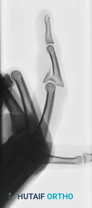
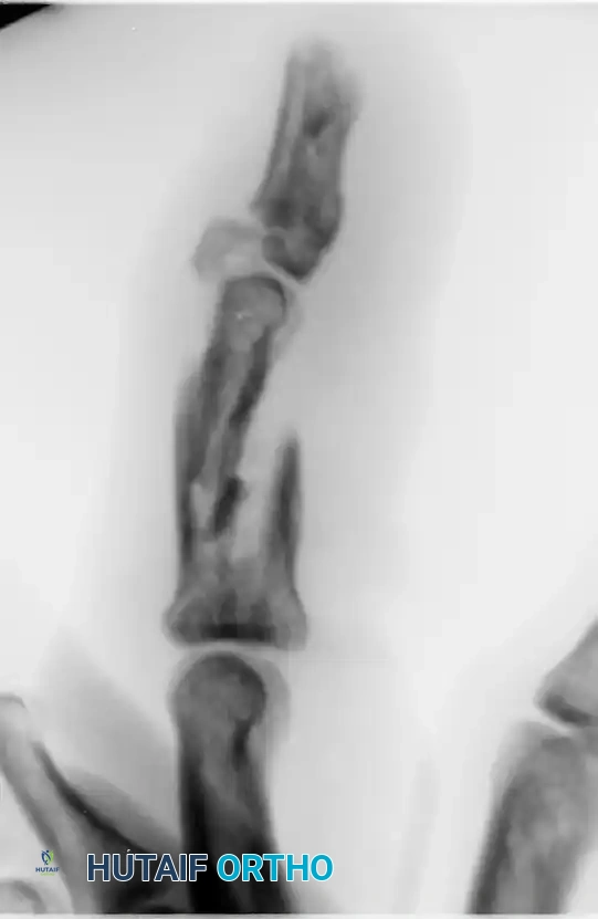
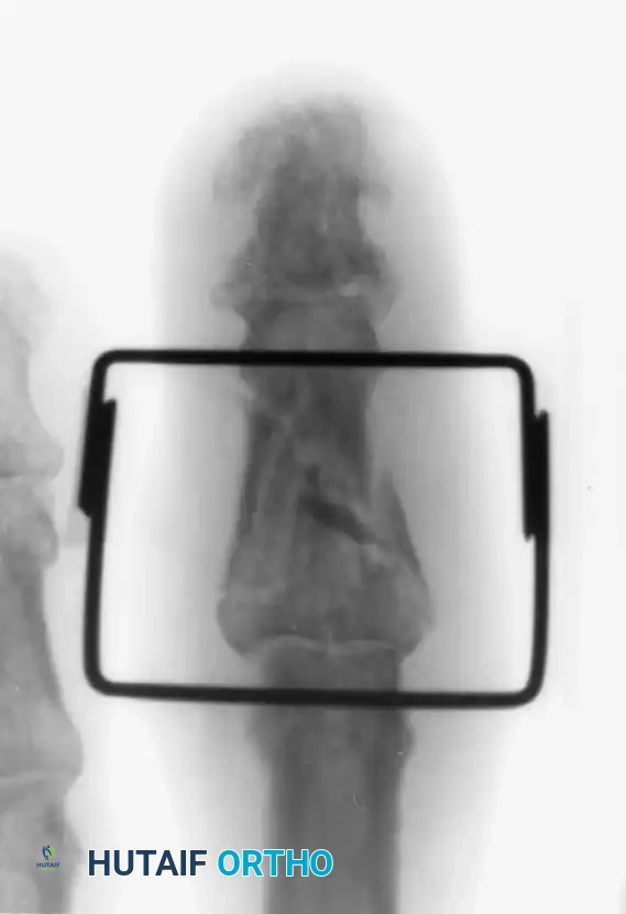
In these specific scenarios, closed reduction is invariably unstable. The joint will reliably subluxate dorsally as the finger is extended, leading to the pathognomonic "V" sign on lateral radiographs. Surgical intervention is therefore absolutely mandated to restore articular congruity, re-establish the ligamentous tethers, and normalize joint kinematics. The distinction of a "noncomminuted" fracture pattern is critical; it implies that the volar fragment is a single, structurally sound osseous piece capable of holding internal fixation, thereby distinguishing the surgical approach from the reconstructive salvage procedures required for highly comminuted, pilon-type injuries.
Detailed Surgical Anatomy and Biomechanics
The osseous architecture of the PIP joint is a highly congruent, bicondylar hinge joint that inherently dictates its primary plane of motion while relying heavily on soft tissue restraints for multiplanar stability. The head of the proximal phalanx features two distinct condyles separated by an intercondylar notch, which perfectly articulate with the biconcave base of the middle phalanx. This middle phalangeal base is bisected by a median ridge that tracks precisely within the proximal intercondylar notch, providing intrinsic resistance to radioulnar translation and rotatory forces. The preservation of this articular geometry is the primary objective of any surgical intervention, as even minor step-offs or incongruities exponentially increase contact stresses, rapidly accelerating cartilage degradation.
The static stability of the PIP joint is conceptualized as a three-dimensional "box" composed of the collateral ligaments laterally, the volar plate volarly, and the extensor mechanism dorsally. The proper collateral ligaments (PCL) originate from the condylar recesses of the proximal phalanx and insert onto the volar-lateral aspect of the middle phalanx base. The accessory collateral ligaments (ACL) fan out more volarly to insert onto the margins of the volar plate. The volar plate itself is a thick, dense ligamentous structure that prevents hyperextension; its distal insertion spans the entire volar width of the middle phalanx base. In a noncomminuted fracture-dislocation involving >50% of the articular surface, this entire volar-lateral footprint is avulsed, effectively destroying the volar and lateral walls of the stabilizing "box."
Dynamically, the PIP joint is influenced by the powerful, opposing forces of the extrinsic and intrinsic musculotendinous units. Dorsally, the central slip of the extensor digitorum communis inserts onto the dorsal base of the middle phalanx. Volarly, the flexor digitorum superficialis (FDS) bifurcates at the level of the proximal phalanx to insert onto the volar-lateral shafts of the middle phalanx, while the flexor digitorum profundus (FDP) glides through Camper's chiasm to reach the distal phalanx. When the volar restraints are fractured and detached, the unopposed extensor tone from the central slip continuously draws the middle phalanx dorsally, perpetuating the subluxation and rendering closed management futile.
Furthermore, the delicate retinacular system enveloping the PIP joint must be intimately understood by the operating surgeon to prevent iatrogenic deformity. The transverse retinacular ligament (TRL) originates from the volar flexor tendon sheath and inserts onto the lateral bands of the extensor mechanism, preventing their dorsal migration during active extension. During the midlateral surgical approach, the TRL must be purposefully divided and meticulously repaired. Failure to reconstruct the TRL allows the lateral bands to subluxate dorsally, altering the moment arm of the intrinsic muscles and precipitating a severe, secondary swan-neck deformity that is exceptionally difficult to salvage.
Exhaustive Indications and Contraindications
The decision to proceed with open reduction and internal fixation (ORIF) of a PIP joint fracture-dislocation requires a nuanced appreciation of both the mechanical instability of the injury and the biological constraints of the patient. The primary, absolute indication for surgical intervention is the inability to maintain concentric joint reduction in a functional position (typically defined as less than 30 degrees of flexion). If the joint demonstrates dorsal subluxation or an asymmetric dorsal opening (the "V" sign) on true lateral radiographs after attempted closed reduction and extension-block splinting, operative stabilization is strictly required to prevent rapid joint destruction.
Specific to the technique of direct ORIF with Kirschner wires or mini-fragment screws, the fracture pattern must meet stringent morphological criteria. The volar fragment must be noncomminuted—a single, solid, contiguous piece of bone. Furthermore, this fragment must be of sufficient size, universally accepted as comprising at least 50% of the volar articular surface. Fragments smaller than this threshold, or those exhibiting significant comminution, lack the structural integrity to withstand the compressive forces of internal fixation and will catastrophically fragment upon drill or wire insertion, necessitating alternative techniques such as volar plate arthroplasty or hemi-hamate autografting.
Contraindications to ORIF are equally critical to recognize to avoid devastating surgical failures. Absolute contraindications include active, uncontrolled local or systemic infection, severe medical comorbidities precluding safe anesthesia, and highly comminuted "pilon-type" fractures where the articular surface is shattered into multiple non-reconstructable fragments. Relative contraindications encompass severe osteopenia or osteoporosis, which may compromise hardware purchase, and profound patient non-compliance or severe cognitive impairment, as the postoperative rehabilitation protocol requires meticulous adherence to complex splinting and motion regimens.
Below is a comprehensive table delineating the indications and contraindications for ORIF of noncomminuted PIP joint fracture-dislocations:
| Category | Specific Criteria | Clinical Rationale |
|---|---|---|
| Absolute Indications | Dorsal instability in >30° flexion | Indicates complete loss of volar tether; joint will subluxate and rapidly degenerate if left unreduced. |
| Absolute Indications | Presence of the radiographic "V" sign | Pathognomonic for lack of concentric reduction; articular hinge is incongruent. |
| Morphological Indications | Fragment size >50% of articular surface | Necessary to ensure the proper collateral ligaments remain attached to the fragment, driving the instability. |
| Morphological Indications | Noncomminuted volar fragment | Fragment must possess sufficient structural integrity to hold a K-wire or screw without iatrogenic shattering. |
| Absolute Contraindications | Severe, multi-part comminution (Pilon) | Hardware cannot secure multiple small fragments; requires dynamic external fixation or arthroplasty. |
| Absolute Contraindications | Active local soft tissue infection | Introduction of hardware into an infected field guarantees osteomyelitis and joint destruction. |
| Relative Contraindications | Presentation delayed >4-6 weeks | Chronic contracture and bone resorption make direct reduction exceptionally difficult; may require osteotomy/grafting. |
| Relative Contraindications | Severe patient non-compliance | Postoperative stiffness is inevitable without rigorous, self-directed hand therapy and splint management. |
Pre-Operative Planning, Templating, and Patient Positioning
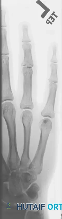
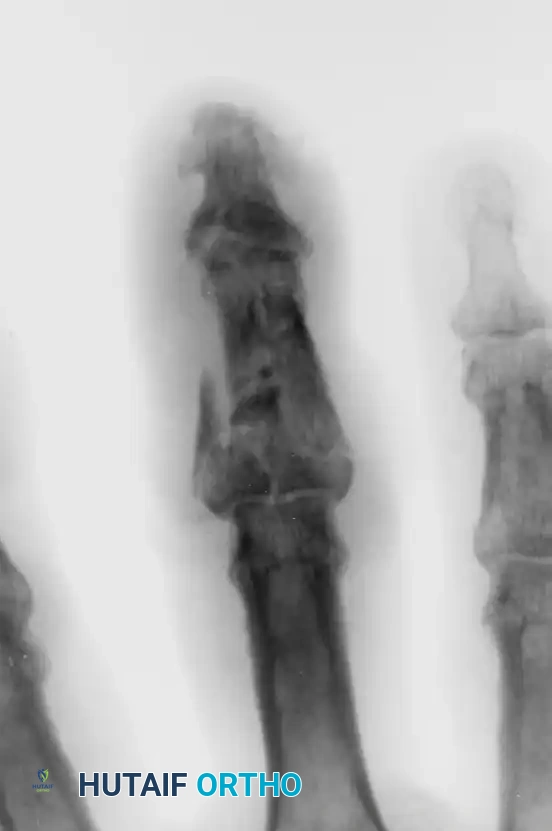
Meticulous pre-operative planning is the cornerstone of a successful surgical outcome in these unforgiving injuries. The foundational step is the acquisition of high-quality, orthogonal radiographs, specifically a true posteroanterior (PA), true lateral, and dedicated oblique views of the affected digit. The true lateral radiograph is the single most critical imaging modality; it must be perfectly superimposed without rotational artifact to accurately quantify the percentage of articular surface involved, the depth of the central depression, and the precise degree of dorsal subluxation.
In cases where the degree of comminution is equivocal on plain radiography, or if the fracture morphology appears unusually complex, a dedicated non-contrast computed tomography (CT) scan with fine (0.5 mm) axial, coronal, and sagittal reconstructions is highly recommended. 3D surface rendering can provide the surgeon with a definitive roadmap of the fracture lines, allowing for precise pre-operative templating. The surgeon must anticipate the exact trajectory of the internal fixation and have a comprehensive array of implants available, including 0.028-inch, 0.035-inch, and 0.045-inch Kirschner wires, as well as a 1.0 mm or 1.2 mm mini-fragment screw set, should the fragment prove large enough to accommodate interfragmentary compression.
Patient positioning and anesthetic selection are tailored to optimize surgical exposure and intraoperative assessment. The patient is positioned supine with the affected upper extremity extended on a radiolucent hand table. A mini C-arm fluoroscopy unit is positioned perpendicular to the table, allowing for seamless transition between PA and lateral imaging without requiring the surgeon to grossly manipulate the unstable digit. While regional anesthesia (such as an axillary or supraclavicular brachial plexus block) combined with an upper arm pneumatic tourniquet is the traditional standard, the Wide Awake Local Anesthesia No Tourniquet (WALANT) technique is increasingly favored by modern hand surgeons.
The WALANT approach utilizes a localized injection of lidocaine with epinephrine, providing profound anesthesia and excellent hemostasis without the need for a proximal tourniquet. The paramount advantage of WALANT in PIP joint reconstruction is the ability to assess the active stability of the joint intraoperatively. Once the internal fixation is placed, the fully awake patient can be instructed to actively flex and extend the digit under direct visualization and fluoroscopy. This real-time, dynamic assessment confirms that the hardware is not impinging on the joint space, that the articular reduction is perfectly concentric throughout the entire arc of motion, and that the fixation construct is robust enough to withstand the physiological forces of early rehabilitation.
Step-by-Step Surgical Approach and Fixation Technique
The Midlateral Approach
The midlateral approach is the gold standard for accessing the PIP joint in the setting of fracture-dislocations, providing unparalleled exposure to the collateral ligaments, the volar plate, and the articular surface while safely avoiding the critical volar neurovascular bundles. The incision is meticulously planned along the midaxial line of the digit. To identify this line, the finger is fully flexed, and a line is drawn connecting the apices of the dorsal flexion creases. The incision extends longitudinally from the mid-portion of the proximal phalanx to the mid-portion of the middle phalanx, remaining strictly on this axis.
Deep dissection proceeds with extreme caution. The neurovascular bundle lies immediately volar to the midaxial line. By maintaining the dissection plane strictly midlateral and dissecting dorsal to Cleland's cutaneous ligaments, the neurovascular bundle is safely protected and naturally falls volarly with the elevated skin flap. The surgeon then encounters the transverse retinacular ligament (TRL), which runs vertically from the volar fascia to the lateral band of the extensor mechanism. The TRL is carefully divided parallel to the lateral band, leaving a sufficient cuff of tissue for later repair. The lateral band is then retracted dorsally with a blunt hook, while the neurovascular bundle is protected volarly, fully exposing the collateral ligament complex and the joint capsule.
To gain access to the intra-articular fracture, the accessory collateral ligament is sharply detached from its distal insertion on the volar-lateral aspect of the middle phalanx. This is a critical maneuver; it allows the surgeon to reflect the ligament dorsally, acting like a book page, thereby exposing the underlying fibrocartilaginous volar plate and the fracture hematoma. The proper collateral ligament is generally left intact if it remains attached to the avulsed volar fragment, preserving its vital stabilizing function once the fragment is anatomically reduced.
Fragment Mobilization and Joint Debridement
Upon entering the joint space, the surgeon's immediate priority is the identification and careful mobilization of the avulsed volar osseous fragment. This fragment is often retracted proximally by the pull of the intact volar plate. The absolute most crucial step in this phase is the strict preservation of all soft tissue and periosteal attachments to the volar fragment. The vascular supply to this small piece of bone is derived entirely from the volar plate insertion. Any aggressive stripping or overzealous skeletonization will completely devascularize the fragment, inevitably leading to avascular necrosis, hardware failure, and catastrophic collapse of the reconstructed articular surface.
Once the fragment is identified, the joint space must be meticulously debrided. A copious amount of sterile saline irrigation is utilized to flush out the organized fracture hematoma and any microscopic osteochondral debris that could interpose between the fracture fragments and block an anatomical reduction. A small dental pick or a fine curette is used to gently clear the fracture bed on the remaining dorsal aspect of the middle phalanx. The surgeon must visually inspect the deep recesses of the joint to ensure no loose bodies remain trapped within the intercondylar notch or the volar recesses.
Following debridement, the surgeon assesses the mobility of the fragment. Using a fine freer elevator or a specialized reduction hook, the fragment is gently manipulated to assess its fit into the anatomical bed. The surgeon must visualize the articular surface directly, ensuring that the cartilage cap of the fragment aligns perfectly with the cartilage of the intact dorsal middle phalanx. Any step-off greater than 1 millimeter is unacceptable and will lead to rapid post-traumatic arthritis.
Anatomical Reduction and Internal Fixation
With the joint debrided and the fragment mobilized, the surgeon proceeds to definitive anatomical reduction. Using a dental pick or a small freer elevator, the volar fragment is gently leveraged and manipulated into its anatomical bed. The reduction must be held perfectly flush under direct visualization. Once congruity is confirmed visually, it is temporarily verified under multi-planar fluoroscopy. The surgeon must ensure that the reduction eliminates the radiographic "V" sign and restores the smooth, continuous biconcave contour of the middle phalangeal base.
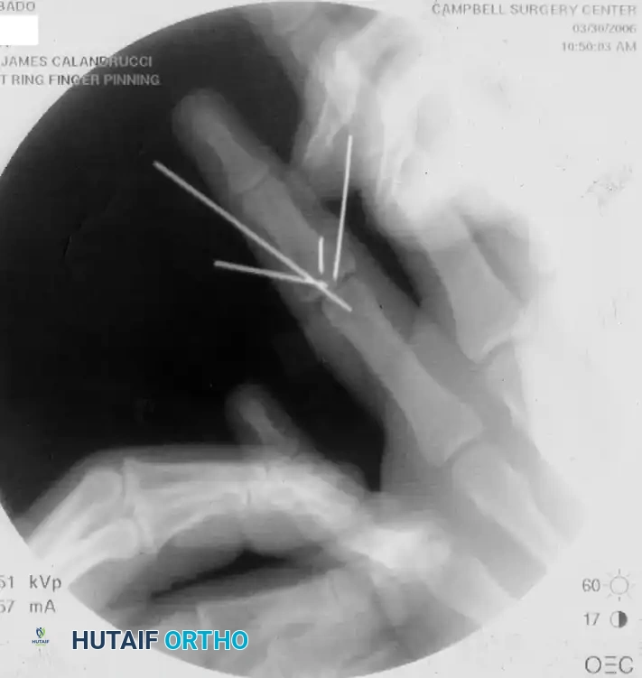
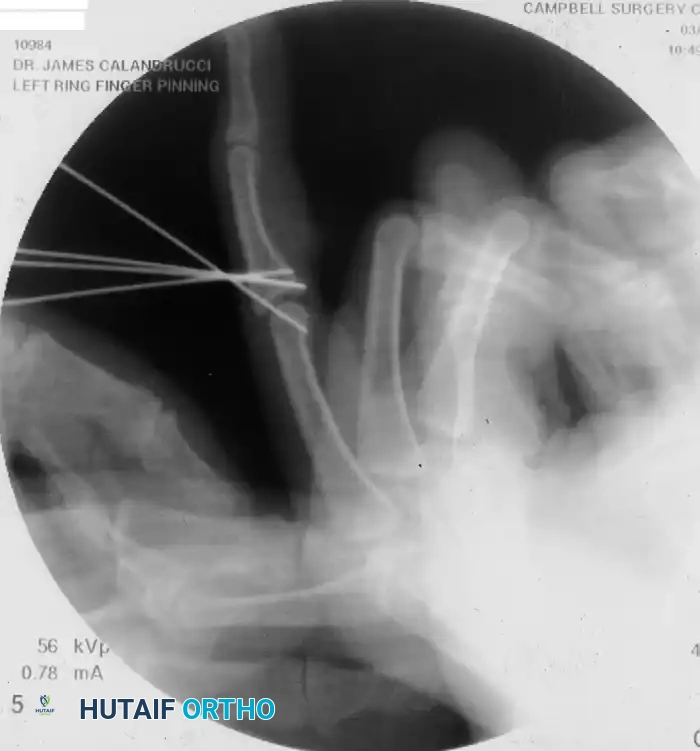
Primary fixation is achieved using a small-diameter Kirschner wire, typically 0.028-inch or 0.035-inch, depending on the absolute size of the fragment. The trajectory of this wire is of paramount importance. The wire must be inserted in a strict dorsal direction, starting from the volar-distal aspect of the fragment and aiming toward the dorsal-proximal cortex of the middle phalanx. This trajectory ensures that the wire captures the maximum volume of the fragment and directs the compression vector precisely across the fracture plane.
Crucially, the wire must be subject to "subchondral burial." The surgeon draws the wire dorsally until its volar, intra-articular end lies just beneath the subchondral bone of the articular surface of the fragment. It is absolutely imperative that the wire does not protrude even a fraction of a millimeter into the joint space. A protruding wire will mechanically gouge the opposing articular cartilage of the proximal phalanx during flexion, causing rapid, irreversible chondrolysis. Once the subchondral position is fluoroscopically confirmed, the dorsal end of the wire is cut flush with the dorsal cortical surface of the middle phalanx to prevent mechanical irritation or rupture of the overlying extensor mechanism.
Following fragment fixation, the reconstructed PIP joint is reduced and placed into a functional position, typically 20 to 30 degrees of flexion. To protect the fragile osteosynthesis from the massive shear forces generated by the flexor and extensor tendons during the early healing phase, the joint is stabilized with a second, obliquely inserted transarticular K-wire. This wire is driven from the dorsal aspect of the middle phalanx, across the joint space, and into the proximal phalanx. Finally, meticulous closure is performed. The accessory collateral ligament is sutured back to its insertion using 4-0 non-absorbable braided suture. The transverse retinacular ligament is precisely repaired to prevent dorsal subluxation of the lateral band, and the skin is closed with non-absorbable monofilament sutures.
⚠️ Surgical Warning: Volar Approach Pitfalls
While a volar approach (using a Brunner zigzag incision) can be utilized for direct visualization of the volar plate, it carries a substantially higher risk of postoperative stiffness and flexor tendon adhesions. Extensive volar dissection disrupts the delicate vincular blood supply and creates a massive bed of scar tissue directly adjacent to the gliding FDS and FDP tendons.
Complications, Incidence Rates, and Salvage Management
Despite flawless surgical execution, the PIP joint remains notoriously unforgiving, and complications following ORIF of fracture-dislocations are frequent and challenging to manage. The most ubiquitous complication is postoperative joint stiffness, which affects virtually all patients to some degree. Stiffness arises from a combination of intra-articular adhesions, capsular contracture, and extra-articular flexor tendon tethering within the surgical scar bed. Prevention is heavily reliant on minimizing surgical trauma, achieving rigid fixation that allows for early motion, and strict adherence to postoperative edema control.
Loss of reduction and hardware failure represent catastrophic mechanical complications that typically occur within the first three weeks postoperatively. This can manifest as K-wire migration, cut-out through osteopenic bone, or fragmentation of the volar piece due to unrecognized intraoperative comminution. If loss of reduction occurs, the joint will rapidly subluxate dorsally. Immediate re-operation is mandated; however, direct re-fixation is rarely possible due to bone damage. Salvage in this acute setting often requires conversion to a dynamic external fixator or a volar plate arthroplasty to restore stability.
Avascular necrosis (AVN) of the volar fragment and subsequent post-traumatic arthritis are devastating late complications. AVN is almost entirely iatrogenic, resulting from aggressive soft tissue stripping of the volar plate insertion during exposure. As the fragment dies and collapses, the articular surface becomes highly incongruent, leading to rapid, painful destruction of the joint cartilage. When severe arthritis develops, salvage options are limited and represent significant functional compromises. For the index and middle fingers, where lateral pinch stability is paramount, PIP joint arthrodesis in a functional position (40-50 degrees of flexion) is the procedure of choice. For the ring and small fingers, where grip wrap is more critical, surface replacement arthroplasty (silicone or pyrocarbon) may be considered in low-demand patients.
Below is a detailed table outlining the primary complications, their estimated incidence rates, and established salvage strategies:
| Complication | Estimated Incidence | Etiology / Risk Factors | Salvage Management Strategy |
|---|---|---|---|
| Joint Stiffness (Flexion Contracture) | 60% - 80% | Capsular scarring, prolonged immobilization, flexor tendon adhesions. | Aggressive hand therapy, dynamic extension splinting; late surgical tenolysis or dorsal capsulectomy. |
| Loss of Reduction / Hardware Failure | 5% - 15% | Unrecognized comminution, osteopenia, premature removal of transarticular pin. | Revision surgery; conversion to dynamic external fixation, volar plate arthroplasty, or hemi-hamate graft. |
| Avascular Necrosis of Fragment | 5% - 10% | Iatrogenic stripping of volar plate attachments; severe initial crush injury. | Observation if asymptomatic; if symptomatic collapse occurs, requires arthrodesis or arthroplasty. |
| Post-Traumatic Osteoarthritis | 20% - 40% | Articular step-off >1mm, chronic subluxation, cartilage necrosis from initial impact. | Conservative management (NSAIDs, splinting); definitive salvage via PIP joint arthrodesis or joint replacement. |
| Pin Tract Infection | 2% - 5% | Poor local hygiene around exposed transarticular K-wires. | Oral antibiotics, local wound care; early pin removal if infection tracks deeply into the bone or joint. |
Alternative and Adjunctive Techniques
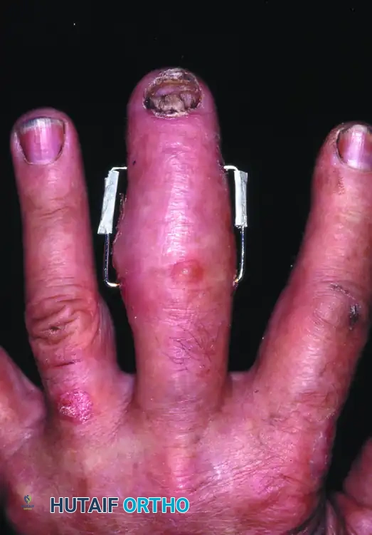
While the primary focus is on noncomminuted fractures, the orthopedic surgeon must be prepared to pivot intraoperatively if severe comminution is discovered. In cases of severe fragmentation where internal fixation is impossible, dynamic external fixation (such as the Suzuki frame) is the treatment of choice. This utilizes the principle of ligamentotaxis to maintain joint space and alignment while allowing early active motion.
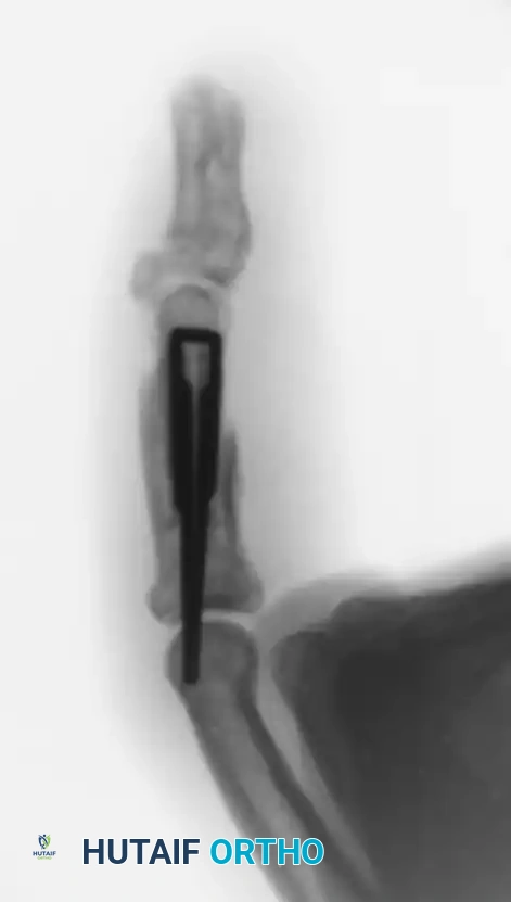
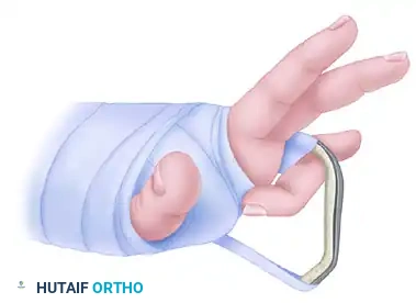
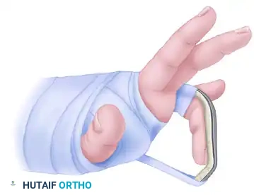
For fractures that are stable in flexion but subluxate in extension, or as a step-down therapy following pin removal, extension-block splinting is highly effective.
Phased Post-Operative Rehabilitation Protocols
The postoperative management of PIP joint ORIF is a highly complex, tightrope-walking endeavor that requires a delicate, continuous balance between protecting the fragile osteosynthesis and preventing debilitating, permanent joint stiffness. The rehabilitation protocol must be phased, meticulously supervised by a certified hand therapist (CHT), and tailored to the patient's individual healing response and compliance level.
Phase I: Maximum Protection (Weeks 0-3)
Immediately following surgery, the digit is placed in a bulky, compressive dressing, and a custom-fabricated dorsal blocking splint is applied. The PIP joint is immobilized in the functional position dictated by the transarticular K-wire (typically 20 to 30 degrees of flexion). The metacarpophalangeal (MCP) joint is splinted in 70 degrees of flexion to maintain the collateral ligaments at their maximum length, preventing MCP extension contractures.
During this initial three-week phase, the absolute priority is the protection of the hardware and the control of digital edema. Swelling is the primary catalyst for secondary stiffness, as protein-rich exudate rapidly organizes into dense fibrotic adhesions. Strict elevation above the level of the heart and the use of compressive wrapping (such as Coban) are mandatory. While the PIP joint is rigidly
