Comprehensive Introduction and Patho-Epidemiology
The management of open fractures and dislocations in the hand presents a profoundly complex surgical challenge that demands a delicate, calculated balance between aggressive infection control and the meticulous preservation of functional anatomy. Unlike major long bone trauma, where substantial soft tissue envelopes can often mask or accommodate minor skeletal discrepancies, the hand possesses a highly compact, unforgiving architecture. Within this specialized distal appendage, bones, gliding tendons, neurovascular bundles, and specialized soft tissues exist in intimate proximity, leaving virtually no dead space. Consequently, massive trauma to the hand—whether from crush injuries, high-velocity ballistics, or industrial avulsions—requires extensive, multi-staged reconstructive efforts to restore not only bony integrity but also the intricate functional kinematics essential for prehension and tactile interaction.
The patho-epidemiology of open hand trauma is heavily skewed toward young, working-age populations, predominantly males, engaging in industrial, agricultural, or high-risk recreational activities. The mechanisms of injury dictate the pathophysiology of the wound bed. Crush injuries, commonly seen in industrial press accidents, impart massive kinetic energy over a broad area, leading to widespread microvascular thrombosis, severe soft tissue contusion, and a high risk of subsequent compartment syndrome or delayed tissue necrosis. Conversely, agricultural injuries frequently involve power take-off (PTO) shafts or combine harvesters, resulting in severe avulsion forces combined with gross contamination by soil-dwelling pathogens, including Clostridium species and mixed Gram-negative bacilli. High-velocity ballistic trauma introduces a unique patho-anatomic profile characterized by temporary cavitation, thermal injury, and severe intercalary bone loss, necessitating highly specialized approaches to both immediate stabilization and delayed reconstruction.
The physiological response of the hand to severe open trauma is characterized by rapid, profound edema. Because the dorsal skin is thin, highly compliant, and loosely attached to the underlying extensor paratenon, edema fluid preferentially accumulates dorsally. This swelling forces the metacarpophalangeal (MCP) joints into extension and the interphalangeal (IP) joints into flexion—the classic "intrinsic-minus" or claw posture. If allowed to persist, the collateral ligaments of the MCP joints rapidly foreshorten and fibrose, leading to devastating, irreversible extension contractures. Furthermore, the disruption of the delicate synovial sheaths and paratenon exposes the relatively avascular tendons to desiccation and surrounding hematoma, setting the stage for dense, restrictive adhesions that can obliterate digital motion even if perfect skeletal union is achieved.
The primary objectives in treating these devastating injuries follow a strict hierarchy: the immediate prevention of sepsis through radical débridement, the restoration of anatomic skeletal stability to permit early motion, the provision of durable, gliding soft tissue coverage, and the facilitation of aggressive, early rehabilitation. The traditional Gustilo-Anderson classification, while universally applied to long bone open fractures, often falls short in accurately prognosticating outcomes in the hand, where a tiny puncture wound communicating with a flexor tendon sheath (e.g., a human bite) can be far more functionally devastating than a large, clean dorsal laceration overlying a metacarpal fracture. Therefore, a comprehensive understanding of the unique patho-anatomy and a customized, tissue-specific approach are paramount for the operating surgeon.
Detailed Surgical Anatomy and Biomechanics
A profound mastery of the surgical anatomy and biomechanics of the hand is the absolute prerequisite for managing complex open fractures and dislocations. The skeletal framework of the hand is organized into three distinct integrated arches: the rigid proximal transverse arch formed by the distal carpal row, the mobile distal transverse arch formed by the metacarpal heads, and the longitudinal arches formed by each individual digital ray. Disruption of these arches by comminuted fractures or fracture-dislocations inevitably results in a loss of cupping ability, diminished grip strength, and severe functional impairment. The second and third carpometacarpal (CMC) joints form the rigid central pillar of the hand, providing a stable post for pinch and grasp, whereas the highly mobile fourth and fifth CMC joints allow the ulnar border of the hand to cup around objects. Failure to anatomically restore the central pillar or respect the mobility of the ulnar rays during fixation will catastrophically alter hand biomechanics.
The soft tissue envelope of the hand exhibits extreme regional variation that directly impacts surgical decision-making. The palmar skin is thick, glabrous, and firmly tethered to the underlying palmar aponeurosis by robust vertical fascial septa, providing a stable, non-slip surface for gripping. However, this lack of compliance means that palmar wounds cannot be closed under tension without risking severe marginal necrosis. In contrast, the dorsal skin is thin, pliable, and mobile, allowing for the extreme flexion required to make a full fist. Beneath the dorsal skin lies a delicate layer of areolar tissue and the paratenon of the extensor tendons. If the dorsal skin and paratenon are avulsed or excised during debridement, the underlying extensor tendons are left completely bare. Tendons rely on a delicate intrinsic blood supply via the vincula and diffusion from surrounding synovial fluid; without paratenon or sheath coverage, they rapidly desiccate, necrose, and rupture.
The neurovascular anatomy of the digits is exquisitely vulnerable in open trauma. The proper palmar digital arteries and nerves travel volar to the mid-axial line, enveloped within a protective fascial canal bounded by Cleland’s ligaments dorsally and Grayson’s ligaments volarly. Cleland’s ligaments are robust, true fascial structures that remain taut during flexion and extension, preventing the neurovascular bundle from bowstringing. When extending traumatic wounds for adequate debridement, the surgeon must meticulously identify and protect these bundles. Furthermore, the venous drainage of the digits is entirely dorsal and highly superficial. Massive dorsal crush injuries or overzealous dorsal debridement can easily obliterate this venous network, leading to profound digital congestion, intractable edema, and eventual venous gangrene, even in the presence of intact arterial inflow.
Biomechanically, the digits operate through an exquisitely balanced tensioning system between the extrinsic flexors, extrinsic extensors, and the intrinsic musculature (lumbricals and interossei). The maintenance of absolute skeletal length is critical to preserving this balance. Even a few millimeters of metacarpal shortening can significantly alter the resting tension of the intrinsic muscles, leading to an intrinsic-plus deformity or a clinically significant lag in active extension. Furthermore, the phalanges are subjected to massive deforming forces. For example, a fracture of the proximal phalanx typically assumes an apex-volar angulation due to the unopposed pull of the intrinsic muscles inserting on the proximal fragment and the central slip pulling the distal fragment dorsally. Recognizing these predictable biomechanical deforming forces dictates the required strength and placement of internal or external fixation constructs.
Exhaustive Indications and Contraindications
The decision-making algorithm for the management of open hand fractures and dislocations is dictated by the degree of soft tissue compromise, the level of contamination, the geometry of the fracture, and the systemic status of the patient. There is no universally applicable fixation method; rather, the surgeon must dynamically adapt their strategy to the specific demands of the injury zone.
Indications for immediate surgical intervention are absolute in the setting of vascular compromise, compartment syndrome, or massive gross contamination (e.g., farmyard slurry, high-velocity ballistics). In these scenarios, the primary goal is life and limb salvage through radical debridement and provisional skeletal stabilization, typically utilizing percutaneous Kirschner wires (K-wires) or low-profile external fixators. Definitive internal fixation with plates and screws is strongly indicated for displaced, unstable, or intra-articular fractures where early mobilization is mandatory to prevent joint stiffness, provided the soft tissue envelope is pristine and primary closure is achievable. Rigid internal fixation is the gold standard for restoring the articular congruity of open fracture-dislocations of the PIP joint, as even minor step-offs will lead to rapid post-traumatic arthrosis.
Contraindications to primary internal fixation with plates and screws include grossly contaminated wounds, injuries presenting with a delay of greater than 24 hours, and scenarios involving massive soft tissue loss where the hardware would be left exposed. Placing bulky hardware in a compromised wound bed is a major technical error that virtually guarantees deep space infection, osteomyelitis, and subsequent hardware failure. Similarly, primary closure is absolutely contraindicated in bite wounds (human or animal), agricultural injuries, and severe crush injuries where the zone of injury is likely to demarcate and expand over the ensuing 48 to 72 hours. In these cases, the wound must be managed open, utilizing negative pressure wound therapy (NPWT) or non-adherent dressings, with planned second-look debridement.
The use of external fixation is highly indicated in the management of highly comminuted fractures with significant intercalary bone loss, where maintaining length and alignment is paramount while allowing unrestricted access to complex soft tissue wounds. However, external fixation is contraindicated in uncooperative patients or those with severe osteopenia where pin purchase is inadequate. Furthermore, spanning external fixators across uninjured joints should be minimized in duration, as prolonged joint immobilization in the hand rapidly leads to irreversible capsular contracture and devastating stiffness.
| Surgical Strategy | Primary Indications | Absolute Contraindications | Relative Contraindications |
|---|---|---|---|
| Percutaneous K-Wires | Minimally comminuted fractures; tenuous soft tissue envelope; pediatric physeal injuries; provisional fixation. | Need for immediate rigid active mobilization; severe non-compliance. | Osteoporotic bone (poor purchase); fractures requiring interfragmentary compression. |
| Mini-Fragment Plating | Unstable, displaced fractures; intra-articular step-offs; clean soft tissue envelope allowing coverage. | Gross contamination; active infection; inadequate soft tissue coverage; severe crush with expected necrosis. | Extreme comminution lacking structural support; significant intercalary bone loss. |
| External Fixation | Massive crush injuries; severe intercalary bone loss; highly contaminated wounds requiring repeated access. | Inadequate bone stock for pin purchase; uncooperative patient. | Simple, closed fractures; injuries where internal fixation is safely achievable. |
| Primary Wound Closure | Clean, sharp lacerations; minimal delay in presentation (<6-8 hours); no tension on skin edges. | Human/animal bites; agricultural injuries; high-velocity ballistic wounds; closure under excessive tension. | Crush injuries with evolving zones of necrosis; delayed presentation. |
| Delayed Primary Closure / NPWT | Contaminated wounds; severe crush injuries; questionable tissue viability requiring "second look". | Exposed bare tendons without paratenon (relative, requires flap); exposed nerves/vessels (NPWT contraindicated directly over them). | Clean wounds amenable to immediate closure; minor lacerations. |
Pre-Operative Planning, Templating, and Patient Positioning
The initial assessment and pre-operative planning for a patient with an open hand injury must be executed with rigorous systematic precision, beginning with advanced trauma life support (ATLS) principles. High-energy mechanisms, such as industrial crush injuries or ballistic trauma, are frequently associated with concomitant life-threatening systemic injuries that must take absolute precedence. Once the patient is stabilized, a meticulous, documented examination of the hand is mandatory. This includes a rigorous assessment of the vascular status utilizing digital Allen tests and capillary refill, neurologic function via static and moving two-point discrimination, and an assessment of musculotendinous integrity through active and passive tenodesis testing.
Antibiotic stewardship is a critical component of pre-operative planning. Intravenous antibiotics must be administered immediately upon presentation. The standard of care dictates a first-generation cephalosporin (e.g., cefazolin) for routine open fractures. However, for highly contaminated wounds, severe crush injuries, or injuries involving standing water, an aminoglycoside (e.g., gentamicin) is added to provide broad-spectrum Gram-negative coverage. Penicillin remains strictly indicated for farm-related injuries or injuries involving soil to cover Clostridium species and prevent gas gangrene. Tetanus prophylaxis must be administered based on the patient's immunization history and the severity of the wound. Notably, superficial swabbing of acute wounds in the emergency department is an obsolete practice; it primarily isolates colonizing skin flora rather than identifying the true deep-tissue pathogens responsible for subsequent osteomyelitis.
Radiographic evaluation is paramount. Standard posteroanterior (PA), true lateral, and specialized oblique radiographs of the hand are mandatory. The true lateral is critical for assessing volar or dorsal comminution and subluxation, particularly at the PIP and MCP joints. In cases of complex intra-articular fractures, particularly involving the metacarpal bases or the carpus, pre-operative computed tomography (CT) with 3D reconstructions is highly recommended to delineate fracture geometry, assess the degree of impaction, and template for specific mini-fragment plates or lag screws. When dealing with segmental bone loss, templating against the uninjured contralateral hand is invaluable for determining the exact length of the intercalary defect, which will guide the sizing of PMMA cement spacers or structural bone grafts.
Patient positioning and operating room setup must be optimized to facilitate complex, potentially prolonged reconstructive procedures. The patient is typically positioned supine with the injured extremity extended on a radiolucent hand table. A pneumatic tourniquet is applied to the upper arm. A critical distinction must be made regarding exsanguination: in the setting of severe open trauma, the limb should be elevated for 3 to 5 minutes to allow for gravity exsanguination rather than using an Esmarch bandage. The compressive force of an Esmarch bandage can inadvertently force gross contaminants, foreign bodies, and infected hematoma proximally along uninjured fascial planes, massively expanding the zone of injury. Regional anesthesia, such as an axillary or supraclavicular brachial plexus block, is highly advantageous, providing excellent intra-operative analgesia and profound post-operative sympathectomy, which maximizes digital perfusion and mitigates vasospasm.
Step-by-Step Surgical Approach and Fixation Technique
The cornerstone of managing open fractures and dislocations is meticulous, systematic surgical debridement combined with copious, low-pressure irrigation. This phase of the operation is arguably more critical to the ultimate outcome than the skeletal fixation itself. The procedure must be performed in a fully equipped operating room under optimal lighting, loupe magnification, strict tourniquet control, and appropriate anesthesia.
Debridement and Wound Preparation
Severely traumatized hands commonly present with significant soft tissue defects, and the traumatic wound itself often provides an adequate window for initial exploration and fracture management. However, if extension is required to adequately visualize the zone of injury or to retrieve retracted tendon ends, incisions must strictly follow Brunner's zigzag principles or mid-axial lines. Crossing flexion creases at a perpendicular angle is a catastrophic error that will inevitably lead to severe, functionally limiting flexion contractures.
Debridement proceeds systematically from superficial to deep. All devitalized skin, crushed subcutaneous fat, and necrotic muscle must be sharply excised until healthy, bleeding tissue is encountered. The viability of muscle is assessed using the classic "4 Cs": color, consistency, contractility, and capacity to bleed. Bone fragments devoid of soft tissue attachments that do not contribute to articular congruity should generally be removed to eliminate nidi for infection. However, large structural diaphyseal fragments may be retained after rigorous mechanical decontamination and chemical sterilization (e.g., soaking in chlorhexidine or betadine, followed by copious saline lavage) if they are absolutely critical for structural stability.
A critical, often overlooked scenario involves the self-reduced open dislocation. If an open dislocation is self-reduced prior to presentation and a contaminant is suspected (e.g., a human "fight bite" over the MCP joint or a fall into agricultural soil), the surgeon must formally redislocate the joint and thoroughly irrigate the intra-articular space. Failure to expose the hidden joint surfaces and clear intra-articular debris will lead to catastrophic septic arthritis, rapid chondrolysis, and irreversible joint destruction.
Skeletal Stabilization Strategies
Following adequate debridement, the fracture or dislocation must be anatomically reduced and stabilized. The choice of fixation is dictated by the fracture pattern, the degree of bone loss, and the absolute requirement to respect the compromised soft tissue envelope. Fixation must permit subsequent wound inspection, delayed closure, or flap coverage without loss of fracture alignment.
Percutaneous Kirschner wires (K-wires) remain the workhorse for minimally comminuted fractures or when the soft tissue envelope is too tenuous to support bulky internal hardware. They can be placed percutaneously or under direct vision through the traumatic wound. While K-wires are technically straightforward, they provide inferior biomechanical stability compared to plates and often necessitate prolonged external splinting, which exacerbates joint stiffness.
Mini-fragment plates and screws (ranging from 1.2mm to 2.4mm systems) provide rigid internal fixation, allowing for immediate, aggressive mobilization. This is particularly advantageous in complex intra-articular fractures where early motion is required to mold the healing cartilage and prevent capsular adhesions. However, plating requires extensive periosteal stripping, which can further devitalize bone fragments, and demands adequate soft tissue coverage. Plating should be strictly avoided if the wound is grossly contaminated or if primary closure is impossible, as exposed hardware rapidly colonizes with biofilm-producing bacteria.
Intraoperative Assessment of Rotational Alignment
Malrotation is a devastating complication of hand fractures, particularly in the metacarpals and proximal phalanges. Because the digits operate in a tight, parallel cascade, even 5 degrees of rotational deformity at the metacarpal level can lead to 1.5 centimeters of digital overlap (scissoring) during full flexion, severely impairing grip function and causing painful interdigital clashing.
Because the hand is draped out and the patient is paralyzed or under regional anesthesia, active motion cannot be used to assess alignment. Therefore, the surgeon must rely on passive tenodesis and anatomic landmarks. To ensure accurate reduction, the fingers must be passively flexed at the MCP, PIP, and DIP joints by applying pressure to the volar forearm (compressing the flexor muscle bellies). The cascade of the injured digit must be meticulously compared with the adjacent uninjured fingers before definitive rotational control is achieved. A universal anatomic landmark is that all fingertips should point toward the scaphoid tubercle during passive flexion. If there is any doubt regarding rotational alignment, the fixation must be revised immediately; rotational malunions do not remodel and require complex, secondary corrective osteotomies.
Clinical Case Example: High-Energy Ballistic Trauma
The following case illustrates the principles of managing severe open fractures with segmental bone loss and significant soft tissue compromise.
FIGURE 67-4 A and B: Comminuted middle finger metacarpal shaft fracture with intercalary bone loss from a self-inflicted handgun injury in a 17-year-old boy.
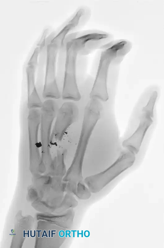
Figure 67-4 A: Anteroposterior (AP) radiograph demonstrating a highly comminuted fracture of the third metacarpal with significant segmental bone loss and retained ballistic fragments.
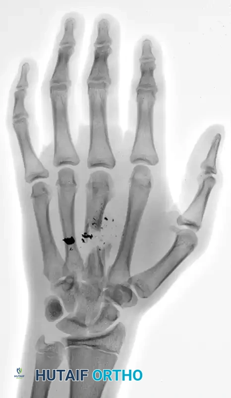
Figure 67-4 B: Oblique radiograph confirming the extent of the intercalary defect and the disruption of the longitudinal arch of the hand.
C and D: Clinical appearance of the hand before débridement.
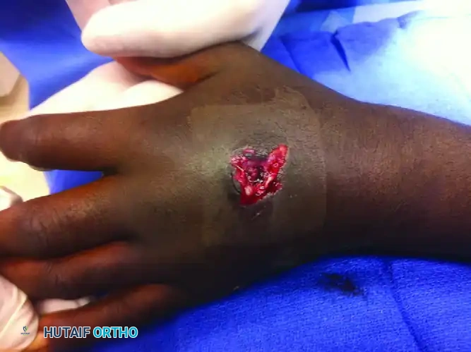
Figure 67-4 C: Dorsal view of the hand demonstrating the entry wound with surrounding soft tissue contusion and thermal injury characteristic of close-range ballistic trauma.
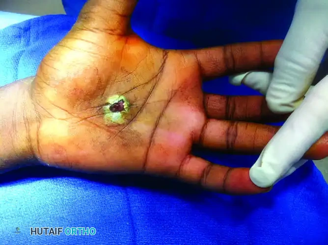
Figure 67-4 D: Volar view demonstrating the exit wound. Note the stellate tearing of the palmar skin and the extrusion of hematoma and devitalized tissue.
In this scenario, the initial procedure must focus on aggressive debridement of the ballistic tract, removal of devitalized bone and debris, and stabilization of the third ray to prevent collapse. High-energy trauma frequently results in intercalary bone loss. Segmental defects of tubular bones must be held to length by wire spacers, intramedullary rods, or spanning external fixators to prevent the collapse of the soft tissue envelope while the wound is healing. Maintaining the anatomic length of the metacarpal is critical to preserve the tension of the intrinsic and extrinsic musculature. In this case, a polymethylmethacrylate (PMMA) cement spacer impregnated with antibiotics (e.g., tobramycin or vancomycin) can be utilized to maintain the defect space, deliver local high-dose antimicrobials, and induce a pseudo-synovial membrane (the Masquelet technique). This membrane secretes osteoinductive growth factors (VEGF, TGF-beta) that will provide a highly vascularized bed for massive structural cancellous bone grafting during the definitive second stage of reconstruction, typically performed 6 to 8 weeks later.
Soft Tissue Management and Wound Closure
The timing and method of wound closure require significant clinical judgment. The surgeon must determine whether the wound is sufficiently clean to permit primary closure or whether it should be left open for repeat debridement. If primary closure is attempted, loose skin-edge approximation is recommended. Soft tissue edema over the ensuing 48 hours will exponentially increase tension on the traumatized tissues; tight sutures will inevitably lead to marginal necrosis, wound breakdown, and catastrophic exposure of underlying hardware or bare tendons.
For contaminated wounds, crush injuries, or wounds with questionable tissue viability, the wound must be left open. It can be packed with non-adherent dressings or managed with negative pressure wound therapy (NPWT) at low continuous pressures (e.g., -75 mmHg) to manage exudate, reduce edema, and promote angiogenesis. At 48 to 72 hours, the wound is reevaluated in the operating room. This "second look" allows the surgeon to identify and excise any tissue that has demarcated and necrosed since the index procedure.
The reconstructive window is narrow. The goal is to achieve definitive soft tissue coverage within the first 4 to 5 days before dense granulation tissues form and fibrotic contractures develop. Exposed tendons devoid of their paratenon or sheath mandate immediate coverage with local, regional (e.g., reverse radial forearm flap), or free vascularized flaps (e.g., anterolateral thigh or lateral arm flaps) to provide a healthy, gliding tissue bed. Simple split-thickness skin grafting over bare tendon or exposed cortical bone is doomed to failure, resulting in graft necrosis and deep space infection.
Complications, Incidence Rates, and Salvage Management
Despite meticulous surgical technique, the complication rates following severe open hand trauma remain notoriously high. The compact anatomy of the hand means that a failure in one tissue system inevitably compromises the function of the entire unit. Anticipating these complications and employing proactive salvage strategies is essential for optimizing long-term outcomes.
Infection, ranging from superficial cellulitis to deep space abscesses and intractable osteomyelitis, is the most dreaded acute complication. Deep infections require immediate return to the operating room for radical re-debridement, copious irrigation, and hardware removal if the fixation is loose or acting as a nidus. Suppressive oral antibiotics are generally ineffective; culture-directed intravenous therapy, often guided by an infectious disease specialist, is mandatory. In cases of chronic osteomyelitis with segmental bone loss, radical resection of the infected bone segment followed by bone transport or vascularized bone grafting (e.g., free fibula flap) may be the only alternative to amputation.
Malunion and nonunion are frequent sequelae of inadequate initial fixation, premature loss of reduction, or severe periosteal stripping. As previously discussed, rotational malunions are functionally devastating and do not remodel. They require formal corrective osteotomies, typically performed at the metacarpal base where the cancellous bone provides a larger surface area for healing and the correction can be dialed in precisely. Nonunions, particularly atrophic nonunions in areas of segmental bone loss, require rigid re-fixation and structural autogenous bone grafting (e.g., iliac crest) once the soft tissue envelope is pristine and free of infection.
Tendon adhesions represent the most ubiquitous complication following open hand fractures, particularly those involving the proximal phalanx (Zone II flexor tendon injuries). The inflammatory cascade triggered by the fracture, combined with surgical trauma and post-operative immobilization, leads to dense scar tissue binding the flexor or extensor tendons to the fracture callus and overlying skin. Prevention through rigid fixation and immediate early active motion is the absolute best defense. Established adhesions that fail to improve after 6 months of aggressive hand therapy may require secondary surgical tenolysis. However, tenolysis is a major undertaking and must only be performed after skeletal union is unequivocally solid, joint contractures have been maximized, and the soft tissues are supple.
| Complication | Estimated Incidence | Primary Etiology | Salvage Management Strategy |
|---|---|---|---|
| Deep Infection / Osteomyelitis | 5% - 15% | Inadequate initial debridement; retained foreign body; primary closure of contaminated wound. | Radical serial debridement; hardware removal; PMMA antibiotic beads; culture-directed IV antibiotics; eventual bone grafting. |
| Rotational Malunion | 10% - 20% | Failure to assess passive tenodesis intra-operatively; loss of provisional K-wire fixation. | Corrective derotational osteotomy (typically at the metacarpal base); rigid internal plating. |
| Atrophic Nonunion | 5% - 10% | Severe periosteal stripping; intercalary bone loss; thermal/ballistic necrosis; inadequate stabilization. | Debridement of fibrous nonunion site; structural autogenous bone grafting (iliac crest); rigid plate fixation. |
| Tendon Adhesions | 30% - 50% | Prolonged immobilization; massive soft tissue trauma; bare tendon exposure. | Aggressive hand therapy; dynamic splinting; secondary surgical tenolysis (post-fracture union). |
| Joint Stiffness / Contracture | 40% - 60% | Splinting in non-functional positions; intra-articular scarring; capsular fibrosis from edema. | Serial static progressive splinting; surgical capsulotomy; collateral ligament release (for MCP extension contractures). |
Phased Post-Operative Rehabilitation Protocols
The ultimate functional success of complex open hand fracture management hinges entirely on the execution of a rigorous, phased post-operative rehabilitation protocol. The hand is uniquely prone to stiffness due to the rapid formation of dense adhesions between the healing fracture callus, disrupted tendon sheaths, and overlying skin. The surgical intervention merely sets the stage; it is the post-operative therapy that dictates the final functional result.
The paradigm of hand rehabilitation has definitively shifted away from prolonged immobilization toward early active motion. If rigid, stable internal fixation was achieved intra-operatively, early active range of motion (AROM) can often commence within 3 to 5 days post-operatively, guided by a specialized, certified hand therapist. Early motion subjects the healing tendons to longitudinal stress, which promotes the parallel alignment of collagen fibers, enhances intrinsic tendon healing, and limits the formation of restrictive extrinsic adhesions to the fracture site.
Post-operative edema is the primary enemy of hand function. Edema fluid is rich in proteins; if allowed to stagnate, it rapidly organizes into dense fibrotic scar tissue. Furthermore, edema increases the diffusion distance for cellular nutrition and mechanically restricts joint flexion by engorging the periarticular tissues. Aggressive edema control is mandatory from day one. This includes strict elevation of the limb above the level of the heart, the application of compressive dressings (e.g., Coban wrapping or customized compression garments), and active muscle pumping exercises to facilitate lymphatic and venous return.
Between active exercise sessions, the hand must be meticulously splinted to prevent contractures. The universally accepted safe position is the "intrinsic-plus" or Edinburgh position. The orthosis should maintain the wrist in 20 to 30 degrees of extension, the metacarpophalangeal (MCP) joints in 70 to 90 degrees of flexion, and the interphalangeal (IP) joints in full, absolute extension. This highly specific posture places the collateral ligaments of the MCP joints at their maximal length, preventing devastating extension contractures, while simultaneously maintaining the volar plates of the PIP joints on stretch, preventing debilitating flexion contractures. As radiographic union progresses (typically 4 to 6 weeks), therapy is advanced to include passive range of motion (PROM), dynamic splinting to overcome specific deficits, and progressive strengthening exercises to restore pre-injury grip and pinch mechanics.
Summary of Landmark Literature and Clinical Guidelines
The evolution of open hand fracture management is deeply rooted in landmark orthopedic literature and continuously refined by modern clinical guidelines. Historically, the Gustilo-Anderson classification, published in 1976, provided the foundational framework for managing open long bone fractures, emphasizing the direct correlation between soft tissue injury severity and infection rates. However, Swanson and colleagues later highlighted the limitations of applying this classification directly to the hand, noting that the unique anatomy of the hand requires a more nuanced approach where even minor puncture wounds communicating with synovial sheaths carry a disproportionately high morbidity.
The timing and duration of antibiotic prophylaxis have been extensively studied and debated. Current clinical guidelines from the American Academy of Orthopaedic Surgeons (AAOS) and the Surgical Infection Society advocate for the immediate administration of systemic antibiotics upon presentation. Landmark studies have demonstrated that delaying antibiotics beyond 3 hours significantly increases the risk of deep infection. Furthermore, recent literature strongly supports limiting prophylactic antibiotics to 24 hours post-closure for Type I and II injuries, and 72 hours for severe Type III injuries, as prolonged administration does not decrease infection rates but significantly increases the risk of multi-drug resistant organism emergence and Clostridium difficile colitis.
The management of massive segmental bone defects in the hand was revolutionized by the work of Alain Masquelet. The two-stage induced membrane technique, first described
