Operative Management of Monteggia and Distal Humerus Nonunions
Key Takeaway
Long-standing nonunions of the proximal ulna associated with radial head dislocations (Monteggia fractures) present complex reconstructive challenges. Surgical management typically involves resection of the dislocated radial head rather than attempted reduction, combined with rigid internal fixation of the ulna using compression plating or intramedullary nailing, supplemented by autologous iliac crest bone grafting. Distal humeral nonunions require meticulous rigid fixation, ulnar nerve neurolysis, and structural grafting to prioritize bone healing over immediate motion.
Comprehensive Introduction and Patho-Epidemiology
The management of long-standing nonunions of the proximal third of the ulna associated with chronic dislocation of the radial head—the classic missed or failed Monteggia fracture-dislocation—alongside nonunions of the distal humerus, represents a formidable and highly specialized challenge in reconstructive orthopaedic surgery. These clinical entities are not merely unhealed fractures; they are complex, multi-planar deformities that disrupt the fundamental kinematics of the entire upper extremity. The proximal ulna dictates the anatomic length and alignment of the forearm. Thus, an ununited, deformed ulna invariably leads to persistent radiocapitellar instability, restricted forearm rotation, and debilitating elbow pain. Similarly, the distal humerus serves as the foundational keystone for the elbow joint, and its failure to unite compromises the transmission of massive mechanical forces during basic activities of daily living.
Epidemiologically, nonunions of the Monteggia variant are frequently the sequelae of missed initial diagnoses or inadequate primary surgical stabilization. The Bado classification categorizes these injuries based on the direction of the radial head dislocation, but in the chronic nonunion setting, the pathoanatomy transcends the initial injury pattern. The radiocapitellar joint undergoes irreversible morphologic changes over time. The radial head becomes hypertrophic or profoundly deformed, the capitellum loses its congruent articular cartilage, and the annular ligament is entirely replaced by dense, unyielding fibrotic scar tissue. Concurrently, the ununited proximal ulna typically assumes an apex-dorsal or apex-radial angular deformity, effectively shortening the forearm and tethering the interosseous membrane.
Distal humerus nonunions, conversely, are profoundly debilitating lesions that most commonly result from biological failure secondary to poor initial fracture fixation, particularly in the supracondylar and intercondylar regions (AO/OTA Type 13-C fractures). The distal humerus is subjected to massive torsional and bending forces during elbow flexion and extension. Inadequate plate constructs, failure to restore the medial and lateral columns, or early aggressive mobilization in the face of tenuous fixation frequently lead to hardware failure and subsequent pseudarthrosis. The incidence of distal humerus nonunion ranges from 2% to 10% following operative intervention, with higher rates observed in cases of high-energy trauma, severe comminution, open fractures, and patient-specific risk factors such as tobacco use and metabolic bone disease.
In both Monteggia and distal humeral nonunions, the surgical strategy must pivot from simple fracture fixation to comprehensive structural reconstruction. The treating surgeon must address not only the osseous discontinuity but also the profound soft-tissue contractures, the compromised neurovascular envelopes (particularly the ulnar and posterior interosseous nerves), and the altered joint mechanics. The overarching goal is the restoration of absolute mechanical stability via rigid internal fixation, combined with aggressive biological augmentation using autologous bone grafting, to provide the necessary osteoinductive and osteoconductive stimuli for union.
Detailed Surgical Anatomy and Biomechanics
A profound understanding of the surgical anatomy and biomechanics of the elbow and forearm is the absolute prerequisite for successfully navigating these complex nonunions. The forearm functions as a highly sophisticated, articulating joint ring composed of the radius, the ulna, the proximal radioulnar joint (PRUJ), the distal radioulnar joint (DRUJ), and the intervening interosseous membrane (IOM). The ulna acts as the stable axis around which the radius rotates during pronation and supination. The proximal ulna possesses a critical anatomic feature known as the proximal ulnar angle (PUA) or the varus bow, which must be anatomically restored to re-tension the IOM and allow the radial head to articulate congruently with the capitellum. Failure to restore the anatomic length and bow of the ulna in a Monteggia nonunion leaves the radiocapitellar joint chronically subluxated or dislocated, leading to rapid articular degeneration.
Biomechanically, the proximal ulna is subjected to significant bending and torsional forces, particularly from the pull of the triceps brachii inserting on the olecranon and the brachialis inserting on the ulnar tuberosity. A nonunion in this region is typically atrophic or oligotrophic due to the relatively poor surrounding soft-tissue envelope and the inherent instability of the initial injury. The vascular supply to the proximal ulna is derived primarily from the nutrient artery arising from the ulnar artery, but in the setting of chronic nonunion and previous surgeries, the periosteal blood supply is often severely compromised, necessitating meticulous soft-tissue handling and decortication to bleeding bone during reconstruction.
The distal humerus is structurally conceptualized as a triangle, comprising the medial and lateral columns converging at the articular segment (the trochlea and capitellum). This "tie-arch" configuration is essential for transferring axial, coronal, and sagittal loads from the forearm to the humeral shaft. The medial column diverges at an angle of approximately 45 degrees, while the lateral column diverges at about 20 degrees. When a nonunion occurs in the supracondylar region, this architectural arch collapses. The biomechanical principles of distal humerus fixation, championed by O'Driscoll, dictate that fixation must maximize stability to resist varus/valgus bending and rotational torque. This is achieved through orthogonal (90-90) or parallel dual plating, where the plates are applied to the distinct columns to lock the articular segment to the metadiaphysis.
The neural anatomy is of paramount concern in both regions. In the proximal forearm, the posterior interosseous nerve (PIN) traverses the supinator muscle and wraps around the radial neck. In a chronic Monteggia nonunion with a dislocated radial head, the PIN is often distorted, encased in scar tissue, and intimately draped over the hypertrophic radial head, placing it at extreme risk during resection. In the distal humerus, the ulnar nerve traverses the cubital tunnel posterior to the medial epicondyle. In the setting of a distal humeral nonunion, the ulnar nerve is frequently tethered by fracture callus, heterotopic ossification, or prior surgical scarring. The biomechanical excursion of the ulnar nerve is significantly impaired, and meticulous neurolysis or formal anterior transposition is an obligatory step in the reconstructive sequence to prevent devastating iatrogenic traction neuropraxia.
Exhaustive Indications and Contraindications
The decision to operate on a chronic Monteggia or distal humerus nonunion requires a meticulous risk-benefit analysis, taking into account the patient's physiologic age, functional demands, bone stock, and the presence of active infection. Operative intervention is generally indicated for all symptomatic nonunions that result in pain, instability, or severe functional impairment, provided the patient is medically optimized for a prolonged surgical procedure and the subsequent rigorous rehabilitation protocol.
In the context of Monteggia nonunions, the primary indication is a painful, unstable forearm with restricted rotation. If the radiocapitellar joint demonstrates irreversible morphologic changes (hypertrophy, cartilage loss), the indication shifts from joint preservation to radial head resection coupled with ulnar reconstruction. For distal humerus nonunions, indications encompass painful pseudarthrosis, progressive deformity, and impending or established hardware failure. Advanced reconstructive techniques, such as free vascularized fibular grafting (FVFG), are indicated for younger, high-demand patients with massive segmental bone loss (>3-4 cm) where conventional autografting would fail. Total Elbow Arthroplasty (TEA) is strictly reserved as a salvage procedure for elderly, low-demand patients with profound intra-articular nonunions where joint reconstruction is impossible.
Contraindications are equally critical to define. Absolute contraindications include active, untreated deep infection (which necessitates a staged approach rather than definitive internal fixation), medically unstable patients who cannot tolerate prolonged anesthesia, and non-ambulatory patients with painless, stable fibrous nonunions who lack functional demands. Relative contraindications include severe osteoporosis that precludes adequate screw purchase, active smoking (which profoundly inhibits osteogenesis and microvascular graft incorporation), and severe, uncorrectable psychiatric or cognitive disorders that would prevent compliance with the strict postoperative weight-bearing and rehabilitation restrictions.
| Parameter | Indications for Operative Intervention | Contraindications (Absolute and Relative) |
|---|---|---|
| Monteggia Nonunion | Symptomatic ulnar pseudarthrosis; Chronic radial head dislocation with pain/stiffness; Progressive ulnar deformity; Ulnar shortening >2mm affecting DRUJ. | Active localized osteomyelitis (requires staging); Painless fibrous nonunion in a low-demand patient; Severe non-compliant psychiatric profile. |
| Distal Humerus Nonunion | Painful supracondylar/intercondylar mobility; Hardware failure/breakage; Progressive varus/valgus collapse; Ulnar neuropathy secondary to tethering. | Active infection (Absolute); Severe osteoporosis precluding fixation (Relative - consider TEA); Medically unfit for prolonged anesthesia. |
| Salvage TEA | Age > 65 years; Low functional demands; Unreconstructible articular comminution/bone loss; Rheumatoid arthritis with nonunion. | Age < 60 years or high manual labor demands (lifetime 5-lb lifting restriction); Active infection; Insufficient triceps or soft-tissue envelope. |
| Vascularized Bone Graft | Young, high-demand patient; Segmental defect > 4 cm; Failed previous autogenous grafting; Avascular or irradiated recipient bed. | Peripheral vascular disease precluding microvascular anastomosis; Elderly, low-demand patient; Active smoking (Relative/Absolute). |
Pre-Operative Planning, Templating, and Patient Positioning
Preoperative evaluation for complex upper extremity nonunions must be exhaustive. Standard orthogonal radiographs (anteroposterior and lateral views) of the elbow and the entire forearm are mandatory, but they are insufficient for comprehensive surgical planning. Computed tomography (CT) with 3D reconstructions is highly recommended, and arguably standard of care, to assess the exact morphology of the nonunion, the precise volume of segmental bone loss, the degree of sclerosis at the fracture margins, and the presence of occult intra-articular extensions or heterotopic ossification. For Monteggia nonunions, CT helps delineate the morphologic changes in the radial head and capitellum, confirming the necessity of radial head resection.
Digital templating is a critical phase of the preoperative workflow. The surgeon must template the anticipated plate lengths, screw trajectories, and the required volume of bone graft. In cases of significant ulnar deformity, radiographs of the contralateral, uninjured forearm should be obtained to serve as an anatomic template for restoring the native proximal ulnar angle and overall ulnar length. The surgeon must calculate the exact amount of ulnar lengthening required to restore the interosseous membrane tension without over-stuffing the radiocapitellar or distal radioulnar joints. Furthermore, preoperative laboratory markers (ESR, CRP, CBC) must be drawn to rule out indolent subclinical infection, which is a frequent cause of aseptic-appearing nonunions.
Patient positioning is dictated by the specific pathology and the planned surgical approach. For isolated Monteggia nonunions, the patient is typically positioned supine with the affected arm draped free on a radiolucent hand table. A sterile tourniquet is applied high on the arm to provide a bloodless field during the critical neurolysis and initial exposure phases. The ipsilateral iliac crest must be meticulously prepped and draped to allow for the harvesting of autologous cancellous or tricortical structural bone graft.
For distal humerus nonunions, the lateral decubitus position is frequently preferred. The patient is secured with a beanbag, and the operative arm is draped over a padded post, allowing the elbow to flex to 120 degrees. This position provides unparalleled access to the posterior aspect of the distal humerus and facilitates gravity-assisted retraction of the triceps. Alternatively, the prone position can be utilized, though it presents distinct challenges for airway management and requires meticulous padding of all bony prominences. Regardless of the position, the entire upper extremity from the axilla to the fingertips must be prepped to allow for intraoperative assessment of elbow range of motion and forearm rotation following definitive fixation.
Step-by-Step Surgical Approach and Fixation Technique
The surgical execution of complex nonunion takedown and reconstruction demands meticulous soft-tissue handling, precise osteology, and rigid biomechanical fixation. The operative sequence must be rigorously adhered to, ensuring that neural structures are protected before any osseous resection or manipulation occurs.
Approach and Exposure
For Monteggia nonunions, a modified Boyd approach or separate incisions (a posterolateral Kocher approach for the radial head and a posterior approach for the ulna) may be utilized. The posterior approach to the ulna utilizes the internervous plane between the extensor carpi ulnaris (supplied by the posterior interosseous nerve) and the flexor carpi ulnaris (supplied by the ulnar nerve). This approach provides extensile access to the entire proximal and middle thirds of the ulna. For distal humerus nonunions, a universal posterior approach is mandatory. Depending on the location of the nonunion and the need for articular exposure, the surgeon may elect a triceps-sparing approach, a Bryan-Morrey triceps-reflecting approach, or an olecranon osteotomy. An olecranon osteotomy provides the gold-standard visualization of the articular surface but introduces the risk of a secondary nonunion at the osteotomy site.
Joint Preparation and Radial Head Resection
In the chronic Monteggia setting, the radiocapitellar joint is accessed via the Kocher interval (between the anconeus and extensor carpi ulnaris). The dislocated, often deformed radial head is identified amidst dense fibrotic scar tissue. A retractor is carefully placed anterior to the radial neck to protect the posterior interosseous nerve (PIN), which may be distorted and tethered due to the chronic anterior or lateral dislocation. No attempt is made to reduce the chronic radial head dislocation, as forcible reduction in this setting leads to severe loss of motion, radiocapitellar arthritis, and high rates of re-dislocation.
Instead, the radial head and a portion of the radial neck are resected using a micro-oscillating saw. The level of resection should be just proximal to the bicipital tuberosity to preserve the insertion of the biceps brachii and maintain active supination strength. The capsule is aggressively released to ensure unhindered forearm pronation and supination.
Figure A: Radiographic demonstration of a chronic nonunion of the ulna following radial head excision. Note the sclerotic margins and the retained wire/suture material from previous failed interventions.
Ulnar Nonunion Takedown and Grafting
Attention is then directed to the ulnar nonunion. The pseudarthrosis is taken down meticulously. All intervening fibrous tissue, devascularized cartilage, and necrotic bone are excised. The sclerotic bone ends are aggressively decorticated using a high-speed burr, rongeurs, or sharp osteotomes. The medullary canals of both the proximal and distal fragments must be opened and reamed until punctate bleeding (the "paprika sign") is observed, indicating healthy, vascularized bone capable of participating in osteogenesis. If an angular deformity exists, the ulnar ends must be completely mobilized, releasing the tethering interosseous membrane as needed, and realigned to restore the anatomic bow and length of the proximal ulna.
The fragments of the ulna are fixed with either a rigid compression plate (typically a 3.5-mm dynamic compression plate or a locking compression plate) or a locked intramedullary nail, depending on the fracture morphology, the presence of segmental defects, and surgeon preference. If plating is chosen, a minimum of six to eight cortices of fixation proximal and distal to the nonunion is required. Absolute compression should be achieved across the nonunion site if the fracture pattern allows, utilizing articulated tensioning devices if necessary. In cases of poor bone quality, osteopenic diaphyses, or specific segmental defects, a locked forearm intramedullary nail can provide excellent load-sharing biomechanics and preserve the periosteal blood supply.
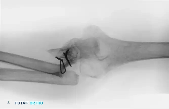
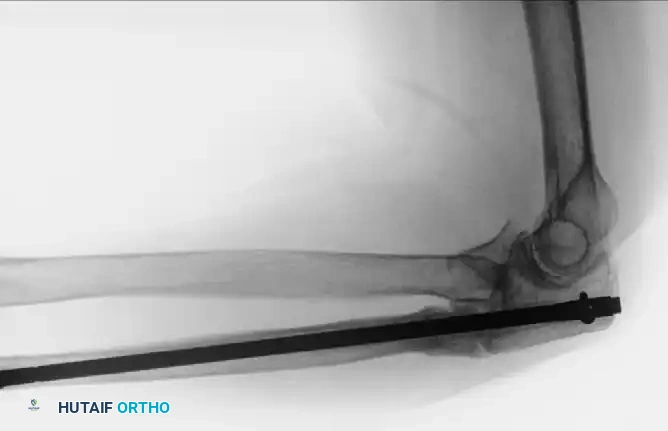
Figure B: Proximal ulnar fracture nonunion successfully treated with a locked forearm intramedullary nail and a tricortical iliac crest bone graft to bridge the defect and restore ulnar length.
Once rigid fixation is achieved, copious autologous iliac crest bone grafts are applied. If a structural defect exists following debridement, a tricortical iliac crest graft is impacted into the defect to restore length and mechanical continuity. Cancellous autograft is then packed circumferentially around the nonunion site to provide massive osteoinductive and osteoconductive stimuli.
Distal Humerus Reconstruction and Ulnar Nerve Management
For distal humerus nonunions, the ulnar nerve is at extreme risk and must be addressed first. The nerve is identified proximally in the triceps interval and traced distally into the cubital tunnel and between the heads of the flexor carpi ulnaris. A meticulous microscopic neurolysis is required to free the nerve from fracture callus and scar tissue. The nerve is handled with vessel loops and mobilized extensively. A formal anterior subcutaneous or submuscular transposition is routinely performed to remove the nerve from the reconstructive bed and prevent postoperative tethering.
The nonunion site is then exposed, debrided, and decorticated as described above. The current gold standard for fixation involves orthogonal (90-90) or parallel dual plating techniques using highly pre-contoured, anatomically specific locking plates. The plates must be long enough to bypass the nonunion by at least three to four screw holes in healthy diaphyseal bone. If there is medial column comminution or a segmental defect, a structural tricortical graft is mandatory to prevent varus collapse. Autologous cancellous bone graft is packed into all remaining interstices.
In infected nonunions, a staged approach is obligatory. Stage 1 involves complete hardware removal, aggressive surgical débridement (often utilizing the Masquelet technique principles), capsulectomy, and the placement of an antibiotic-impregnated cement spacer to sterilize the bed and induce a pseudo-synovial membrane. Stage 2, performed weeks later after normalization of inflammatory markers, involves removal of the spacer, massive bone grafting, and stabilization, occasionally utilizing a ring fixator (Ilizarov frame) to avoid placing large metallic implants into a previously infected, compromised soft-tissue envelope.
Complications, Incidence Rates, and Salvage Management
The operative management of Monteggia and distal humerus nonunions is fraught with potential complications, even in the hands of highly experienced reconstructive surgeons. The local biology is compromised, the soft-tissue envelope is scarred, and the mechanical forces across the elbow and forearm are immense. Patients must be extensively counseled preoperatively regarding the high likelihood of residual morbidity, including permanent stiffness, weakness, chronic pain, and the potential need for secondary or salvage operations.
Hardware failure and persistent nonunion represent the most devastating mechanical complications. In the distal humerus, inadequate plate length, failure to achieve bi-columnar stability, or premature weight-bearing can lead to catastrophic pullout of screws from the osteopenic distal fragment or fracture of the plates at the nonunion level. The incidence of persistent nonunion following primary revision surgery ranges from 10% to 20%, heavily dependent on the adequacy of the bone grafting and the rigidity of the fixation. Infection is another profound risk, particularly in cases with a history of open fractures or multiple prior operations. The poor soft-tissue coverage over the olecranon and proximal ulna makes hardware prominence and subsequent wound breakdown a frequent clinical challenge, sometimes necessitating soft-tissue coverage with regional rotational flaps (e.g., anconeus or flexor carpi ulnaris flaps) or free tissue transfer.
Neurologic complications, specifically ulnar neuropathy and posterior interosseous nerve (PIN) palsy, occur with distressing frequency. Iatrogenic traction neuropraxia of the ulnar nerve during distal humerus reconstruction can occur despite meticulous neurolysis and transposition, with transient dysfunction reported in up to 15% to 30% of cases. Heterotopic ossification (HO) is a profound biological complication that severely limits postoperative range of motion. The extensive surgical dissection, decortication of bone, and trauma to the brachialis and triceps muscles create a highly osteogenic environment. Prophylaxis with non-steroidal anti-inflammatory drugs (NSAIDs) such as indomethacin, or single-dose localized radiation therapy, is frequently employed in high-risk patients.
When conventional ORIF and bone grafting fail irrevocably, salvage management strategies must be deployed. In younger, high-demand patients with massive segmental bone loss or recalcitrant aseptic nonunions, a free vascularized fibular graft (FVFG) is the salvage procedure of choice. This highly technically demanding microsurgical technique provides a living, structurally robust strut of bone that bypasses the hostile local biology, demonstrating union rates exceeding 80% in salvage scenarios. Conversely, in elderly, low-demand patients (typically over 65 years of age) with profound intra-articular nonunions, severe osteopenia, or failed previous reconstructions, Total Elbow Arthroplasty (TEA) is the definitive salvage. TEA provides immediate stability and excellent pain relief, but subjects the patient to a lifetime 5-lb lifting restriction to prevent aseptic loosening and catastrophic mechanical failure of the implant.
| Complication | Estimated Incidence | Etiology / Risk Factors | Salvage / Management Strategy |
|---|---|---|---|
| Persistent Nonunion | 10% - 20% | Inadequate fixation; Poor bone graft incorporation; Smoking; Infection. | Revision ORIF with dual plating; Free Vascularized Fibular Graft (FVFG); Bone morphogenetic proteins (BMP). |
| Hardware Failure | 5% - 15% | Single plate use in distal humerus; Premature weight-bearing; Severe osteoporosis. | Revision to orthogonal/parallel locking plates; Total Elbow Arthroplasty (TEA) in elderly. |
| Ulnar Neuropathy | 15% - 30% | Traction during exposure; Inadequate release; Scar tethering; Hardware impingement. | Meticulous microscopic neurolysis; Anterior submuscular transposition; Corticosteroid therapy. |
| Heterotopic Ossification | 10% - 25% | Extensive soft-tissue trauma; Head injury; Multiple prior elbow surgeries. | Prophylactic Indomethacin/Radiation; Delayed open arthroscopic/open capsular release and HO excision (at 6-9 months). |
| Deep Infection | 3% - 8% | Poor soft-tissue envelope; Prior open fracture; Prolonged operative time. | Staged management: Hardware removal, antibiotic spacer (Masquelet), IV antibiotics, delayed reconstruction/Ilizarov. |
Phased Post-Operative Rehabilitation Protocols
The postoperative rehabilitation following the reconstruction of complex forearm and elbow nonunions represents a delicate, often frustrating balance between protecting the tenuous osseous fixation and preventing irreversible joint stiffness. The surgical warning must be reiterated: bone healing takes absolute precedence over motion when attempting to unite a nonunion. The surgeon must not compromise the rigidity of the fixation or risk hardware failure in an attempt to allow early aggressive rehabilitation. Restoration of motion can be, and often must be, addressed after solid bony union has been achieved via physical therapy, dynamic splints, and delayed capsular releases.
Phase 1: Immediate Postoperative Period (Weeks 0-2)
Postoperatively, the arm is placed in a well-padded posterior splint or a custom orthosis at 90 degrees of elbow flexion with the forearm in neutral rotation. This period is dedicated strictly to soft-tissue healing, edema control, and pain management. The arm is kept elevated. Active range of motion (ROM) of the digits and shoulder is encouraged immediately to prevent dependent edema and distal stiffness. The surgical incisions are monitored meticulously for any signs of dehiscence or superficial infection.
Phase 2: Early Mobilization and Protection (Weeks 2-8)
Following suture removal at 10 to 14 days, the rehabilitation protocol diverges based on the surgeon's intraoperative assessment of fixation stability. If rigid, bi-columnar dual plating was achieved in a distal humerus nonunion, or if a robust compression plate was applied to an ulnar nonunion, gentle active and active-assisted ROM of the elbow and forearm is initiated. The patient is transitioned to a hinged elbow brace locked in a functional arc of motion. Aggressive passive stretching, forceful manipulation by therapists, and any form of weight-bearing or lifting are strictly prohibited. The goal during this phase is to allow the articular surfaces to glide and prevent dense intra-articular adhesions, without imparting bending or torsional stresses to the healing bone graft.
Phase 3: Consolidation and Strengthening (Weeks 8-16+)
Progression to Phase 3 is contingent upon radiographic evidence of bridging callus and graft incorporation, typically observed between 8 and 12 weeks. Once clinical and radiographic union is progressing satisfactorily, the hinged brace is discontinued. Progressive resistance exercises and strengthening protocols are initiated. Dynamic or static progressive splinting (e.g., turnbuckle splints) may be prescribed to address residual flexion or extension contractures.
If profound stiffness persists after solid bony union is definitively achieved (typically assessed at 6 to 9 months postoperatively), the patient may be offered an open or arthroscopic capsular release, combined with excision of any heterotopic ossification, to optimize final functional outcomes. This delayed approach ensures that the structural integrity of the limb is never compromised in the pursuit of early motion.
Summary of Landmark Literature and Clinical Guidelines
The evolution of operative strategies for Monteggia and distal humerus nonunions is heavily documented in the orthopaedic literature, reflecting a historical shift from single-plate constructs and conservative grafting to aggressive dual-plating, structural bone grafting, and precise neurovascular management.
For distal humeral nonunions, the foundational work by Sanders and Sackett established the modern standard of care, recommending rigid internal fixation with a plate and screws combined with an aggressive bone graft. They recognized that the biological failure of the nonunion could only be overcome by overwhelming mechanical stability. This concept was further refined by O'Driscoll, who codified the biomechanical principles of parallel plating for the distal humerus, demonstrating superior resistance to torsional forces compared to traditional single-plate or crossed-screw constructs.
Mitsunagase, Bryan, and Linscheid provided a landmark series demonstrating the efficacy of open reduction, internal fixation, and bone grafting, obtaining union in 25 of 32 patients. Their work highlighted that union of the fractures resulted in significant relief of pain and good functional motion of the elbow. However, the complexity of these cases was underscored by their complication rate, with a significant subset requiring secondary procedures for repeat bone grafting or revision of the fixation device. Their series also delineated the role of Total Elbow Arthroplasty (TEA), treating seven elderly patients with massive bone loss with TEA, firmly establishing it as the salvage procedure of choice for low-demand individuals where ORIF is deemed impossible.
In the realm of massive segmental bone loss, Beredjiklian et al. pioneered the utilization of free vascularized bone grafts (typically from the fibula) in patients with recalcitrant nonunions of the distal humerus and proximal ulna. Their work demonstrated that while highly technically demanding, FVFG is the most reliable method for bridging massive defects in younger, high-demand patients for whom a TEA would yield an unacceptably poor long-term outcome due to lifting restrictions.
Current clinical guidelines synthesize these landmark studies into a cohesive algorithmic approach: absolute mechanical stability via modern locking plate technology, aggressive biological augmentation via autologous bone grafting, meticulous protection of the ulnar and posterior interosseous nerves, and a rigorous, phased postoperative rehabilitation protocol that prioritizes bone healing over early aggressive motion.
