Introduction & Epidemiology
Injuries to the coccyx and distal sacrum, often resulting from a fall onto the buttocks or direct trauma, are commonly encountered in orthopedic and emergency medicine practices. These injuries are classified within the broader spectrum of pelvic ring trauma, although A1 injuries, specifically coccygeal/sacral compression or ligamentous avulsion fractures, represent a distinct and generally stable subset. The Young-Burgess classification, while primarily focused on the stability of the posterior pelvic arch, broadly categorizes these as stable injuries (Type A), where the pelvic ring remains intact or has minimal displacement, not involving weight-bearing structures. Specifically, coccygeal fractures, often considered extra-pelvic or peripheral to the true pelvic ring stability, typically do not compromise the load-bearing capacity of the sacrum and ilia.
The coccyx, a vestigial caudal remnant of the spine, articulates with the sacrum via the sacrococcygeal joint. Injuries here can manifest as direct fractures of the coccygeal segments, avulsion fractures of the ligaments attaching to the coccyx (e.g., sacrococcygeal ligaments, anococcygeal raphe), or dislocations of the sacrococcygeal joint. Compression injuries to the distal sacrum, while less common than coccygeal fractures, may also fall into this A1 designation if they are stable and non-displaced. The estimated incidence of coccygeal injuries, though difficult to ascertain precisely due to variable reporting and often self-managed cases, is significant. They are more prevalent in females, potentially due to differences in pelvic anatomy, and in individuals with lower body mass index (BMI) due to reduced soft tissue padding. While most A1 injuries inherently possess a strong propensity for nonoperative healing, the associated pain and functional limitations can be substantial, underscoring the importance of judicious management strategies.
Surgical Anatomy & Biomechanics
The coccyx is typically composed of three to five fused vertebral segments, although variations exist. It articulates superiorly with the fifth sacral segment (S5) forming the sacrococcygeal joint, a symphysis with an intervertebral disc that can ossify with age. This joint allows for limited flexion-extension movements, which are critical during defecation and parturition. Key ligamentous structures providing stability to the sacrococcygeal region include:
- Anterior and Posterior Sacrococcygeal Ligaments: These are direct continuations of the anterior and posterior longitudinal ligaments, reinforcing the sacrococcygeal joint capsule.
- Lateral Sacrococcygeal Ligaments: Extending from the lateral sacral cornua to the transverse processes of the coccyx, providing robust lateral stability.
- Anococcygeal Raphe (Ligament): A fibromuscular band extending from the anterior coccyx to the external anal sphincter, providing support to the anorectal region.
- Sacrotuberous and Sacrospinous Ligaments: While not directly attaching to the coccyx, these robust ligaments originating from the sacrum contribute significantly to the stability of the posterior pelvic outlet and indirectly influence coccygeal mechanics by anchoring surrounding musculature (e.g., gluteus maximus, piriformis, coccygeus, levator ani).
The coccyx serves as an attachment point for various muscles of the pelvic floor, including the coccygeus (ischiococcygeus) and portions of the levator ani muscle group (iliococcygeus, pubococcygeus, puborectalis). These muscles collectively form the pelvic diaphragm, supporting pelvic viscera and playing a role in continence. Injury to the coccyx can lead to disruption of these muscular attachments, contributing to pain and potential functional deficits.
Biomechanically, the coccyx acts as a minor weight-bearing structure when sitting, particularly during backward tilting of the pelvis. Direct impact, such as a fall onto the buttocks, concentrates significant force on this region, leading to fracture or dislocation. Coccygeal fractures can be transverse, oblique, or comminuted. Sacrococcygeal dislocations typically involve anterior displacement of the coccyx relative to the sacrum, often with significant angulation. A fracture-dislocation can also occur. Ligamentous avulsions, by definition, involve the tearing away of a ligamentous attachment, potentially with a small bone fragment, representing a form of tensile failure.
A1 coccygeal/sacral compression or ligamentous avulsion fractures are characterized by their inherent stability. The surrounding dense ligamentous and muscular attachments, combined with the often-minimal displacement, ensure that these injuries do not typically compromise the integrity of the pelvic ring's load-bearing function. The primary sequela is localized pain, often exacerbated by sitting, defecation, and direct pressure. Chronic pain, or coccydynia, is a common and often debilitating outcome if initial management is inadequate or if there is persistent instability or malunion leading to impingement. Neurological structures in close proximity include the terminal portions of the sacral plexus, specifically the pudendal nerve, which courses near the sacrotuberous and sacrospinous ligaments and provides innervation to the perineum, external anal sphincter, and external urethral sphincter. While direct nerve injury from A1 fractures is rare, chronic inflammation or scar tissue formation can occasionally lead to pudendal neuralgia.
Indications & Contraindications
The management of A1 coccygeal/sacral compression or ligamentous avulsion fractures is predominantly nonoperative, given their intrinsic stability and high potential for healing. However, specific circumstances may necessitate a shift toward operative intervention.
Non-Operative Indications
- Stable Coccygeal Fractures: The vast majority of transverse, oblique, or comminuted fractures with minimal or no displacement, or without significant angulation.
- Stable Sacrococcygeal Dislocations: Dislocations with minimal displacement and without significant anterior angulation.
- Ligamentous Avulsion Fractures: Generally stable injuries, often without significant displacement of the main bony segments.
- Mild to Moderate Pain: Symptoms that are manageable with conservative measures and improve over weeks to months.
- Absence of Neurological Deficits: No signs of pudendal nerve irritation or other neurological compromise.
- Patient Preference: When conservative management aligns with the patient's wishes and functional expectations, particularly in the absence of absolute operative indications.
Operative Indications
Operative intervention for A1 coccygeal/sacral injuries is reserved for a select subset of patients who fail conservative management or present with specific, severe deformities.
- Failed Nonoperative Management (Chronic Coccydynia): Persistent, debilitating pain (coccydynia) for at least 6-12 months despite comprehensive nonoperative treatment, including activity modification, specialized cushions, pain medication, physical therapy, and local injections. This is the most common indication for surgery.
-
Significant and Irreducible Displacement:
- Anterior Angulation/Subluxation: A coccyx that is significantly angulated anteriorly, particularly when impacting the anorectal region, causing functional issues with sitting or defecation, and refractory to closed reduction attempts.
- Lateral Displacement with Impingement: Rare, but if lateral displacement causes nerve root impingement or functional obstruction.
- Nonunion or Malunion with Symptomatic Impingement: A painful pseudarthrosis or grossly malunited coccyx causing mechanical impingement or chronic pain, unresponsive to conservative care.
- Neurological Deficits: Though exceedingly rare with A1 injuries, documented and persistent pudendal neuralgia directly attributable to bone impingement or instability that is refractory to conservative measures.
- Open Fractures: While rare in this region, any open injury requires surgical debridement and management.
Contraindications
Contraindications to Operative Treatment (e.g., Coccygectomy):
- Acute Fractures/Dislocations: Unless there is severe, irreducible displacement causing functional impairment, operative intervention is generally contraindicated in the acute phase due to high rates of infection and wound healing complications, and the strong potential for spontaneous healing.
- Active Infection: Localized soft tissue or bone infection.
- Poor General Health/Co-morbidities: Patients with significant medical co-morbidities that preclude safe anesthesia or surgery.
- Psychogenic Pain/Secondary Gain: Careful psychological evaluation is crucial, as chronic pain in this region can have significant psychosocial components. Operative intervention in patients with significant psychological overlay without clear organic pathology may lead to unsatisfactory outcomes.
- Unrealistic Patient Expectations: Patients must understand that surgery is not a guaranteed cure for pain and may carry its own set of complications.
- Inadequate Pre-Operative Assessment: Lack of appropriate imaging (dynamic flexion-extension views, MRI) to confirm the source of pain.
- Lack of Surgical Expertise: Coccygectomy requires careful dissection in a contaminated field, and should only be performed by surgeons experienced with pelvic floor and perianal anatomy.
Table: Operative vs. Non-Operative Indications
| Feature | Non-Operative Treatment (Primary) | Operative Treatment (Secondary) |
|---|---|---|
| Fracture/Dislocation Type | Most A1 coccygeal/sacral compression or ligamentous avulsion fractures, stable sacrococcygeal dislocations. | Failed nonoperative management for >6-12 months (chronic coccydynia), significant irreducible anterior coccygeal angulation/dislocation with functional impingement, painful nonunion/malunion, rare documented neurological impingement. |
| Displacement/Angulation | Minimal or no displacement; slight angulation. | Significant anterior angulation (often >25-30 degrees on lateral dynamic views) refractory to closed reduction, causing functional issues. |
| Pain Duration/Severity | Acute pain, mild-to-moderate chronic pain responding to conservative measures. | Persistent, debilitating pain (coccydynia) for >6-12 months, refractory to all conservative modalities. |
| Functional Impairment | Tolerable difficulty with sitting, defecation, activities of daily living. | Severe impairment in sitting, defecation, or activities of daily living due to mechanical impingement or intractable pain. |
| Neurological Symptoms | Absent or transient. | Documented, persistent pudendal neuralgia directly attributable to a bony lesion, refractory to conservative measures. |
| Imaging Findings | Stable fracture pattern, minimal displacement, minimal angulation on static/dynamic films. | Significant anterior hypermobility, posterior subluxation, or marked angulation on dynamic films. Callus formation consistent with painful nonunion. MRI showing inflammation/edema. |
Pre-Operative Planning & Patient Positioning
When surgical intervention, most commonly coccygectomy, is deemed necessary following failed nonoperative management of A1 injuries, meticulous pre-operative planning is essential.
Pre-Operative Planning
-
Diagnostic Confirmation:
- Radiographs: Anteroposterior and lateral radiographs are standard. Crucially, dynamic lateral radiographs (sitting and standing/supine positions) are mandatory to assess coccygeal hypermobility, posterior subluxation of the coccyx, or excessive anterior angulation, which are often the underlying causes of chronic coccydynia. Measurements of sacrococcygeal angle and intercoccygeal angle can be performed.
- MRI: Magnetic Resonance Imaging can assess soft tissue pathology, inflammation, disc herniation (rare but possible at sacrococcygeal joint), nerve impingement, and rule out other causes of pelvic pain. It is particularly useful for identifying edema or inflammation around the coccyx or sacrococcygeal joint.
- CT Scan: Computed Tomography provides detailed bony anatomy, useful for characterizing fracture patterns, nonunions, or complex malunions.
- Conservative Treatment Record Review: Thorough review of all failed nonoperative treatments, including types of analgesics, physical therapy, specialized cushions, and particularly the number and location of local anesthetic/steroid injections. Pain relief following successful injections into the sacrococcygeal joint or adjacent ganglia often predicts a better outcome from coccygectomy.
- Psychological Assessment: As chronic pain often has a significant psychological component, especially in this region, pre-operative psychological evaluation can be beneficial to set realistic expectations and identify any significant contributing psychosocial factors.
- Infection Prophylaxis: Given the proximity to the perianal region, pre-operative bowel preparation is often considered to minimize bacterial load. This typically involves a low-residue diet and oral antibiotics (e.g., metronidazole, neomycin) for 1-2 days prior to surgery, although its efficacy in reducing surgical site infection for coccygectomy is debated in literature. Intravenous broad-spectrum antibiotics (e.g., cefazolin) are administered pre-incision.
- Patient Counseling: Detailed discussion with the patient regarding potential risks (infection, wound dehiscence, persistent pain, nerve injury, rectal injury) and expected recovery. Emphasize that pain relief is not guaranteed and full recovery can be protracted.
Patient Positioning
- Prone Position: This is the preferred position for coccygectomy to provide optimal exposure of the sacrococcygeal region.
- Padding: Adequate padding is crucial to prevent pressure neuropathies, especially ulnar and common peroneal nerve palsies. Chest rolls are used to facilitate respiration and abdominal compression should be avoided.
- Legs: The hips and knees can be slightly flexed, with pillows under the shins, to reduce tension on the gluteal muscles and facilitate access.
- Draping: The perianal region must be meticulously cleaned and isolated from the surgical field using sterile drapes. Some surgeons may perform a sterile rectal examination and place a sterile rectal pack or foley catheter to aid in identifying the anterior wall of the rectum during dissection.
Detailed Surgical Approach / Technique
While the title emphasizes "Nonoperative Healing," this section will detail the surgical interventions that are considered when nonoperative management of A1 coccygeal/sacral compression or ligamentous avulsion fractures fails, or when a severe deformity necessitates intervention. This primarily focuses on coccygectomy for chronic coccydynia and, to a lesser extent, closed reduction for acute, significantly displaced coccygeal dislocations.
Closed Reduction of Acute Coccygeal Dislocation
This is typically performed for acute dislocations with significant anterior angulation or subluxation that are causing acute, severe pain and functional limitation. It is rarely indicated for chronic conditions.
- Anesthesia: General or regional (e.g., spinal) anesthesia is preferred to ensure complete muscle relaxation and pain control. Local anesthesia with sedation can be used for less complex cases.
- Patient Positioning: Prone or lateral decubitus position.
-
Technique:
- A sterile glove is applied to one hand for intra-anal manipulation.
- The index finger is inserted into the rectum, palpating the anterior surface of the displaced coccyx.
- Simultaneously, the thumb of the same hand (or the surgeon's other hand) is placed externally over the skin, posterior to the coccyx.
- With gentle but firm pressure, the coccyx is manipulated to reduce the dislocation. The rectal finger pushes the anteriorly displaced coccyx posteriorly, while the external thumb applies counter-pressure or guides the coccyx into alignment.
- Care is taken to avoid excessive force, which could cause rectal perforation.
- Fluoroscopic Guidance: Real-time fluoroscopy can be invaluable to confirm reduction and assess stability.
- Post-Reduction Assessment: Palpate the coccyx to ensure stable reduction. Assess for any signs of rectal injury.
- Post-Operative Care: Activity modification, donut cushion for sitting, stool softeners to prevent straining during defecation.
Coccygectomy (for Failed Nonoperative Management)
Coccygectomy involves the surgical excision of the coccyx. This procedure is considered a salvage operation for refractory chronic coccydynia following exhaustive nonoperative treatment of A1 injuries.
-
Incison: A longitudinal midline incision is made directly over the coccyx, typically extending from the tip of the coccyx superiorly for approximately 5-7 cm, centered over the sacrococcygeal joint. The incision is deepened through the subcutaneous fat to the lumbosacral fascia. Some surgeons advocate for an elliptical incision to remove a small amount of skin, which may reduce dead space and aid in primary closure.
-
Dissection:
- The superficial fascia (containing fat and cutaneous nerves) is carefully incised.
- The gluteus maximus attachments to the coccyx are identified laterally and divided with electrocautery or sharp dissection.
- The surgeon then proceeds superiorly, dissecting through the fat and connective tissue, staying directly on the midline to expose the posterior aspect of the coccyx and the sacrococcygeal joint.
- Internervous Planes: While not a true internervous plane in the classical sense, the dissection aims to stay in the relatively avascular plane between the gluteal musculature laterally and the pelvic floor musculature (coccygeus, levator ani) anteriorly/inferiorly, directly over the bony coccyx.
- Rectal Protection: This is the most critical step. The anterior surface of the coccyx is intimately associated with the anococcygeal raphe and the posterior rectal wall. The surgeon must carefully dissect anteriorly, staying meticulously close to the bone to avoid rectal injury. A finger in the rectum (placed pre-operatively and draped, or introduced sterilely during the procedure) can help guide the dissection and identify the rectal wall. Blunt dissection with a periosteal elevator along the anterior coccygeal surface is often employed.
- Ligamentous Division: The sacrococcygeal ligaments (posterior, lateral) and the anococcygeal raphe are carefully divided using a scalpel or electrocautery, staying close to the bone.
-
Coccyx Excision:
- The sacrococcygeal joint is identified. If it is fused or significantly arthritic, a small osteotome or oscillating saw can be used to osteotomize the coccyx from the distal sacrum. If the joint is mobile, the ligaments are divided, and the coccyx is disarticulated. The level of excision is typically at the sacrococcygeal junction (S5-Co1) or through the most proximal coccygeal segment.
- Some surgeons advocate for partial coccygectomy, removing only the hypermobile or fractured segments, while others prefer total coccygectomy. The extent of resection should be dictated by the pathology identified pre-operatively and intra-operatively.
- After excision, inspect the resected bone for any spurs, osteophytes, or unusual pathology.
-
Hemostasis: Meticulous hemostasis is paramount. The area is highly vascular. Bipolar electrocautery is used to control bleeding from small vessels. Large vessel injury is rare but should be anticipated.
-
Wound Closure:
- Irrigation: The wound is thoroughly irrigated with copious amounts of saline.
- Drainage: A closed suction drain (e.g., Penrose or Blake drain) is often placed to prevent hematoma formation and reduce the risk of seroma, which can be particularly problematic in this area due to sitting pressures. The drain is typically brought out through a separate stab incision.
-
Layered Closure:
- The fascia and muscle attachments are reapproximated with absorbable sutures to reduce dead space. This often involves approximating the gluteus maximus fascia and the remnants of the anococcygeal raphe/pelvic floor musculature, taking care to not tension the perineum.
- Subcutaneous tissue is closed with interrupted absorbable sutures.
- Skin is closed with non-absorbable sutures (e.g., nylon) or staples.
- Dressing: A sterile, pressure dressing is applied.
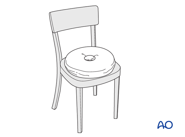
Fig. 1: Example of nonoperative management illustration. While the primary focus here is nonoperative, the image underscores the context of coccygeal injuries, where stability and comfort during recovery are paramount, regardless of ultimate treatment pathway.
Complications & Management
Both nonoperative and operative treatments for A1 coccygeal/sacral compression or ligamentous avulsion fractures carry potential complications.
Nonoperative Complications
- Chronic Coccydynia: The most common complication, defined as persistent coccygeal pain for more than 2 months. This is often the primary reason patients seek further intervention, including surgery. Management involves continued conservative care, pain management specialist referral, various injection therapies (corticosteroid, local anesthetic, prolotherapy, botulinum toxin, ganglion impar block), physical therapy including pelvic floor rehabilitation, and psychological support.
- Malunion/Nonunion: While coccygeal fractures often heal, malunion with persistent angulation or a painful nonunion (pseudarthrosis) can occur, leading to chronic pain and impingement. Initial management is conservative, but if refractory, it can become an indication for coccygectomy.
- Functional Limitations: Difficulty with sitting, walking, defecation, and sexual activity due to pain. Managed with activity modification, ergonomic aids, and physical therapy.
- Skin Breakdown: Prolonged pressure from sitting on the injured coccyx can lead to localized pressure sores, especially in bed-bound or debilitated patients. Preventative measures include pressure-relieving cushions and regular position changes.
Operative Complications (Coccygectomy)
| Complication | Incidence (%) | Salvage Strategies |
|---|---|---|
| Surgical Site Infection (SSI) | 5-20 | Prophylactic antibiotics. If superficial, local wound care, oral antibiotics. If deep, intravenous antibiotics, surgical debridement, potential drain placement, or wound vacuum-assisted closure (VAC). Early detection is key. |
| Wound Dehiscence | 5-15 | Risk increased by infection, hematoma, tension, poor nutrition, radiation. Managed with wound care (secondary intention, VAC therapy, debridement), re-suturing if possible, or flap coverage in severe cases. |
| Hematoma/Seroma Formation | 5-10 | Prevented by meticulous hemostasis and drain placement. Managed by aspiration, drain re-insertion, or surgical evacuation if large or symptomatic. |
| Persistent Pain (Failed Surgery) | 10-30 | Multi-factorial. Requires thorough re-evaluation: repeat imaging (MRI, dynamic X-rays) to rule out residual bone fragment, new pathology, nerve entrapment (pudendal neuralgia). Referral to pain management specialists, psychological support. |
| Rectal Injury/Fistula | <1 | Rare but severe. Meticulous dissection is key. If recognized intra-operatively, primary repair in layers, diverting colostomy may be necessary in high-risk or delayed presentations. Intensive antibiotic therapy. |
| Nerve Injury (Pudendal) | <1 | Rare. Due to direct trauma, traction, or entrapment in scar tissue. Diagnosis via nerve conduction studies/EMG. Management involves conservative measures, nerve blocks, or surgical neurolysis if persistent and severe. |
| Sacral Instability | Very Rare | If too much of the sacrum is resected, potentially leading to pelvic instability. More likely with extensive sacral trauma. Managed by conservative care, activity restrictions, or internal fixation if severe. |
Post-Operative Rehabilitation Protocols
The post-operative rehabilitation protocol varies depending on whether the patient underwent closed reduction or coccygectomy, but the overarching goal is pain management, wound healing, and gradual return to function.
Nonoperative Management Rehabilitation (Initial Injury or Failed Closed Reduction)
This protocol is initiated immediately for A1 coccygeal/sacral compression/avulsion fractures.
-
Pain Management:
- Pharmacological: Nonsteroidal anti-inflammatory drugs (NSAIDs), acetaminophen, muscle relaxants (if spasm present), and short-term opioids for severe acute pain. Neuropathic pain agents (gabapentin, pregabalin) may be considered for chronic coccydynia.
- Local Injections: Corticosteroid and local anesthetic injections into the sacrococcygeal joint, coccygeal periosteum, or ganglion impar can provide temporary relief and are diagnostic/prognostic.
-
Activity Modification:
- Sitting: Avoid direct pressure on the coccyx. Use specialized donut cushions, wedge cushions, or gel pads to offload the coccyx during sitting. Avoid prolonged sitting.
- Positioning: Encourage lying on the side or stomach.
- Defecation: Stool softeners and high-fiber diet to prevent straining, which can exacerbate pain.
-
Physical Therapy:
- Initial Phase (Acute): Focus on pain reduction and gentle mobilization. Ice/heat application.
-
Subacute/Chronic Phase:
Pelvic floor physical therapy is often crucial.
- Manual Therapy: External and internal manual release of tight pelvic floor muscles (levator ani, coccygeus) and fascial restrictions.
- Myofascial Release: Addressing trigger points in the gluteal and piriformis muscles.
- Stretching: Gentle stretching of hip flexors and extensors, piriformis, and hamstrings to improve pelvic mechanics.
- Postural Training: Education on proper sitting posture and body mechanics to reduce coccygeal stress.
- Core Strengthening: Strengthening of abdominal and deep spinal muscles to improve overall trunk stability.
- Biofeedback: For pelvic floor muscle dysfunction.
- Education: Patients are educated on the self-limiting nature of many coccygeal injuries and the importance of adherence to activity modification.
Post-Coccygectomy Rehabilitation Protocol
-
Immediate Post-Operative Phase (Days 0-7):
- Pain Control: Aggressive multimodal analgesia (opioids, NSAIDs, acetaminophen, nerve blocks).
- Wound Care: Daily wound inspection for signs of infection, hematoma, or dehiscence. Dressing changes.
- Drain Management: Monitor drain output. Drain typically removed when output is minimal (<30-50 mL/24h), usually within 2-5 days.
- Activity: Strict avoidance of direct sitting on the buttocks. Encourage lying supine or prone, or side-lying. Ambulation is encouraged from post-op day 1, but with careful attention to posture and avoiding coccygeal pressure.
- Bowel Management: Stool softeners are crucial to prevent straining.
-
Early Rehabilitation Phase (Weeks 1-6):
- Wound Healing: Continue meticulous wound care until fully healed. Sutures/staples typically removed at 2-3 weeks.
- Gradual Sitting: Begin very gradually with a donut or wedge cushion, for short periods. Increase duration as tolerated, avoiding pain.
- Activity: Light activities of daily living. Avoid heavy lifting, strenuous exercise, or activities that place direct pressure on the surgical site.
-
Physical Therapy:
- Gentle Mobilization: Focusing on lumbar spine and hip mobility.
- Pelvic Floor Relaxation: If pelvic floor muscles became hypertonic pre-op.
- Soft Tissue Mobilization: Gentle massage around the incision once healed to prevent scar contracture.
-
Intermediate Rehabilitation Phase (Weeks 6-12):
- Progressive Weight Bearing: Increase sitting tolerance and duration.
- Strengthening: Gradual introduction of core and gluteal strengthening exercises.
- Flexibility: Continue stretching to maintain flexibility around the hips and lumbar spine.
- Return to Activity: Gradual return to light work and recreational activities, guided by pain and functional tolerance. Avoid high-impact activities initially.
-
Advanced Rehabilitation Phase (Months 3-6+):
- Full Functional Recovery: Aim for return to pre-injury activity levels.
- Addressing Residual Pain: For any persistent discomfort, consider additional targeted physical therapy, pain management modalities, or psychological counseling.
- Long-term Monitoring: Patients should be counseled that complete pain resolution can take many months, and some degree of residual discomfort may persist in a minority of cases.
Summary of Key Literature / Guidelines
The existing literature consistently supports a nonoperative approach as the first-line treatment for most A1 coccygeal/sacral compression or ligamentous avulsion fractures and stable dislocations. Operative intervention, primarily coccygectomy, is considered a last resort for chronic, debilitating coccydynia refractory to prolonged conservative management.
Nonoperative Management Success:
Multiple retrospective studies and case series have demonstrated high success rates (ranging from 70% to 90%) with nonoperative management. Conservative measures typically include activity modification, specialized seat cushions, NSAIDs, physical therapy (including pelvic floor rehabilitation), and local corticosteroid injections. A study by Kim et al. (2014) showed that sacrococcygeal joint injection with steroids provided significant pain relief in a large cohort of patients with coccydynia, highlighting the efficacy of targeted interventions. Maigne and Doursounian (2012) emphasized the role of dynamic radiographs in identifying cases with hypermobility or posterior subluxation, which are often good candidates for conservative care but may represent a subset more prone to chronic pain.
Indications for Coccygectomy:
The primary indication for coccygectomy remains chronic coccydynia refractory to at least 6-12 months of comprehensive nonoperative treatment. Maigne et al. (2000) reported that coccygectomy was effective in 80% of carefully selected patients with intractable coccydynia. The strongest predictors for successful surgical outcome often include:
1. Clear objective pathology identified on dynamic radiographs (e.g., hypermobility, posterior subluxation, spicules, or fracture fragment).
2. Temporary relief following diagnostic local anesthetic injections.
3. Absence of significant psychological overlay.
Outcomes of Coccygectomy:
Success rates for coccygectomy in alleviating chronic pain vary in the literature, generally ranging from 60% to 90%. Factors influencing outcomes include patient selection, duration of symptoms, and surgical technique. Ramieri et al. (2010) reported a good or excellent outcome in 85% of patients undergoing coccygectomy for coccydynia caused by hypermobility. However, it is crucial to acknowledge that a significant minority (10-30%) may experience persistent pain or complications.
Complications of Coccygectomy:
Surgical site infection and wound dehiscence are the most frequently cited complications, due to the anatomical location near the perianal region and the constant pressure from sitting. Literature indicates infection rates between 5-20% and wound dehiscence rates similarly high. Rectal injury is rare but devastating. Persistent pain post-coccygectomy, although not a complication of the surgery itself, represents a failure to achieve the primary goal and can occur in a notable percentage of patients, underscoring the importance of careful patient selection and counseling.
Guidelines:
While no specific universal orthopedic guidelines exist solely for A1 coccygeal/sacral injuries, the general consensus among orthopedic surgeons, pain specialists, and spine societies (e.g., North American Spine Society - NASS) aligns with a conservative-first approach. The AO Foundation, in its principles for pelvic and acetabular trauma, broadly classifies coccygeal fractures as Type A injuries (stable), reinforcing the nonoperative bias. The decision to proceed with surgery is highly individualized, based on the severity of symptoms, objective findings, duration of failed conservative care, and patient expectations. Current best practice emphasizes a multidisciplinary approach for chronic coccydynia, involving orthopedic surgeons, pain management specialists, and physical therapists.
