Comprehensive Introduction and Patho-Epidemiology
The human thumb represents the functional epicenter of the hand, accounting for approximately 40% to 50% of overall prehensile capability. It serves as the critical, opposable post for precision pinch, power grasp, and complex fine motor manipulation. However, the mechanical utility of the thumb is severely, if not entirely, compromised without the presence of protective and tactile sensation. An insensate thumb, often referred to in hand surgery literature as a "blind thumb," renders the digit highly susceptible to unrecognized thermal and mechanical trauma, ultimately leading to trophic ulcerations, deep infections, and progressive functional amputation. In the complex reconstructive algorithm for the mutilated hand, restoring stereognosis and two-point discrimination to the volar thumb pulp is paramount.
In clinical scenarios involving irreparable median nerve damage, severe crush injuries, extensive soft tissue avulsions, or deep electrical burns where primary epineurial repair or interpositional nerve grafting is anatomically unfeasible, the Neurovascular Island Graft Transfer emerges as a gold-standard reconstructive modality. Historically pioneered by J. William Littler in the mid-20th century, this sophisticated procedure involves the pedicled transposition of a sensate, vascularized composite island of glabrous skin and subcutaneous tissue from a less functionally critical donor digit (typically the ulnar aspect of the ring finger) to the volar aspect of the thumb.
The patho-epidemiology of thumb pulp loss and sensory deprivation frequently involves high-energy industrial accidents, agricultural machinery trauma, or severe avulsion injuries (such as degloving or ring avulsion injuries extending to the thumb). Additionally, failed replantations or devastating localized infections can result in composite tissue defects requiring both structural coverage and neural restoration. By transferring tissue on its intact, native neurovascular pedicle, the surgeon provides immediate, durable, and sensate coverage to the thumb pulp. This elegant technique bypasses the prolonged, unpredictable, and often incomplete process of Wallerian degeneration and subsequent axonal regeneration associated with conventional nerve grafts or bioabsorbable conduits, ensuring immediate viability and the preservation of specialized sensory end-organs (Meissner's and Pacinian corpuscles).
Detailed Surgical Anatomy and Biomechanics
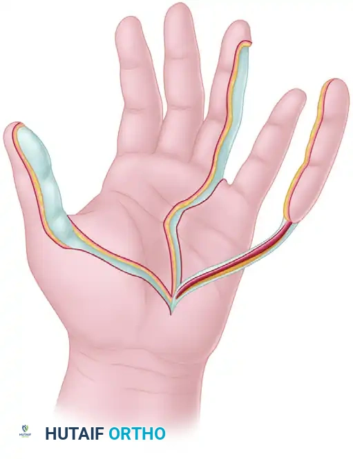
The enduring success of the neurovascular island graft transfer relies entirely upon an intimate, profound understanding of palmar microvascular and neural anatomy. The vascular supply to the digits is derived primarily from the superficial palmar arch, which is formed predominantly by the ulnar artery with a variable contribution from the superficial palmar branch of the radial artery. From this arch, the common palmar digital arteries arise and travel distally within the protective environs of the lumbrical canals, deep to the palmar aponeurosis. At the level of the web spaces, specifically proximal to the natatory ligaments, these common arteries bifurcate into the proper palmar digital arteries that supply the adjacent sides of the fingers.
The venous drainage of the palmar digital integument is notoriously delicate and constitutes the most critical anatomical consideration in island flap survival. Unlike the robust dorsal venous system of the hand, the palmar venous outflow relies entirely on microscopic venae comitantes that intimately accompany the proper digital artery, as well as a fragile, diffuse subcutaneous venous plexus. During surgical dissection, it is an absolute anatomical imperative that the digital artery is not skeletonized. A generous cuff of perivascular areolar tissue must be meticulously preserved around the artery to maintain these microscopic venous channels; failure to do so invariably results in catastrophic venous congestion and subsequent flap necrosis.
Neurologically, the common palmar digital nerves arise from the median and ulnar nerves, lying volar to the arteries within the palm. The fourth web space, which is the primary donor site for this procedure, is typically innervated by the superficial sensory branch of the ulnar nerve. The proper digital nerve supplying the ulnar aspect of the ring finger must be carefully separated from the common digital nerve via precise intraneural dissection under magnification. Biomechanically, the transferred glabrous skin is uniquely suited for the thumb pulp. Unlike non-glabrous skin grafts or distant flaps (e.g., groin or radial forearm flaps), the palmar digital skin possesses dense fibrous septa connecting the dermis to the underlying periosteum and tendon sheaths. This structural microanatomy prevents shear forces during forceful pinch and grasp, providing a stable, non-sliding contact surface essential for the biomechanics of opposition.
Exhaustive Indications and Contraindications
The decision to proceed with a neurovascular island graft transfer requires rigorous patient selection and a thorough evaluation of the injury pattern. The procedure is technically demanding and permanently alters the anatomy of a healthy donor digit; therefore, it is reserved for specific, debilitating deficits where simpler reconstructive ladder options are inadequate.
Indications
The primary indication is permanent, irreparable loss of median nerve sensation to the thumb pulp where proximal nerve reconstruction has failed or is anatomically impossible. This includes extensive segmental nerve loss, severe crush injuries with extensive zone of injury scarring, or late presentations where motor endplate and sensory receptor atrophy preclude successful nerve grafting. Furthermore, severe traumatic pulp loss—such as deep burns, avulsions, or composite tissue defects requiring both durable soft tissue coverage and sensory restoration—are prime indications. The procedure is also heavily favored in cases of failed previous nerve reconstructions, where epineurial repairs, autologous nerve grafts (e.g., sural nerve), or bioabsorbable conduits (polyglycolic acid or collagen tubes) have failed to restore functional two-point discrimination (defined as greater than 10-15 mm).
Contraindications
Contraindications must be strictly respected to prevent devastating ischemic complications in the hand. Absolute contraindications include severe peripheral vascular disease, advanced diabetic microangiopathy, active Raynaud's phenomenon, or any condition compromising the microcirculation of the digits. A positive or equivocal Allen's test indicating an incomplete superficial palmar arch is an absolute contraindication, as sacrificing a proper digital artery could induce profound ischemia in the donor or adjacent digits. Widespread nerve damage, such as concomitant ulnar nerve injury rendering the potential donor sites insensate, nullifies the purpose of the operation. Previous trauma, severe scarring, or Dupuytren's contracture along the volar aspect of the intended donor digit or the fourth web space precludes safe pedicle dissection.
| Category | Specific Condition | Clinical Rationale |
|---|---|---|
| Absolute Indication | Irreparable Median Nerve Deficit | Restores critical protective sensation to the "blind thumb" pulp. |
| Absolute Indication | Composite Volar Thumb Defect | Provides durable, glabrous skin with intact sensory end-organs. |
| Absolute Contraindication | Incomplete Superficial Palmar Arch | Risk of catastrophic digital ischemia upon ligation of the digital artery. |
| Absolute Contraindication | Concomitant Ulnar Nerve Injury | Donor site (ring finger) is already insensate; transfer offers no benefit. |
| Relative Contraindication | Advanced Age / Cognitive Decline | Inability to participate in the rigorous post-operative sensory re-education program. |
| Relative Contraindication | Heavy Tobacco Use | Significant risk of pedicle vasospasm, thrombosis, and flap failure. |
Pre-Operative Planning, Templating, and Patient Positioning
Meticulous preoperative planning is the cornerstone of a successful neurovascular island graft transfer. The initial clinical assessment must include a rigorous vascular examination. The traditional Allen's test is mandatory, but modern preoperative protocols dictate the use of digital Doppler ultrasonography or digital plethysmography to precisely map the superficial palmar arch and confirm robust collateral flow to the middle, ring, and little fingers. The surgeon must definitively prove that ligating the proper digital artery to the radial side of the little finger will not compromise its perfusion.
The hierarchy of donor site selection is strictly defined to minimize donor site morbidity while maximizing functional gain. The ulnar aspect of the ring finger is the undisputed primary choice. It resides outside the primary radial pinch mechanism (which involves the thumb, index, and middle fingers) and is innervated by the ulnar nerve, making it the perfect donor for median nerve deficits. If the ring finger is traumatized or unavailable, the radial aspect of the little finger serves as the secondary alternative. The ulnar aspect of the middle finger is considered a salvage option, utilized exclusively if the median nerve is completely intact and the sensory deficit is strictly localized to the thumb pulp; however, this is generally avoided as it risks compromising a primary pinch digit.
Patient positioning and operating room setup must facilitate uninterrupted microsurgical workflow. The patient is positioned supine with the operative arm extended on a radiolucent hand table. A pneumatic upper arm tourniquet is applied over generous cast padding. Regional anesthesia, specifically an axillary or supraclavicular brachial plexus block, is highly preferred over general anesthesia, as the sympathectomy effect of the regional block induces profound vasodilation, maximizing pedicle perfusion and reducing the incidence of postoperative vasospasm. The surgeon and assistant should be seated comfortably, utilizing high-quality loupe magnification (minimum 3.5x to 4.5x) or an operating microscope for the intraneural dissection phase.
Step-by-Step Surgical Approach and Fixation Technique
Recipient Site Preparation and Excision
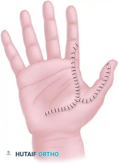
The operative limb is exsanguinated by elevation or via the application of an Esmarch bandage, and the pneumatic tourniquet is inflated to 250 mmHg. Utilizing a sterile skin marker, the exact area of sensory deficit, scarring, or tissue loss on the volar thumb is accurately outlined. It is critical to excise all pathological, insensitive, or heavily scarred tissue back to healthy, well-vascularized margins. If the excised skin is structurally intact but merely insensate, it should be meticulously defatted and preserved in moist saline to be utilized later as a full-thickness skin graft (FTSG) for the donor site defect. A zigzag (Bruner) incision is then designed across the palm, conforming to the natural skin creases, connecting the proximal aspect of the thumb recipient site to the mid-palm to accommodate the transposed pedicle.
Donor Site Marking and Incision Design
The template of the thumb defect is transferred to the ulnar aspect of the chosen donor digit (typically the ring finger). The flap geometry must be designed with extreme precision. It should encompass the majority of the ulnar side of the finger but must crucially incorporate V-shaped darts extending toward the volar and dorsal midlines between the interphalangeal joints. These darts are essential to break up the linear scar and prevent severe postoperative flexion contractures of the donor digit. The proximal incision is designed as a continuous zigzag (Bruner) pattern extending from the base of the flap, across the fourth web space, and proximally into the palm to meet the recipient site incision.
Microvascular and Neural Dissection
This phase represents the most technically demanding portion of the procedure, requiring absolute hemostasis and microscopic precision. The palmar skin flaps are elevated, and the palmar aponeurosis is carefully divided to expose the common volar digital artery and nerve within the fourth web space. The proper digital artery supplying the radial aspect of the little finger is identified, securely ligated with micro-clips or fine silk sutures, and divided. This critical maneuver allows the common digital artery to be mobilized exclusively with the ulnar proper digital artery of the ring finger.
Simultaneously, the neural anatomy must be addressed. The proper digital nerve to the ulnar side of the ring finger must be separated from the common volar digital nerve. Utilizing micro-scissors and an operating microscope, an intraneural dissection is performed, longitudinally splitting the epineurium proximally well into the palm. This proximal mobilization is essential to allow adequate length for the pedicle transfer without tethering or tension.
Flap Elevation and Pedicle Mobilization
With the proximal pedicle secured, the dissection proceeds distally to elevate the skin island. The flap is raised from proximal to distal, remaining superficial to the flexor tendon sheath to preserve the paratenon for subsequent grafting. As the flap is elevated in continuity with its neurovascular bundle, the surgeon must exercise extreme caution to preserve a generous cuff of perivascular areolar tissue. This tissue houses the delicate venae comitantes and subcutaneous venous plexus. Skeletonization of the pedicle is strictly contraindicated. Hemostasis of small, unnamed arterial branches must be achieved exclusively with bipolar electrocautery; monopolar cautery is absolutely forbidden in the vicinity of the neurovascular bundle due to the risk of thermal necrosis and subsequent thrombosis.
Flap Transfer, Insetting, and Tension Management
Once fully mobilized, the composite island graft and its elongated neurovascular pedicle are gently passed through the open palmar incision to the recipient site on the thumb. The pedicle must be meticulously inspected to ensure a gentle, sweeping curve without any longitudinal stretch, kinking, or torsional twisting. The island graft is then inset into the thumb defect. The placement should cover the primary contact area for pinch (the ulnar aspect of the thumb pulp) but must deliberately avoid extending to the extreme distal edge of the nail bed to prevent postoperative nail matrix deformity and dystrophic nail growth. The flap is secured using interrupted non-absorbable monofilament sutures (e.g., 5-0 or 6-0 nylon), ensuring precise epidermal approximation without strangulating the tissue.
Donor Site Management and Grafting
The resultant soft tissue defect on the ulnar aspect of the ring finger must be immediately addressed. The preferred method is utilizing the defatted full-thickness skin graft (FTSG) harvested from the thumb. If this tissue is unavailable or unsuitable, a thick split-thickness skin graft (STSG) or a fresh FTSG is harvested from the hypothenar eminence, proximal volar forearm, or groin crease. The graft is meticulously sutured into the donor defect and secured with a tie-over bolster (stent) dressing utilizing long silk sutures. This bolster ensures intimate contact between the graft and the recipient bed, obliterates dead space, and prevents hematoma formation, which is the primary cause of graft failure.
Tourniquet Release and Reperfusion Assessment
Prior to palmar closure, the pneumatic tourniquet is deflated. The wrist is positioned in 20° to 30° of slight flexion, and the thumb is placed in palmar abduction to completely eliminate any residual tension on the transferred neurovascular bundle. The surgeon must carefully observe the island graft for the return of capillary refill and normalization of turgor. The flap should rapidly transition from pale to pink. Transient vascular spasm is common immediately following tourniquet release; if the flap remains pale, the pedicle should be bathed in warm saline or topical vasodilators (e.g., 2% lidocaine without epinephrine, or papaverine). If deep cyanosis or intractable pallor persists, the palmar incision must be immediately reopened to explore the pedicle for mechanical kinking, extrinsic compression from a tight fascial band, or an expanding hematoma.
Complications, Incidence Rates, and Salvage Management
Despite meticulous surgical technique, the neurovascular island graft transfer carries a distinct profile of potential complications. The margin for error in microvascular tissue handling is exceedingly narrow. Venous congestion represents the most frequent and threatening early postoperative complication, directly resulting from inadequate preservation of the perivascular areolar tissue cuff during dissection. Arterial insufficiency is less common but can occur due to pedicle kinking, excessive tension, or severe vasospasm.
Long-term complications are predominantly neurological and functional. Cortical misdirection, where the patient perceives sensory stimuli on the thumb as originating from the donor ring finger, is an expected physiological phenomenon that requires extensive sensory re-education. Cold intolerance is nearly universal in the early postoperative period and may persist for years, significantly impacting patients in colder climates. Donor site morbidity, including flexion contractures of the proximal interphalangeal (PIP) joint, hypertrophic scarring, and graft hyperpigmentation, requires aggressive hand therapy and dynamic splinting.
| Complication | Estimated Incidence | Pathophysiology & Presentation | Salvage & Management Strategy |
|---|---|---|---|
| Venous Congestion | 10% - 15% | Stripping of venae comitantes; presents as a swollen, deep blue/purple flap with brisk, dark capillary refill. | Immediate removal of restrictive sutures; elevation; medicinal leeches (Hirudo medicinalis) for venous offloading; systemic anticoagulation. |
| Arterial Thrombosis | 2% - 5% | Pedicle kinking, tension, or intimal injury; presents as a pale, cool flap with absent capillary refill. | Emergent surgical re-exploration; release of mechanical compression; topical vasodilators; possible microvascular vein graft interposition if segmentally thrombosed. |
| Cortical Misdirection | 100% (Initially) | Sensory input from the thumb is mapped to the ring finger in the somatosensory cortex. | Aggressive, structured Phase III sensory re-education programs utilizing visual-tactile feedback loops. |
| Donor Site Contracture | 15% - 20% | Linear scar contracture or poor graft take over the PIP/DIP joints of the donor finger. | Preventative V-dart flap design; rigorous postoperative AROM; dynamic extension splinting; surgical Z-plasty if refractory. |
| Cold Intolerance | 60% - 80% | Sympathetic denervation and microvascular hyper-reactivity to thermal stimuli. | Patient education; thermal protection (gloves); biofeedback therapy; typically improves gradually over 12-24 months. |
Phased Post-Operative Rehabilitation Protocols
The postoperative rehabilitation following a neurovascular island graft transfer is as critical to the ultimate functional outcome as the surgical execution itself. The protocol is rigidly phased to balance the competing demands of protecting the fragile microvascular pedicle and preventing debilitating joint stiffness and tendon adhesions.
Phase I: Protection and Viability (Days 0 to 14)
Immediately postoperatively, the hand is immobilized in a bulky, non-compressive soft dressing reinforced with a custom-molded dorsal plaster or fiberglass blocking splint. The wrist is immobilized in 20° to 30° of flexion, the thumb in wide palmar abduction, and the fingers in the intrinsic-plus (safe) position (metacarpophalangeal joints flexed 60° to 70°, interphalangeal joints fully extended). This specific posture maximizes pedicle laxity. Strict, continuous elevation of the operative extremity above heart level is mandatory for the first 4 to 5 days to utilize gravity in assisting delicate venous drainage and minimizing interstitial edema. Flap viability checks (assessing color, capillary refill time, tissue turgor, and temperature) are performed by trained nursing staff every 2 hours for the first 24 hours, and every 4 hours thereafter.
Phase II: Early Motion and Graft Maturation (Weeks 2 to 4)
At approximately 10 to 14 days postoperatively, the initial dressings are taken down, and skin sutures are carefully removed, provided the island flap and the donor site skin grafts demonstrate complete incorporation and epithelialization. The tie-over bolster on the donor digit is removed. While the dorsal blocking splint may be maintained for an additional week for protection, gentle, protected active range of motion (AROM) exercises are initiated under the direct supervision of a certified hand therapist. Passive range of motion (PROM) is strictly avoided at this stage to prevent traction injury to the healing pedicle. Scar massage utilizing silicone gel sheets and emollients is initiated to soften the palmar incisions and prevent tethering of the underlying flexor tendons.
Phase III: Sensory Re-education and Functional Integration (Weeks 4 and Beyond)
Sensory re-education is the absolute cornerstone of long-term functional recovery and represents the most challenging phase for the patient. Because the transferred digital nerve remains anatomically connected to its original cortical map in the somatosensory cortex, the patient will initially experience profound cortical misdirection (e.g., touching the thumb is felt as touching the ring finger). A highly structured program of cortical reorientation is instituted.
Therapy begins with moving touch, progressing to constant touch, and finally to object identification (stereognosis) without visual input. Patients are instructed to watch the therapist or themselves touch the thumb flap, consciously forcing the brain to associate the visual input of the thumb being touched with the sensory input arriving from the "ring finger" neural pathway. This visual-tactile feedback loop induces neuroplasticity. Over a period of 6 to 12 months, the majority of highly motivated patients achieve functional cortical integration, successfully localizing stimuli to the thumb. However, surgeons must counsel patients that under times of extreme stress, fatigue, or cold, a transient return of dual perception or sensory confusion may occur.
Summary of Landmark Literature and Clinical Guidelines
The evolution of the neurovascular island graft transfer is deeply rooted in the foundational literature of hand surgery. J. William Littler's landmark 1960 publication in Plastic and Reconstructive Surgery firmly established the anatomical feasibility and immense clinical utility of transferring sensate tissue on a neurovascular pedicle, fundamentally altering the approach to the insensate thumb. Subsequent modifications by Foucher and Rose emphasized the critical importance of incorporating V-shaped darts into the flap design to mitigate the high incidence of donor digit flexion contractures that plagued early iterations of the procedure.
Modern clinical guidelines and long-term outcome studies, such as those published in the Journal of Hand Surgery, focus heavily on the durability of two-point discrimination (2PD) and the success rates of cortical sensory re-education. Current literature indicates that while the transferred flap maintains its intrinsic neural architecture, the ultimate static 2PD often degrades slightly from the donor baseline (e.g., from 4 mm to 6-8 mm), primarily due to the challenges of cortical remapping rather than peripheral nerve failure. Furthermore, recent advancements in peripheral nerve surgery, including the use of collagen conduits or muscle-in-vein nerve guides to cap the resected proximal nerve stumps of the injured thumb, have significantly reduced the incidence of painful postoperative neuromas, a complication that previously compromised the functional utilization of the newly sensate thumb.
Ultimately, the neurovascular island graft transfer remains a highly demanding but immensely rewarding procedure. When executed with uncompromising microsurgical precision and followed by rigorous, specialized hand therapy, it reliably restores the critical protective and tactile sensation necessary for a functional, opposable thumb, rescuing the hand from the devastating consequences of severe sensory deprivation.
