Nerves of the upper extremity
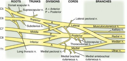
1. Brachial plexus ( Fig. 2.15)
- Formed from the ventral primary rami of C5 to T1
- Exits neck between the anterior and middle scalene muscles
- Dorsal rami of C5 to T1 innervate the dorsal neck musculature and skin.
- From proximal to distal: roots, trunks, divisions, cords, and branches (mnemonic: “ R eal T exans d rink c old b eer”).
- Five roots (C5–T1), although contributions from C4 (pre-fixed) and T2 (post-fixed) can be small
- Dorsal scapular nerve (C5): to levator scapula, rhomboid major/minor
- Long thoracic nerve (C5–7): to serratus anterior
- Three trunks (upper, middle, lower)
- Suprascapular nerve (upper trunk, C5, 6)
- Nerve to subclavius (upper trunk, C5, 6)
- Six divisions (anterior and posterior; two limbs from each trunk)
- No terminal branches at this level
- Three cords (lateral, medial, and posterior) named for their anatomic relationship to the axillary artery ( Table 2.15)
Table 2.14
Carpal Tunnel Borders BORDER
|
STRUCTURES
| ---|---|
Roof
| Transverse carpal ligament
Floor
| Carpal bones
Radial
| Scaphoid tubercle/trapezium
Ulnar
| Hook of hamate

---
FIG. 2.15 Brachial plexus.
From Miller MD et al:
Orthopaedic surgical approaches,
Philadelphia, 2008, Saunders, Figure SA-7.
1. Main terminal branches (musculocutaneous, axillary, radial, median, ulnar)
2. Other terminal branches
1. Lateral cord
1. Lateral pectoral nerve (C5–7): to pectoralis major
2. Posterior cord
1. Upper subscapular nerve (C5, 6): to
subscapularis
2. Lower subscapular nerve (C5, 6): to subscapularis, teres major
3. Thoracodorsal nerve (C6–8): to latissimus dorsi
3. Medial cord
1. Medial pectoral nerve (C8, T1): to pectoralis minor/major
2. Medial brachial cutaneous nerve (T1)
3. Medial antebrachial cutaneous nerve (C8, T1)
All minor medial and lateral cord branches have medial or lateral in their names. Posterior cord branches do not.
1. 1. 1. 2. Preclavicular branches (from roots and upper trunk)
- Dorsal scapular nerve
- Long thoracic nerve
- Suprascapular nerve
- Nerve to the subclavius
- Innervation of all rotator cuff muscles is derived from C5 and C6 of the brachial plexus ( Table 2.16 ). 4. Brachial plexus injury
- Preganglionic brachial plexus injuries
- Proximal to dorsal root ganglion: CNS injury with little potential for recovery
- Preclavicular nerve involvement
-
Medial scapular winging (long thoracic nerve to serratus anterior), rhomboid paralysis (dorsal scapular nerve), Horner syndrome (disruption of stellate ganglion/sympathetic chain at C8, T1), rotator cuff dysfunction (suprascapular nerve to
supraspinatus/infraspinatus), latissimus dorsi paralysis (thoracodorsal nerve), elevated hemidiaphragm (phrenic nerve) - Nerve conduction study: absent motor but intact sensory conduction
- Postganglionic brachial plexus injuries
- Less preclavicular nerve involvement (no scapular winging, rhomboid paralysis, etc.)
- Characteristic obstetric brachial plexus palsies ( Table 2.17)
- Scapular winging
- Medial winging: long thoracic nerve (C5–7) injury leading to serratus anterior dysfunction
- Superior elevation with the scapula translated medially and the inferior angle rotated medially
- Lateral winging: spinal accessory nerve (cranial nerve XI) injury leading to trapezius dysfunction
- Shoulder depression with the scapula translated laterally and the inferior angle rotated laterally because of the unopposed pull of the serratus anterior
Usually a history of ipsilateral neck surgery (i.e., thyroidectomy)
1. 1. 2. Major brachial plexus branches ( Table 2.18)
-
Suprascapular nerve (upper trunk, C5, 6)
BRANCH
|
ORIGIN
|
ROOTS
|
MOTOR INNERVATION
|
SENSORY INNERVATION
| ---|---|---|---|---|
Dorsal scapular
| C5 root
| C5
| Rhomboid major, rhomboid minor
| Long thoracic | C–7 roots
| C5–7
| Serratus anterior
| Suprascapular | Superior trunk
| C5, 6
| Supraspinatus, infraspinatus
| Nerve to subclavius | Superior trunk
| C5, 6
| Subclavius
| Lateral pectoral | Lateral cord
| C5–7
| Pectoralis major
| Upper subscapular | Posterior cord
| C5, 6
| Subscapularis
| Table 2.15 Brachial Plexus Lower subscapular | Posterior cord
| C5, 6
| Subscapularis, teres major
| ---|---|---|---|--- Thoracodorsal | Posterior cord
| C6–8
| Latissimus dorsi
| Medial pectoral | Medial cord
| C8, T1
| Pectoralis major, pectoralis minor
| Medial brachial cutaneous | Medial cord
| C8, T1
| |
Medial arm Medial antebrachial cutaneous | Medial cord
| C8, T1
| |
Medial forearm Musculocutaneous | Lateral cord
| C5–7
| All muscles in the anterior compartment of the arm
| Lateral forearm Axillary | Posterior cord
| C5, 6
| Deltoid, teres minor
| Lateral shoulder Radial | Posterior cord
| C5–T1
| All muscles in the posterior compartments of arm and forearm
| Posterior
arm/forearm, dorsum of radial hand Median | Medial/lateral cords
| C5–T1
| All muscles in the anterior compartment of the forearm (except FCU and ulnar FDP),
| opponens pollicis, FPB, APB, and two lateral lumbrical muscles | Radial 3½ digits, radial palm Ulnar | Medial cord | C8, T1 | FCU, ulnar FDP, all intrinsic muscles of the hand (except opponens pollicis, FPB, APB, and two lateral | Ulnar 1½ digits, ulnar palm/wrist | | | lumbricals) |
| --- | --- | --- | --- | --- |
Table 2.16
Rotator Cuff Muscle Innervation Muscles Innervated Nerves ---
External Rotators Supraspinatus | Suprascapular nerve (C5, 6) Infraspinatus | Suprascapular nerve (C5, 6) Teres minor | Axillary nerve (C5, 6)
Internal Rotator Subscapularis | Upper (C5) and lower (C5, 6) subscapular nerves
Table 2.17
Obstetric Brachial Plexus Palsies Palsy Type Roots Deficit Prognosis --- Erb- Duchenne | C5, 6
| Weakness of deltoid, rotator cuff, elbow flexors, and wrist and hand extensors
“Waiter’s tip”
| Best Klumpke | C8, T1
| Weakness of wrist flexors and intrinsic apparatus, Horner syndrome
| Poor Total plexus | C5–T1
| Flaccid arm
| Worst
Table 2.18
Major Brachial Plexus Branches Nerves Muscles Innervated --- Musculocutaneous (lateral cord) | Coracobrachialis, biceps, brachialis Axillary (posterior cord) | Deltoid, teres minor
Radial Nerve Radial (posterior cord) | Triceps, brachioradialis, extensor carpi radialis longus, extensor carpi radialis brevis Posterior interosseous | Supinator, extensor carpi ulnaris, extensor digitorum, extensor digiti minimi, abductor pollicis longus, extensor pollicis longus, extensor pollicis brevis, extensor indicis proprius
Median Nerve Median (medial and lateral cord) | Pronator teres, flexor carpi radialis, palmaris longus, flexor digitorum superficialis, abductor pollicis brevis, superficial head of flexor pollicis brevis, opponens pollicis, first and second lumbrical muscles Anterior interosseous | Flexor digitorum profundus (first and second), flexor pollicis longus, pronator quadratus
Ulnar Nerve Ulnar (medial cord) | Flexor carpi ulnaris, flexor digitorum profundus (third and fourth), pollicis brevis, abductor digiti minimi, opponens digiti minimi, flexor digiti minimi, third and fourth lumbrical muscles, interossei, adductor pollicis, deep head of flexor pollicis brevis


FIG. 2.16 Nerves in shoulder and arm (lateral cord).
From Drake RL et al, editors: Gray’s atlas of anatomy, ed 2, Philadelphia, 2015, Churchill Livingstone. - Passes through scapular notch beneath superior transverse scapular ligament, sending a branch to the supraspinatus before traveling through the spinoglenoid notch to innervate the infraspinatus
- Musculocutaneous nerve (lateral cord, C5–7) ( Fig. 2.16)
- Pierces the coracobrachialis 5 to 8 cm distal to the coracoid
- Branches supply the coracobrachialis, biceps, and brachialis.
-
Gives off a sensory branch to the elbow joint before it becomes the lateral antebrachial cutaneous nerve of the forearm, which is located deep to the cephalic vein


 FIG. 2.17 Nerves in shoulder and arm (posterior cord).
From Drake RL et al, editors:
Gray’s atlas of anatomy,
ed 2, Philadelphia, 2015, Churchill Livingstone.
FIG. 2.17 Nerves in shoulder and arm (posterior cord).
From Drake RL et al, editors:
Gray’s atlas of anatomy,
ed 2, Philadelphia, 2015, Churchill Livingstone.
- Axillary nerve (posterior cord, C5, 6)
Passes anterior to subscapularis muscle and inferior to shoulder capsule, traveling from anterior to posterior through quadrangular space
1. Anterior branch supplying deltoid and skin over lateral shoulder passes around humerus in deep deltoid fascia approximately 5–7 cm distal to acromion.
- Posterior branch supplies teres minor and posterior deltoid.
- Radial nerve (posterior cord, C5–T1) ( Fig. 2.17)
- Passes through triangular interval and then spirals around the humerus (medial to lateral) in the spiral groove. Approximately 20 cm from medial epicondyle and 14 cm from lateral epicondyle
- Emerges on the lateral side of the arm after piercing the lateral intermuscular septum approximately 7.5 cm above the trochlea between the brachialis and brachioradialis anterior to the lateral epicondyle (where it supplies the anconeus muscle)
- Passes anterior to the lateral epicondyle between the brachialis and brachioradialis and divides into the superficial and deep (posterior interosseous nerve [PIN]) branches approximately 1–3 cm distal to lateral epicondyle
PIN splits the supinator and supplies all of the extensor muscles except the mobile wad (brachioradialis, ECRB, ECRL).
1. 1. 1. Terminal sensory branch to dorsal wrist joint in floor of fourth extensor compartment
Superficial branch of the radial nerve emerges through antebrachial fascia approximately 6–9 cm proximal to the radial styloid. Runs between the brachioradialis and ERCL to supply
sensation to the dorsal radial surface distal forearm and hand.
1. 1. 2. Median nerve (medial and lateral cords, C5–T1) ( Fig. 2.18)
- Accompanies brachial artery in the arm, crossing it during its course (lateral to medial) approximately 15 cm from the medial epicondyle
- Supplies some branches to the elbow joint but has no branches in the arm itself
- Medial to brachial artery and superficial to brachialis muscle as it passes
- In forearm, the median nerve splits the two heads of the pronator teres and then runs between the FDS and FDP. Supplies all the superficial flexor muscles of the forearm except the FCU.
Anterior interosseous nerve branches 4 cm distal to elbow and runs between the FPL and FDP; supplies all the deep flexors except the ulnar half of the FDP. Terminates in the pronator quadratus (PQ).
1. Palmar cutaneous branch arises approximately 6 cm proximal to radial styloid and passes superficial to the flexor retinaculum to innervate the thenar skin.
- Median nerve passes through the carpal tunnel between FDS and flexor carpi radialis (FCR) to supply the radial lumbricals, thenar musculature via a deep recurrent branch, and sensation to the volar aspect of thumb, index, long, and radial half of the ring fingers.
- Ulnar nerve (medial cord, C8, T1) ( Fig. 2.19)
- Posteromedial to brachial artery in upper arm and then passes posterior to the medial intermuscular septum at the arcade of Struthers (8–10 cm from medial epicondyle)
- Crosses elbow posterior to medial epicondyle at elbow through the cubital tunnel
- Cubital tunnel: Osborne ligament (roof), medial collateral ligament (MCL) (floor)
- No branches in arm, but supplies articular branch to elbow joint
Enters the forearm between the two heads of the FCU (humeral and ulnar)
1. Runs between the FCU and FDP, innervating the
ulnar half of this muscle (FDP to ring and small fingers)


 FIG. 2.18 Nerves in shoulder and arm (medial and lateral cords).
From Drake RL et al, editors:
Gray’s atlas of anatomy,
ed 2, Philadelphia, 2015, Churchill Livingstone.
FIG. 2.18 Nerves in shoulder and arm (medial and lateral cords).
From Drake RL et al, editors:
Gray’s atlas of anatomy,
ed 2, Philadelphia, 2015, Churchill Livingstone.
2. Runs radial to FCU at distal forearm and wrist
3. Dorsal cutaneous nerve branches approximately 7 cm proximal to ulnar styloid and provides sensation to dorsoulnar forearm and wrist.
4. Ulnar nerve enters hand superficial to TCL through Guyon canal. Divides into a superficial sensory branch and a deep motor branch.
1. Deep branch travels around the hook of the hamate between the abductor digiti minimi and flexor digiti minimi brevis, providing motor innervation to intrinsic muscles of hand.
2. Sensory branch supplies ulnar half of the ring and the small fingers.


 FIG. 2.19
Nerve regions in shoulder and arm. (From Drake RL et al, editors:
Gray’s atlas of anatomy,
ed 2, Philadelphia, 2015, Churchill Livingstone.)
FIG. 2.19
Nerve regions in shoulder and arm. (From Drake RL et al, editors:
Gray’s atlas of anatomy,
ed 2, Philadelphia, 2015, Churchill Livingstone.)
1.
Cutaneous innervation to upper extremity (
Figs. 2.20
through
2.22)
1. Supraclavicular nerve (C3 and C4) supplies the upper shoulder.
2. Axillary nerve supplies the shoulder joint and the overlying skin.
3. Medial, lateral, and dorsal brachial cutaneous nerves supply the balance of cutaneous innervation of the arm.
4. Lateral antebrachial cutaneous nerve: continuation of the musculocutaneous nerve that passes lateral to the cephalic vein after emerging laterally from between the biceps and brachialis at the elbow
5. Medial antebrachial cutaneous nerve: a branch from the medial cord of the brachial plexus
6. Posterior antebrachial cutaneous nerve: a branch of the radial nerve given off in the arm
7. Sensation to thumb
1. Provided by five branches: lateral antebrachial cutaneous nerve, superficial and dorsal digital branches of the radial nerve, and digital and palmar branches of the median nerve

 FIG. 2.20 Nerve regions in arm and hand.
From Drake RL et al, editors:
Gray’s atlas of anatomy,
ed 2, Philadelphia, 2015, Churchill Livingstone.
FIG. 2.20 Nerve regions in arm and hand.
From Drake RL et al, editors:
Gray’s atlas of anatomy,
ed 2, Philadelphia, 2015, Churchill Livingstone.
8.
Neuroanatomic relationships in the forearm are outlined in
Table 2.19


 FIG. 2.21
Nerve regions in arm and hand (anterior view). (From Drake RL et al, editors:
Gray’s atlas of anatomy,
ed 2, Philadelphia, 2015, Churchill Livingstone.)
FIG. 2.21
Nerve regions in arm and hand (anterior view). (From Drake RL et al, editors:
Gray’s atlas of anatomy,
ed 2, Philadelphia, 2015, Churchill Livingstone.)
Table 2.19 Neuroanatomic Relationships in Forearm Nerve Relationship --- Radial | Between brachialis and brachioradialis Posterior interosseous | Splits supinator Superficial radial | Between brachioradialis and extensor carpi radialis longus Median | Medial to brachial artery at elbow Anterior interosseous | Splits pronator teres and runs between flexor digitorum superficialis and flexor digitorum profundus
Between flexor pollicis longus and flexor digitorum profundus
Ulnar
| Between flexor carpi ulnaris and flexor digitorum profundus


 FIG. 2.22 Nerve regions in arm and hand (posterior view).
From Drake RL et al, editors:
Gray’s atlas of anatomy,
ed 2, Philadelphia, 2015, Churchill Livingstone
