Comprehensive Introduction and Patho-Epidemiology
The medial calcaneal displacement osteotomy (MCDO) represents a monumental paradigm shift in the joint-sparing surgical management of adult-acquired flatfoot deformity (AAFD), which is increasingly referred to in contemporary literature as progressive collapsing foot deformity (PCFD). Historically, the surgical approach to the collapsing medial longitudinal arch relied heavily on isolated soft tissue reconstructions, such as isolated flexor digitorum longus (FDL) transfers and spring ligament plications. However, these isolated soft tissue procedures were plagued by unacceptably high failure rates due to the relentless, uncorrected biomechanical forces exerted on the hindfoot during the stance phase of gait. The popularization of the MCDO by Myerson and others provided a robust, extra-articular structural correction that fundamentally alters the biomechanical axis of the hindfoot, thereby protecting the medial soft tissue reconstruction and ensuring long-term construct survivorship.
Pathophysiologically, Stage II PCFD is characterized by a flexible pes planovalgus deformity driven by the insidious degeneration, elongation, and eventual incompetence of the posterior tibial tendon (PTT) and the crucial static stabilizers of the medial column, primarily the calcaneonavicular (spring) ligament complex. As these medial restraints fail, the talus plantarflexes and rotates medially, while the calcaneus drifts into severe valgus. This multiplanar collapse disrupts the normal kinematic chain of the lower extremity. The resultant valgus alignment of the hindfoot laterally translates the insertion of the Achilles tendon relative to the subtalar joint axis. Consequently, the massive power of the gastrocsoleus complex, which normally acts as a primary plantarflexor and secondary invertor, is pathologically converted into a potent evertor, exacerbating the deformity with every step.
Epidemiologically, PCFD predominantly affects women in their fifth to seventh decades of life, with a well-documented correlation to systemic comorbidities such as obesity, hypertension, and diabetes mellitus. The chronic, repetitive microtrauma to a hypovascular zone of the posterior tibial tendon distal to the medial malleolus initiates a cascade of tenosynovitis, tendinosis, and eventual macroscopic tearing. The insidious nature of this condition often leads to delayed presentation, by which time the deformity has progressed from a purely tendinopathic process (Stage I) to a structurally significant, yet flexible, osseous malalignment (Stage II). The MCDO intervenes precisely at this critical juncture, offering a powerful, joint-preserving intervention before the onset of rigid, irreversible arthrosis of the triple joint complex.
The overarching philosophy of the MCDO is to physically relocate the posterior calcaneal tuberosity—and thereby the insertion of the Achilles tendon—medially. By translating the tuberosity by approximately 1 centimeter, the surgeon effectively medializes the mechanical vector of the gastrocsoleus complex, restoring its physiological role as an invertor of the hindfoot. This structural modification not only corrects the valgus thrust but also significantly unloads the medial column, dramatically reducing the tensile stress placed on the transferred FDL and the reconstructed spring ligament. This synergistic harmony between osseous realignment and soft tissue reconstruction is the cornerstone of modern PCFD management.
Detailed Surgical Anatomy and Biomechanics
A profound understanding of the complex osteology, vascularity, and neuroanatomy of the hindfoot is absolutely paramount for the safe and effective execution of the medial calcaneal displacement osteotomy. The calcaneus is the largest of the tarsal bones, featuring a dense cortical shell enveloping a highly vascular, robust cancellous core. The posterior tuberosity serves as the insertion site for the Achilles tendon superiorly and the origin of the plantar fascia inferiorly. The lateral wall of the calcaneus is relatively flat, providing an excellent surface for the surgical approach, while the medial wall is deeply concave, forming the lateral boundary of the tarsal tunnel. The sustentaculum tali projects medially, supporting the middle facet of the subtalar joint and serving as a crucial pulley for the flexor hallucis longus (FHL) tendon.
The neurovascular anatomy surrounding the calcaneus dictates the surgical approach and the parameters of safe osteotomy execution. Laterally, the sural nerve courses posterior to the peroneal tendons, providing sensory innervation to the lateral hindfoot and midfoot. The branching pattern of the sural nerve—specifically the lateral calcaneal nerve—is highly variable, necessitating meticulous, blunt subcutaneous dissection to avoid iatrogenic injury and the subsequent development of debilitating neuromas. Medially, the posterior tibial artery and the tibial nerve bifurcate into the medial and lateral plantar bundles within the tarsal tunnel. These critical structures lie in close proximity to the medial cortex of the calcaneus. When completing the medial cortical breach during the osteotomy, the surgeon must exercise extreme caution, utilizing precise saw control and protective retractors to prevent catastrophic neurovascular injury.
Biomechanically, the normal subtalar joint acts as a complex, multi-axial hinge, often likened to a mitered joint. The axis of the subtalar joint runs obliquely from posterolateral-inferior to anteromedial-superior. In a physiologically normal foot, the insertion of the Achilles tendon lies slightly medial to this axis, creating an inversion moment during the heel-rise phase of gait. This inversion locks the transverse tarsal joint (calcaneocuboid and talonavicular joints), converting the foot into a rigid lever arm for efficient push-off. In the pathologic setting of PCFD, the calcaneus abducts and everts, shifting the Achilles insertion lateral to the subtalar axis. The Achilles tendon now acts as a deforming evertor, unlocking the transverse tarsal joint and perpetuating the collapse of the medial longitudinal arch.
The MCDO directly addresses this biomechanical derangement. By performing a transverse osteotomy and translating the posterior tuberosity medially by 8 to 10 millimeters, the surgeon physically shifts the Achilles vector back to a position medial to the subtalar axis. This translation restores the inversion moment of the gastrocsoleus complex. Furthermore, the medial translation of the heel pad shifts the ground reaction force medially, decreasing the valgus moment at the subtalar joint. Crucially, unlike lateral column lengthening procedures (such as the Evans osteotomy), the MCDO does not increase contact pressures across the calcaneocuboid joint, making it an ideal choice for patients with pre-existing lateral column pain or those at risk for iatrogenic calcaneocuboid arthrosis.
Exhaustive Indications and Contraindications
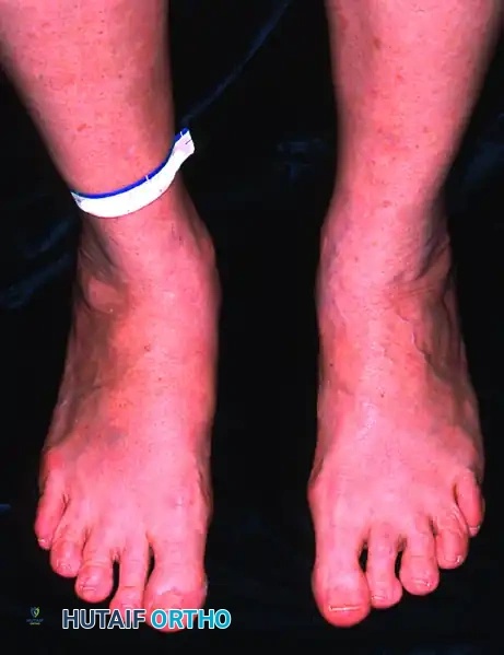
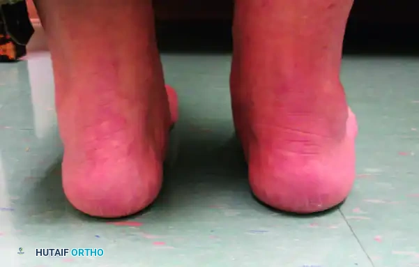
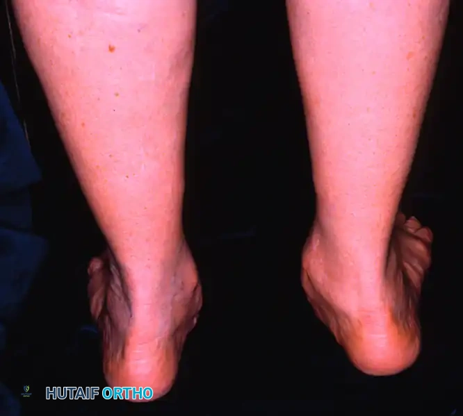
The decision to perform a medial calcaneal displacement osteotomy hinges on a meticulous clinical and radiographic evaluation, ensuring the patient's pathology aligns with the biomechanical benefits of the procedure. The quintessential indication for an MCDO is Stage II Adult-Acquired Flatfoot Deformity (flexible pes planovalgus), where the primary driver of the deformity is hindfoot valgus rather than severe forefoot abduction. Clinically, these patients present with medial hindfoot pain localized to the posterior tibial tendon, an inability to perform a single-limb heel rise, the classic "too many toes" sign viewed from posteriorly, and a passively correctable hindfoot. The flexibility of the deformity is paramount; if the subtalar joint cannot be manually reduced to a neutral or slight varus position, an extra-articular osteotomy will fail to correct the alignment, and an arthrodesis must be considered.
Patient selection requires a nuanced approach, often weighing the MCDO against, or combining it with, a lateral column lengthening (Evans osteotomy). The MCDO is particularly advantageous in heavier patients or those with a high Body Mass Index (BMI). The broad cancellous surfaces of the calcaneal tuberosity heal with a high degree of predictability and are highly resistant to displacement or subsidence, whereas the structural bone graft utilized in an Evans osteotomy is prone to crushing or nonunion under excessive axial loads. Furthermore, in patients whose standing anteroposterior radiographs demonstrate only mild to moderate talonavicular uncoverage (midfoot abduction), the MCDO alone, combined with medial soft tissue reconstruction, provides sufficient correction without the inherent risks of lateral column lengthening.
Conversely, absolute contraindications to the MCDO must be strictly respected to avoid catastrophic surgical failures. Stage III PCFD, defined by a rigid, fixed hindfoot valgus deformity with or without advanced arthrosis of the subtalar or talonavicular joints, is an absolute contraindication. Attempting an extra-articular osteotomy in the presence of a rigid intra-articular deformity will not restore alignment and will leave the patient with persistent pain. Stage IV disease, characterized by deltoid ligament incompetence and valgus tilt of the talus within the ankle mortise, also precludes isolated MCDO, as the primary pathology has ascended to the tibiotalar joint. Severe peripheral neuropathy (e.g., Charcot neuroarthropathy), active local or systemic infection, and critical limb ischemia are standard absolute contraindications.
| Category | Indications for MCDO | Contraindications for MCDO |
|---|---|---|
| Primary Pathology | Stage II PCFD (Flexible pes planovalgus) | Stage III PCFD (Rigid/fixed hindfoot valgus) |
| Joint Status | Passively correctable subtalar joint | Subtalar or talonavicular arthrosis |
| Ankle Alignment | Normal tibiotalar alignment | Stage IV PCFD (Ankle valgus / Deltoid failure) |
| Patient Factors | High BMI (where Evans graft may subside) | Severe peripheral neuropathy (Charcot) |
| Deformity Type | Predominant hindfoot valgus | Severe, isolated forefoot abduction |
| Vascular Status | Intact pedal pulses / good capillary refill | Critical limb ischemia / severe vasculopathy |
Pre-Operative Planning, Templating, and Patient Positioning
Thorough pre-operative planning is the foundation of a successful medial calcaneal displacement osteotomy. The clinical examination must include a precise assessment of the gastrocnemius-soleus complex utilizing the Silfverskiöld test. A tight Achilles tendon or isolated gastrocnemius contracture is a near-universal finding in PCFD. If the ankle cannot be dorsiflexed past neutral with the knee extended but corrects with the knee flexed, a gastrocnemius recession (Strayer or Baumann procedure) is indicated. If the contracture persists with the knee flexed, a percutaneous Achilles tendon lengthening (TAL) is required. Addressing this equinus contracture is non-negotiable; failure to do so will result in excessive strain on the medial reconstruction and persistent midfoot collapse. Additionally, the surgeon must assess for a fixed forefoot supinatus. If the forefoot remains supinated upon reduction of the hindfoot to neutral, an adjunctive medial column stabilization (e.g., Cotton osteotomy or first tarsometatarsal arthrodesis) must be incorporated into the surgical plan.
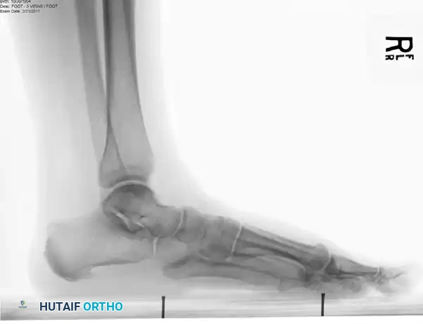
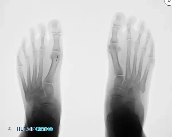
Radiographic evaluation demands high-quality, weight-bearing imaging of the foot and ankle. The weight-bearing anteroposterior (AP) view is utilized to assess talonavicular uncoverage and the talocalcaneal (Kite's) angle. The weight-bearing lateral view is critical for evaluating the longitudinal arch, specifically measuring Meary's angle (the intersection of the longitudinal axes of the talus and the first metatarsal) and the calcaneal pitch. The hindfoot alignment view (Saltzman view) is perhaps the most crucial imaging modality for the MCDO, as it allows for the direct quantification of the valgus moment arm of the calcaneal tuberosity relative to the mechanical axis of the tibia. Advanced imaging, such as Magnetic Resonance Imaging (MRI), is routinely obtained to definitively assess the integrity of the posterior tibial tendon, the spring ligament complex, and the deltoid ligament, as well as to rule out occult osteochondral lesions or early degenerative changes within the subtalar joint.
In the operating theater, patient positioning is a critical logistical consideration, as the procedure requires access to both the lateral and medial aspects of the foot. The preferred technique involves a staged positional approach. The patient is initially placed in the lateral decubitus position on a radiolucent beanbag, ensuring all bony prominences (peroneal nerve at the fibular head, greater trochanter) are meticulously padded. A well-padded thigh tourniquet is applied. This lateral position provides unparalleled, ergonomic access for the calcaneal osteotomy and hardware placement. Once the osteotomy is rigidly fixed and the lateral wound closed, the beanbag is deflated, and the patient is carefully rolled into the supine position. A bump is placed under the ipsilateral hip to allow for external rotation of the leg, providing optimal exposure for the medial soft tissue reconstruction (FDL transfer, spring ligament repair) and any necessary medial column osseous procedures.
Alternatively, some surgeons prefer to perform the entire procedure in the supine position with a large bump under the ipsilateral hip to internally rotate the leg for the lateral approach, subsequently removing the bump for the medial work. While this avoids the need for intraoperative repositioning and re-draping, it can make the lateral calcaneal approach ergonomically challenging and may compromise the surgeon's ability to achieve a perfectly perpendicular saw cut. Regardless of the chosen positioning strategy, the entire lower extremity from the knee down must be prepped and draped free into the sterile field to allow for the intraoperative assessment of overall hindfoot alignment and ankle range of motion.
Step-by-Step Surgical Approach and Fixation Technique
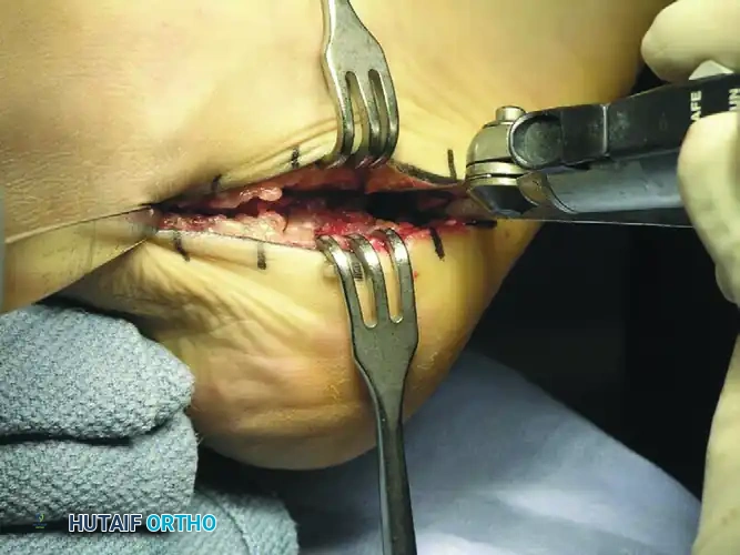
The surgical execution of the MCDO demands precision, anatomical respect, and rigid biomechanical fixation. With the patient in the lateral decubitus position and the tourniquet inflated, the anatomical landmarks—the tip of the lateral malleolus, the course of the peroneal tendons, and the plantar aspect of the heel—are palpated and marked. An oblique incision, approximately 4 to 5 centimeters in length, is made over the lateral wall of the calcaneus. The incision is oriented posteroinferior to the peroneal tendons, following the natural resting skin tension lines. The dissection is carried through the subcutaneous tissue using meticulous blunt spreading with a hemostat. This blunt technique is critical to identify and protect the variable branches of the sural nerve, which frequently traverse the operative field. Once identified, the nerve is gently retracted, usually superiorly, and protected throughout the procedure.
The deep dissection proceeds sharply down to the periosteum of the lateral calcaneal wall. A periosteal elevator is utilized to reflect the periosteum dorsally and plantarly, creating a subperiosteal envelope for the osteotomy. Broad Hohmann retractors or specialized calcaneal retractors are placed superiorly (protecting the peroneal tendons and sural nerve) and inferiorly (protecting the plantar fascia and intrinsic musculature). The trajectory of the osteotomy is then planned. Using a broad oscillating saw blade, a transverse cut is initiated. The orientation of this cut is of paramount importance: it must be exactly perpendicular (90 degrees) to the lateral wall of the calcaneus in the coronal plane, and inclined posteriorly at an angle of approximately 45 degrees to the plantar plane of the foot in the sagittal plane. This 45-degree inclination is a critical biomechanical safeguard; it ensures that the pull of the Achilles tendon on the posterior fragment compresses the osteotomy site rather than causing it to migrate dorsally, which would result in a disastrous loss of calcaneal pitch.

As the saw blade traverses the dense cancellous bone and approaches the medial cortex, the surgeon must exercise extreme tactile feedback. The medial cortex is carefully breached, ensuring the saw blade does not plunge into the medial soft tissues, thereby protecting the neurovascular bundle, the FHL, and the FDL tendons. Once the osteotomy is complete, a toothless lamina spreader or a broad osteotome is inserted into the osteotomy site. The spreader is gently opened to stretch and relax the stout medial periosteum and retinacular attachments. This mobilization is crucial; attempting to translate the tuberosity without adequate soft tissue release will result in a hinged, incomplete displacement or an iatrogenic varus tilt.
Following adequate mobilization, the lamina spreader is removed. Using a specialized calcaneal displacement tool or a robust elevator, the posterior tuberosity is translated medially by 8 to 10 millimeters. The surgeon must palpate the medial and lateral borders to confirm a pure translational shift, strictly avoiding any varus or valgus angulation. Once the desired position is achieved, the fragments are provisionally stabilized with a stout, smooth Steinmann pin driven from the posteroinferior aspect of the tuberosity across the osteotomy site into the anterior calcaneal body.

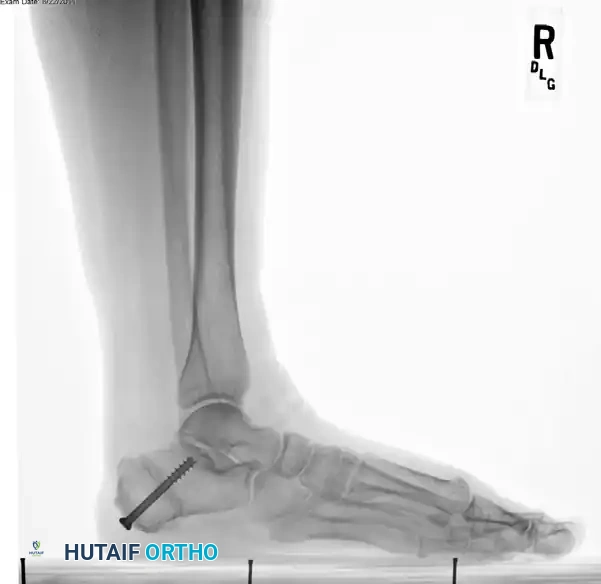
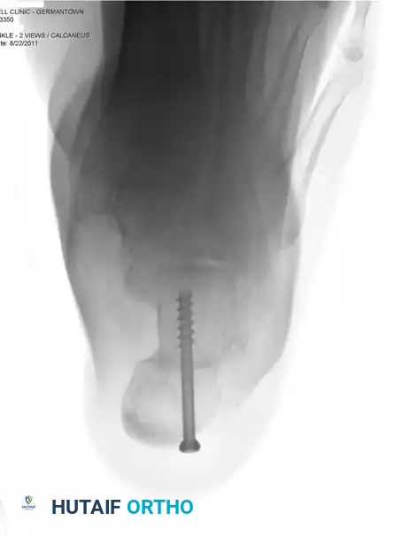
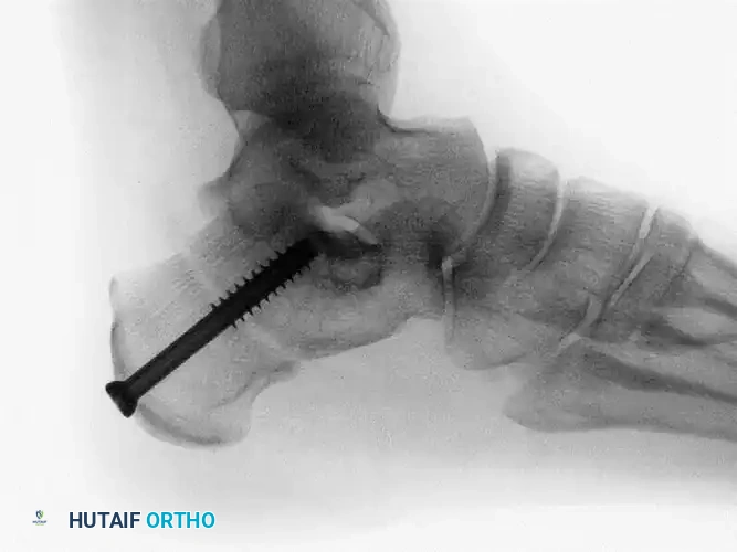
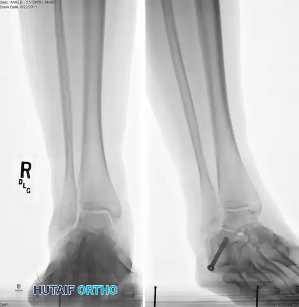
Definitive fixation is achieved utilizing a single, large-diameter (6.5 mm to 7.3 mm), partially threaded, cannulated cancellous screw. The guidewire is introduced from the posteromedial and inferior aspect of the displaced tuberosity, directed anterolaterally and superiorly toward the anterior process of the calcaneus, aiming roughly toward the sinus tarsi. Intraoperative fluoroscopy is mandatory at this stage. Lateral, axial (Harris), and Broden's views are obtained to confirm the trajectory of the guidewire, verify the 10 mm medial translation, and, most importantly, ensure that the hardware has not penetrated the posterior facet of the subtalar joint. Once positioning is confirmed, the outer cortex is drilled, and the screw is advanced over the wire, achieving rigid, dynamic compression across the broad cancellous surfaces. The wound is copiously irrigated and closed in layered fashion, paying special attention to a meticulous, tension-free skin closure to prevent dehiscence.
Complications, Incidence Rates, and Salvage Management
While the medial calcaneal displacement osteotomy is a highly reliable and transformative procedure, it is not without potential complications. The surgeon must be acutely aware of these risks, employ meticulous surgical technique to minimize their occurrence, and possess the requisite knowledge to manage them effectively should they arise. The most frequent complication is injury to the sural nerve or its lateral calcaneal branches, resulting in postoperative paresthesias, hyperesthesia, or the formation of a painful neuroma. The incidence of sural neuritis is reported to be between 5% and 15%. Prevention relies entirely on blunt subcutaneous dissection and gentle retraction. If a neuroma develops and is refractory to conservative management (gabapentinoids, targeted corticosteroid injections), surgical intervention involving neuroma excision and implantation of the proximal nerve stump deep into the lateral musculature or bone may be required.
A far more devastating biomechanical complication is the overcorrection of the osteotomy, resulting in an iatrogenic varus hindfoot. A rigid varus hindfoot is exceptionally poorly tolerated by patients, leading to severe lateral column overload, intractable pain along the fifth metatarsal, and rapid degeneration of the subtalar and calcaneocuboid joints. This typically occurs due to an angular tilt of the tuberosity during translation rather than a pure medial slide. If recognized intraoperatively, it must be immediately corrected. If identified postoperatively and the patient is symptomatic, a revision lateralizing calcaneal osteotomy is mandatory. Conversely, undercorrection (failure to translate the full 10 mm or failure to address a concomitant forefoot supinatus) will leave the patient with residual valgus and continued strain on the medial soft tissue reconstruction, potentially leading to recurrent deformity.
Hardware-related complications are relatively common, given the thin soft tissue envelope over the posterior heel pad. The prominent head of the large-diameter cannulated screw can cause local irritation, bursitis, and pain with shoewear. Symptomatic hardware is reported in 10% to 20% of patients. Management is straightforward: once complete bony consolidation is confirmed via radiography or CT (typically between 6 and 12 months postoperatively), the screw can be safely removed in a minor outpatient procedure. Delayed union or nonunion of the osteotomy is exceedingly rare (<1%) due to the massive cancellous surface area and robust vascularity of the calcaneus. However, in patients with severe vasculopathy, uncontrolled diabetes, or those who continue to use tobacco products, the risk is elevated. Smoking cessation is an absolute prerequisite for this surgery.
| Complication | Estimated Incidence | Etiology / Risk Factors | Salvage / Management Strategy |
|---|---|---|---|
| Sural Neuritis / Neuroma | 5% - 15% | Direct laceration, traction injury, entrapment in scar tissue. | Gabapentinoids, steroid injections. Surgical excision and deep muscular implantation if refractory. |
| Symptomatic Hardware | 10% - 20% | Prominent screw head irritating the posterior heel pad / Achilles insertion. | Outpatient hardware removal after confirmation of complete bony union (6-12 months). |
| Overcorrection (Varus) | 1% - 3% | Iatrogenic varus tilt during medial translation. Failure to assess global alignment. | Revision surgery: Lateralizing calcaneal osteotomy to restore neutral hindfoot alignment. |
| Undercorrection (Residual Valgus) | 5% - 10% | Insufficient translation (<8mm). Failure to address forefoot supinatus (Cotton osteotomy needed). | Custom orthotics. If severe, revision osteotomy or progression to arthrodesis. |
| Delayed Union / Nonunion | < 1% | Smoking, severe vasculopathy, inadequate rigid compression. | Prolonged immobilization, bone stimulators. Revision with bone grafting if frank nonunion. |
| Wound Dehiscence / Infection | 2% - 5% | Poor soft tissue handling, excessive retraction, smoking, uncontrolled diabetes. | Local wound care, oral/IV antibiotics. Surgical debridement for deep infections. |
Phased Post-Operative Rehabilitation Protocols
The success of the MCDO, particularly when performed in conjunction with a medial soft tissue reconstruction (FDL transfer), is highly dependent on strict adherence to a phased, meticulously controlled postoperative rehabilitation protocol. The primary goals are to protect the osseous fixation until clinical and radiographic union is achieved, safeguard the delicate tendon transfer, and ultimately restore the strength, proprioception, and functional kinematics of the lower extremity. The rehabilitation timeline spans approximately 9 to 12 months, requiring significant patient compliance and dedicated physical therapy.
Phase I: Maximum Protection (Weeks 0 to 2)
Immediately following surgery, the patient is placed in a bulky, well-padded short leg plaster or fiberglass splint. The ankle is immobilized in slight plantarflexion and inversion to completely remove tension from the transferred FDL and the reconstructed spring ligament. The patient is designated strictly non-weight-bearing (NWB) on the operative extremity. Crutches, a walker, or a knee scooter are utilized for mobility. Strict elevation of the limb above the level of the heart is absolutely critical during this phase to minimize postoperative edema, which is the primary driver of incisional pain and the greatest threat to wound healing on the lateral calcaneal wall. Deep vein thrombosis (DVT) prophylaxis is initiated based on patient-specific risk factors.
Phase II: Immobilization and Early Healing (Weeks 2 to 6)
At the two-week postoperative mark, the initial splint is removed, and the surgical incisions are meticulously inspected. Suture or staple removal is performed if the wounds are completely coapted and healing appropriately. The patient is then transitioned into a short leg fiberglass cast or a locked controlled ankle motion (CAM) boot. The ankle remains locked in a neutral or slightly plantarflexed and inverted position. The patient remains strictly non-weight-bearing. During this phase, patients are encouraged to perform active range of motion exercises for the knee and hip to maintain proximal muscle tone, as well as isometric quadriceps and hamstring contractions.
Phase III: Progressive Weight-Bearing and Early Mobilization (Weeks 6 to 12)
At six weeks postoperatively, new weight-bearing radiographs (AP, lateral, and axial views) are obtained. The surgeon evaluates the osteotomy site for signs of early bony consolidation, characterized by the blurring of the osteotomy lines and the presence of bridging trabecular bone. If clinical and radiographic progression is satisfactory, the patient is permitted to initiate progressive partial weight-bearing in the CAM boot. Weight-bearing is advanced by approximately 25% of body weight per week, utilizing an assistive device, until full weight-bearing in the boot is achieved. Concurrently, formal physical therapy is initiated. The focus is on active and active-assisted range of motion of the ankle and subtalar joints, avoiding passive stretching into eversion or dorsiflexion, which could jeopardize the tendon transfer. Intrinsic foot muscle strengthening and gentle isometric inversion exercises are introduced.
Phase IV: Advanced Strengthening and Return to Function (Months 3 to 12)
Between 10 and 12 weeks, assuming complete radiographic union and pain-free weight-bearing in the boot, the patient is transitioned into a supportive athletic shoe fitted with a custom-molded medial longitudinal arch orthotic or an over-the-counter rigid insert. Physical therapy intensifies, shifting focus to closed-kinetic-chain exercises, proprioceptive retraining (e.g., balance board activities), and progressive strengthening of the transferred FDL. A critical component of this phase is the eccentric strengthening of the gastrocsoleus complex to restore push-off power. Patients must be counseled that while they may resume activities of daily living by 3 to 4 months, maximum medical improvement, complete resolution of swelling, and the return of explosive lower extremity power may take up to a full year.
