Comprehensive Introduction and Patho-Epidemiology
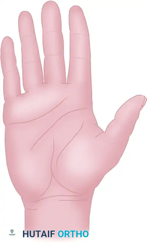
The hand is a highly specialized, dynamic, and exquisitely sensitive organ where the integumentary system is inextricably linked to underlying musculoskeletal function. Unlike other anatomical regions where the skin serves primarily as a passive, elastic envelope accommodating underlying bulk, the skin of the hand—particularly the glabrous volar surface—is a critical, active component of the gripping mechanism. Consequently, surgical incisions in the hand and wrist demand meticulous, preoperative geometric planning. The margin for error is virtually nonexistent; a poorly placed incision can inexorably transform a technically perfect tendon repair or fracture fixation into a catastrophic functional failure due to secondary scar contracture.
Historically, the evolution of hand incisions has been driven by a deeper understanding of cutaneous biomechanics and the patho-epidemiology of wound healing. Early surgical approaches frequently utilized straight longitudinal incisions across flexion creases, leading to an epidemic of iatrogenic bowstringing and severe flexion contractures. The natural biological process of wound contraction, mediated by alpha-smooth muscle actin-expressing myofibroblasts, applies a linear vector of tension along the axis of a healing scar. When this axis crosses a joint's center of rotation perpendicularly, the resulting tether limits excursion and permanently alters the kinematics of the digit or wrist. This patho-epidemiological reality necessitated the development of non-linear, extensile approaches, such as the Bruner zig-zag and the midlateral incisions, which distribute contractile forces obliquely.
The epidemiology of hand pathologies dictating these surgical approaches is vast, encompassing acute high-energy industrial trauma, complex intra-articular fractures, stenosing tenosynovitis, compressive neuropathies, and fibroproliferative disorders such as Dupuytren’s contracture. In each of these scenarios, the primary objective of the surgical incision is to provide extensile, atraumatic exposure to deep structures without compromising the delicate microvascular perfusion of the elevated skin flaps. The surgeon must constantly balance the need for adequate visualization against the biological cost of soft tissue dissection.
As long as fundamental biomechanical, vascular, and anatomical principles are strictly observed, skin incisions can be safely executed anywhere on the hand. The modern orthopedic hand surgeon must view the skin not merely as a barrier to be breached, but as a complex organ system requiring precise geometric manipulation. The ultimate goal is to facilitate profound deep structural repair while simultaneously preventing the formation of restrictive scar contractures that could tether gliding tendons, limit joint excursion, or produce debilitating neuropathic pain.
Detailed Surgical Anatomy and Biomechanics
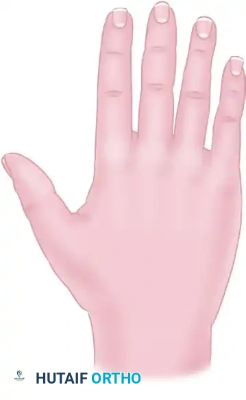
The surgical anatomy of the hand’s integumentary system is distinctly dichotomous, divided into the highly specialized volar (palmar) skin and the more forgiving dorsal skin. The volar skin is thick, heavily keratinized, glabrous (hairless), and densely populated with mechanoreceptors (Meissner and Pacinian corpuscles). Biomechanically, it is engineered to resist shear forces during grip. This is achieved via a complex network of dense vertical fibrous septa that firmly anchor the dermis to the underlying palmar aponeurosis and digital flexor sheaths. While this unique anatomy provides a stable friction surface for prehension, it renders surgical elevation exceptionally challenging, requiring sharp dissection to mobilize full-thickness dermo-fat flaps.

Conversely, the skin on the dorsum of the hand and digits is thin, highly mobile, and possesses a robust subdermal venous and lymphatic network. Because of this inherent mobility and elasticity, shorter incisions frequently suffice on the dorsum, as the skin can be easily retracted to expose adjacent structures. However, this thinness also dictates that dorsal incisions must be handled with extreme delicacy; aggressive retraction easily tears the delicate dermal plexus, and post-operative edema can rapidly compromise venous outflow, leading to flap congestion and marginal necrosis.
A paramount biomechanical concept in hand surgery is the "offset incision" technique. The placement of the skin incision applies strictly to the cutaneous surface. Entries into deeper structures—such as the investing deep fascia, tendon sheaths, or joint capsules—are dictated by their specific underlying anatomy and should frequently be offset, or even perpendicular, to the skin incision. By undermining a full-thickness flap on one side, the deep fascial approach is made parallel to, but physically offset from, the cutaneous wound. This creates a barrier of healthy, vascularized subcutaneous tissue between the two healing layers, preventing the skin scar from adhering directly to the underlying healing tendon or bone, thereby preserving independent glide.
The vascular supply to the skin of the hand relies on a delicate, redundant network of perforating vessels arising from the digital and palmar arteries. Parallel or nearly parallel incisions that are placed too closely together must be universally avoided. Narrow bipedicled flaps created by closely spaced parallel incisions are mechanically isolated from their primary perforator supply and are at an exceptionally high risk of ischemic necrosis. Furthermore, incisions placed directly within deep flexion creases are strictly contraindicated. In these specific anatomical zones, the subcutaneous fat layer is virtually absent, and the dermis is densely tethered to the underlying deep fascia. Moisture naturally accumulates within these deep palmar and digital creases, rendering incisions placed directly within these folds highly susceptible to maceration, wound dehiscence, and secondary bacterial colonization.
Exhaustive Indications and Contraindications
The selection of a specific surgical approach in the hand is dictated by the underlying pathology, the anatomical structures requiring exposure, and the patient's intrinsic soft tissue envelope. The surgeon must synthesize these variables to select an incision that provides maximal utility with minimal iatrogenic morbidity. Absolute and relative indications must be carefully weighed against strict anatomical contraindications.
The midlateral digital approach is highly indicated for extensive exposure of the flexor tendon sheath, particularly during flexor tendon grafting, tenolysis, or the repair of complex Zone II lacerations. It is also the approach of choice for exposing the phalangeal shafts for fracture fixation, as it keeps the hardware and subsequent scar tissue completely off the tactile volar surface. Conversely, the volar zig-zag (Bruner) incision is indicated when bilateral exposure of the digital neurovascular bundles is required, or when addressing volar pathologies such as Dupuytren's cords, where the skin itself may need to be rearranged or excised.
Dorsal approaches are primarily indicated for extensor tendon repairs, dorsal capsulotomies, and arthrodesis of the interphalangeal joints. A straight dorsal longitudinal incision is frequently utilized over the metacarpals, but it must be modified into a curved, lazy-S, or inverted-V configuration when crossing the proximal interphalangeal (PIP) or distal interphalangeal (DIP) joints to prevent linear contractures that would impede joint flexion.
Contraindications in hand surgical incisions are largely absolute and anatomically driven. Crossing any flexion crease at a right angle is strictly contraindicated due to the guaranteed development of a bowstring contracture. Incising directly into the apex of a web space is similarly contraindicated, as scar contracture here will result in severe adduction contractures of the digits. Furthermore, creating distally based flaps with a length-to-width ratio exceeding 1:1 on the volar surface, or 2:1 on the dorsal surface, is a relative contraindication due to the high probability of ischemic tip necrosis.
| Surgical Approach / Incision | Primary Indications | Absolute / Relative Contraindications |
|---|---|---|
| Midlateral Digital | Flexor tendon tenolysis/grafting; Phalangeal shaft fractures; Digital nerve repair. | Pre-existing lateral scarring; Need for bilateral neurovascular bundle exposure. |
| Volar Zig-Zag (Bruner) | Primary flexor tendon repair; Dupuytren's fasciectomy; Purulent flexor tenosynovitis. | Narrow angle flaps (<90 degrees) which risk tip necrosis; Crossing creases perpendicularly. |
| Dorsal Lazy-S / Curved | Extensor tendon central slip repair; PIP/DIP arthrodesis; Dorsal capsulotomy. | Straight longitudinal incisions directly over the exact dorsal midline of the PIP joint. |
| Unilateral Longitudinal (Pulp) | Felon decompression; Distal phalanx osteomyelitis debridement. | "Fish-mouth" incisions; Incisions on the primary tactile pinch surfaces (radial index/middle, ulnar thumb). |
| Offset Volar Wrist | Carpal tunnel release combined with proximal forearm compartment decompression. | Transverse incisions that do not allow proximal/distal extensile extension if needed. |
Pre-Operative Planning, Templating, and Patient Positioning
Flawless execution of a hand surgical approach begins long before the scalpel touches the skin. Pre-operative planning requires meticulous templating and marking of the surgical incisions while the patient is awake and cooperative. Marking the skin prior to the induction of anesthesia or the infiltration of local anesthetics is critical, as tissue distortion from edema or local volume expansion can significantly alter the apparent location of flexion creases and anatomical landmarks. The surgeon must actively flex and extend the patient's joints to precisely identify the apices of the digital and palmar creases, ensuring that planned incisions cross these axes at appropriate oblique angles.
Patient positioning is standardized but requires rigorous attention to detail. The patient is typically positioned supine with the operative extremity extended onto a radiolucent hand table. The shoulder is abducted to approximately 90 degrees, and the arm is secured to prevent intraoperative shifting. A well-padded pneumatic tourniquet is applied to the proximal arm or forearm, depending on the anticipated duration and proximal extent of the procedure. Proper exsanguination using an Esmarch bandage prior to tourniquet inflation is mandatory to provide a bloodless surgical field, which is an absolute prerequisite for safely navigating the dense, microscopic neurovascular anatomy of the hand.
The advent of Wide Awake Local Anesthesia No Tourniquet (WALANT) techniques has revolutionized pre-operative planning for specific procedures, such as tenolysis or tendon transfers. By utilizing a mixture of lidocaine and epinephrine injected directly into the surgical site, the surgeon achieves both profound anesthesia and hemostasis without the need for a pneumatic tourniquet. This allows for intraoperative active movement assessment, permitting the surgeon to dynamically evaluate tendon glide and the extensibility of the skin flaps in real-time. However, WALANT requires the incision plan to account for the substantial volume of local anesthetic injected, which can temporarily distort the subdermal vascular plexus.
Intraoperative equipment is an extension of the pre-operative plan. The use of loupe magnification (minimum 2.5x to 3.5x) is non-negotiable for identifying cutaneous perforators and protecting the digital neurovascular bundles during flap elevation. Skin edges must be handled with exquisite care; the use of fine, single-prong skin hooks or delicate Senn retractors is preferred over heavy-toothed forceps, which crush the dermal edges and precipitate marginal necrosis. The principle of extensile exposure dictates that the incision must be of adequate length to expose deep structures without necessitating excessive, traumatic retraction of the skin edges.
Step-by-Step Surgical Approach and Fixation Technique
The Midlateral Digital Approach
The midlateral approach is the undisputed workhorse incision for exposing the flexor tendon sheath, phalangeal shafts, and digital neurovascular bundles. It is strategically placed along the neutral line of the finger—the precise longitudinal axis that neither stretches during digital flexion nor buckles during extension. To accurately identify this midlateral line, the surgeon connects the apices of the digital flexion creases when the finger is held in maximum flexion.
The skin is sharply incised along this exact midlateral line using a #15 blade. Once the dermis is breached, the surgeon faces a critical anatomical decision regarding the management of the digital neurovascular (NV) bundle. For access to the volar structures (e.g., the flexor tendon sheath), volar retraction is preferred. Dissection is carried carefully dorsal to the NV bundle. The surgeon must identify and sharply divide Cleland’s ligaments, which are robust fascial bands lying dorsal to the bundle, tethering the skin to the phalangeal periosteum. Once Cleland's ligaments are released, the entire NV bundle is mobilized and carried volarward within the full-thickness volar skin flap. This maneuver safely sequesters the bundle out of the operative field and provides unparalleled, unobstructed exposure of the flexor apparatus.

If the surgical objective requires dorsal or lateral access (e.g., for a lateral band repair or collateral ligament reconstruction), superficial dissection is employed. The dissection plane is established superficial to the NV bundle, dividing Grayson’s ligaments (which lie volar to the bundle). Extreme caution is paramount here; the surgeon must maintain a thick, uniform dermo-fat flap to avoid devascularizing the skin, which inevitably leads to catastrophic flap necrosis.
Volar Zig-Zag (Bruner) and Palmar Approaches
The Bruner zig-zag incision is designed to expose the volar surface of the digits and palm without crossing the transverse flexion creases perpendicularly. The incision is composed of a series of V-shaped flaps, with the apices of the flaps extending to the midlateral lines. The angles of these flaps must be kept broad (ideally 90 degrees or greater) to ensure a robust vascular base; acutely angled tips are highly susceptible to ischemic necrosis.
When elevating a Bruner flap, the dissection must be deep to the subdermal vascular plexus, lifting the subcutaneous fat en bloc with the dermis. The digital neurovascular bundles are frequently encountered at the apices of the flaps and must be meticulously identified and protected. In the palm, incisions must carefully navigate the complex neurovascular anatomy, including the superficial palmar arch and the common digital nerves. When extending a palmar incision into the wrist (such as for an extended carpal tunnel release or volar compartment syndrome), the incision must cross the wrist flexion creases obliquely or in a zig-zag fashion to prevent a debilitating volar flexion contracture.
Dorsal Hand, Wrist, and Digital Approaches
Dorsal digital incisions require careful geometric planning due to the extreme thinness of the skin and the proximity of the extensor apparatus. When exposing the central slip of the extensor tendon over the PIP joint, a curved or lazy-S incision is mandatory. A straight longitudinal incision over the PIP joint is strictly avoided, as the resulting scar contracture will severely limit PIP flexion.
For extensive exposure of the distal interphalangeal (DIP) joint, such as in preparation for arthrodesis, an inverted-V (or Y-shaped) incision is highly effective. This design creates a robust, distally based flap that can be reflected distally to provide panoramic exposure of the joint articular surfaces, while safely preserving the germinal matrix of the nail and the terminal extensor tendon insertion. On the dorsal wrist, a straight longitudinal or a lazy-S incision provides excellent, extensile exposure. By elevating full-thickness dermo-fat flaps directly off the extensor retinaculum, the surgeon can access structures from the extreme radial aspect (first dorsal compartment) to the extreme ulnar aspect (sixth dorsal compartment) through a single, versatile incision.
Specialized Distal Pulp and Felon Decompression
Infections of the distal pulp space (felons) represent a unique surgical challenge. The distal pulp is partitioned into multiple, rigid closed compartments by dense vertical fibrous septa radiating from the periosteum of the distal phalanx to the dermis. Accumulation of purulence within these unyielding compartments rapidly elevates interstitial pressure, leading to ischemic necrosis of the pulp fat and subsequent osteomyelitis of the distal phalanx.
Surgical drainage must decompress these septal compartments without compromising the tactile pinch surface. The historically taught "fish-mouth" incision, which circumscribes the entire fingertip, is completely obsolete and must be strictly avoided; it consistently results in an unstable, painful, and insensate fingertip scar that severely impairs prehension. Instead, a unilateral longitudinal incision is the gold standard. It should be strategically placed on the non-contact surface: the ulnar aspect of digits II, III, and IV, and the radial aspect of the thumb and small finger. The incision is placed slightly dorsal to the tactile pad, and the scalpel is swept transversely across the deep pulp space, intentionally dividing the vertical fibrous septa to ensure complete, multi-compartmental decompression.
Complications, Incidence Rates, and Salvage Management
Despite meticulous planning and execution, complications related to hand surgical incisions can and do occur. The unique anatomical constraints of the hand amplify the functional consequences of even minor wound healing issues. The surgeon must be acutely aware of these potential pitfalls, understand their pathophysiology, and possess the surgical armamentarium to manage them effectively.
Ischemic flap necrosis is a devastating complication, typically resulting from technical errors during flap elevation. Creating flaps that are too thin (violating the subdermal plexus), designing flaps with acute angles (poor length-to-width ratio), or applying excessive intraoperative retraction all contribute to microvascular thrombosis and subsequent necrosis. When marginal necrosis occurs, it often leads to delayed healing by secondary intention, which exponentially increases the risk of severe, deep scar contracture tethering the underlying tendons.
Scar contracture (bowstringing) is the direct consequence of violating the cardinal rule of hand surgery: never cross a flexion crease perpendicularly. When a linear scar contracts across a joint, it creates a rigid tether that physically blocks excursion. The incidence of this complication is nearly 100% when longitudinal incisions are incorrectly placed across the volar digital or wrist creases. Salvage management of established contractures is highly complex, frequently requiring multiple Z-plasties to redirect the scar tension lines, or the excision of the scar entirely followed by full-thickness skin grafting or local transposition flaps.
Iatrogenic nerve injury and the formation of painful neuromas represent another significant complication. The superficial branches of the radial nerve and the palmar cutaneous branch of the median nerve are particularly vulnerable during dorsal and volar wrist approaches, respectively. A painful neuroma in the hand can be completely disabling, preventing the patient from utilizing the hand even if the underlying orthopedic repair is mechanically sound.
| Complication | Estimated Incidence | Prevention Strategy | Salvage Management |
|---|---|---|---|
| Ischemic Flap Necrosis | 2 - 5% | Maintain full-thickness flaps; Avoid acute angles (<90°); Use skin hooks instead of forceps. | Debridement of necrotic tissue; Healing by secondary intention; Full-thickness skin grafting if deep structures are exposed. |
| Scar Contracture (Bowstringing) | 5 - 15% (High if creases crossed) | Strictly utilize oblique, zig-zag (Bruner), or midlateral incisions; Avoid crossing creases perpendicularly. | Aggressive scar massage/silicone therapy; Surgical release with multiple Z-plasties or local transposition flaps (e.g., cross-finger flap). |
| Iatrogenic Neuroma | 1 - 3% | Loupe magnification; Meticulous identification and protection of superficial sensory branches. | Excision of neuroma and burying the proximal nerve stump into deep muscle or bone; Targeted Muscle Reinnervation (TMR). |
| Infection / Maceration | 2 - 4% | Avoid incisions directly within deep flexion creases; Meticulous hemostasis prior to closure. | Suture removal and wound drainage; Directed antibiotic therapy; Serial debridement if deep space infection develops. |
Phased Post-Operative Rehabilitation Protocols
The success of a meticulously planned and flawlessly executed hand incision is heavily dependent on rigorous, phased postoperative care. The hand is uniquely prone to rapid, profound stiffness following surgical trauma. The inflammatory cascade initiated by the incision leads to the accumulation of protein-rich interstitial edema, which, if not aggressively managed, rapidly organizes into dense fibrotic adhesions binding the skin, tendons, and joint capsules into a single, immobile unit.
Phase I (0-14 days) focuses on wound healing, hemostasis, and edema control. Prior to skin closure, the pneumatic tourniquet must be deflated to ensure meticulous hemostasis. Postoperative hematoma formation is a primary precursor to excessive fibrosis and must be avoided at all costs. The skin is typically closed with non-absorbable monofilament sutures (e.g., 4-0 or 5-0 nylon) using a precise, tension-free technique. The hand is then immobilized in a bulky, non-compressive dressing. Unless dictated otherwise by a specific tendon or nerve repair, the hand is placed in the "intrinsic-plus" or "safe" position: the wrist in 20-30 degrees of extension, the metacarpophalangeal (MCP) joints in 70-90 degrees of flexion, and the interphalangeal (IP) joints in full extension. This specific posture maintains the collateral ligaments of the MCP and IP joints at their maximal length, preventing secondary joint contractures. Strict elevation above the level of the heart is mandatory for the first 48-72 hours.
Phase II (2-4 weeks) commences with suture removal, typically at 10-14 days postoperatively, once the wound is fully epithelialized. Early active range of motion (ROM) is initiated as soon as the specific underlying surgical procedure allows. Tendon gliding exercises are critical during this phase to prevent the healing skin scar from adhering to the underlying deep fascia and tendons. The offset incision technique, employed intraoperatively, proves its value during this phase by allowing the tendon to glide beneath a bed of healthy subcutaneous tissue rather than directly beneath the healing dermal scar.
Phase III (4-8 weeks and beyond) is dedicated to aggressive scar remodeling and maximizing functional excursion. Once the incision is completely healed and robust, scar massage is initiated to mechanically break down cross-linking collagen fibers. Silicone gel sheeting is highly effective in softening the scar and reducing hypertrophy. If a mild scar contracture begins to develop, dynamic or static-progressive splinting is employed to apply a prolonged, low-load stretch to the maturing collagen, redirecting the vectors of scar contraction to favor joint mobility. The close collaboration between the orthopedic surgeon and a certified hand therapist is absolutely essential throughout all phases of rehabilitation.
Summary of Landmark Literature and Clinical Guidelines
The foundational principles of hand surgical incisions are deeply rooted in landmark orthopedic literature. Sterling Bunnell, widely considered the founding father of modern hand surgery, first articulated the catastrophic consequences of poorly planned incisions in his seminal texts. Bunnell coined the term "no man's land" to describe the critical Zone II of the flexor tendon sheath, emphasizing that the surgical approach to this area must be as atraumatic as the tendon repair itself to prevent restrictive adhesions.
In 1967, J.M. Bruner revolutionized volar hand approaches with the introduction of the zig-zag volar incision. Bruner’s landmark paper provided the geometric and biomechanical rationale for avoiding perpendicular crease crossings, demonstrating that widely angled, full-thickness flaps could provide extensile exposure of the flexor apparatus without inducing the bowstring contractures that plagued earlier straight longitudinal incisions. The "Bruner incision" remains a cornerstone of modern hand surgery guidelines.
More recently, the widespread adoption of the Wide Awake Local Anesthesia No Tourniquet (WALANT) technique, pioneered and extensively published by Donald Lalonde, has forced a paradigm shift in how incisions are planned and executed. Clinical guidelines from the American Society for Surgery of the Hand (ASSH) now frequently incorporate WALANT principles, highlighting that the ability to assess active, voluntary tendon glide and flap tension intraoperatively allows the surgeon to dynamically modify the incision and closure to optimize functional outcomes.
Current guidelines from the American Academy of Orthopaedic Surgeons (AAOS) and the ASSH consistently emphasize the absolute necessity of respecting the cutaneous microcirculation. Modern literature strongly supports the use of loupe magnification, the preservation of the subdermal venous plexus, and the utilization of the offset incision technique as mandatory standards of care. Mastery of these evidence-based principles ensures that the surgical approach facilitates, rather than hinders, the ultimate functional recovery of the traumatized hand.
