Comprehensive Introduction and Patho-Epidemiology
By any epidemiological, clinical, or economic measure, intervertebral disc disorders represent a monumental challenge in modern orthopaedics and neurosurgery. Musculoskeletal conditions in the United States incur staggering direct and indirect costs exceeding $254 billion annually. While these conditions are rarely fatal, they are overwhelmingly chronic and functionally debilitating, with musculoskeletal injuries remaining the primary driver for ambulatory physician visits. Axial spine pain—whether cervical, thoracic, or lumbar—is the most frequent musculoskeletal complaint encountered in clinical practice and is frequently, though sometimes erroneously, attributed to disc degeneration. It is paramount for the practicing spine surgeon to distinguish meticulously between age-related physiological disc degeneration, internal disc derangement (IDD), and frank disc herniation. Each of these pathological entities presents with unique clinical findings, distinct biomechanical alterations, and highly specific therapeutic indications.
The contemporary understanding of disc degeneration has undergone a profound transformation, shifting away from a purely mechanical "wear-and-tear" model to a highly complex interplay of genetics, biochemistry, and biomechanics. The genetic influence on disc degeneration is profound and heavily driven by multiple gene loci. Specific allelic variations and polymorphisms in the aggrecan gene, matrix metalloproteinase-3 (MMP-3) gene, and genes encoding collagen type IX (specifically the alpha 2 and 3 forms, COL9A2 and COL9A3) have been definitively associated with premature disc pathology. These genetic polymorphisms fundamentally alter the structural integrity of the annulus fibrosus and the crucial hydrophilic properties of the nucleus pulposus. This genetic predisposition sets the stage for the functional spinal unit to undergo premature desiccation, loss of hydrostatic pressure, and subsequent mechanical failure under normal physiological loads.
The socioeconomic burden of low back pain is nothing short of staggering, with total costs in the United States alone exceeding $100 billion per year. Notably, only one-third of this figure represents direct medical costs (imaging, surgery, rehabilitation); the vast remainder stems from decreased workplace productivity, lost wages, and absenteeism. A highly disproportionate mere 5% of patients account for approximately 75% of these total societal costs. Return-to-work statistics powerfully dictate the absolute urgency of early, effective intervention and accurate diagnosis. Approximately 90% of patients experiencing an acute episode of radiculopathy or back pain return to work within three months. However, the chronicity curve is unforgiving: if a patient remains off work for six months, the probability of ever returning drops to 50%; at one year, it plummets to a dismal 25%.
Furthermore, the seminal work by Boos et al. definitively demonstrated that nonanatomical factors—specifically work perception, job dissatisfaction, and psychosocial distress—are intimately intertwined with physical complaints and surgical outcomes. Socioeconomic factors, low educational attainment, and active workers' compensation litigation status are powerful, independent predictors of prolonged disability. This highlights the absolute necessity of a comprehensive biopsychosocial approach to patient evaluation. The surgeon must recognize that operating on a structurally abnormal disc in a patient with overwhelming psychosocial distress or secondary gain motives is the primary genesis of Failed Back Surgery Syndrome (FBSS).
Detailed Surgical Anatomy and Biomechanics
The intervertebral disc is a highly specialized, complex, and predominantly avascular structure designed to transmit tremendous axial loads while simultaneously providing multidirectional flexibility to the spinal column. It consists of three distinct, yet seamlessly integrated, anatomical components. The first is the nucleus pulposus, a gelatinous, centrally located core rich in proteoglycans (predominantly aggrecan) and type II collagen. This matrix binds water avidly, allowing the nucleus to resist immense compressive forces via the generation of hydrostatic pressure. The second component is the annulus fibrosus, composed of 15 to 25 concentric lamellae of highly organized type I collagen. These fibers are oriented at approximately 30-degree angles to the horizontal plane, alternating in direction with each successive layer, perfectly engineered to resist tensile, shear, and torsional forces. The third component comprises the cartilaginous endplates, thin layers of hyaline cartilage that firmly anchor the disc to the adjacent osseous vertebral bodies and critically facilitate the passive diffusion of nutrients and oxygen to the avascular inner annulus and nucleus.
The innervation and vascularity of the intervertebral disc are highly relevant to the pathophysiology of discogenic pain. In a healthy adult, only the outer one-third of the annulus fibrosus is innervated and vascularized. The sensory innervation is supplied primarily by the sinuvertebral nerve of Luschka, which arises from the ventral ramus and the sympathetic trunk, re-entering the spinal canal through the intervertebral foramen. This nerve supplies the posterior annulus, the posterior longitudinal ligament (PLL), and the ventral dura. The anterior and lateral portions of the disc are innervated by branches from the gray rami communicantes and the sympathetic chain. In severely degenerated discs, neo-innervation and neo-vascularization occur, with nociceptive nerve fibers penetrating deep into the inner annulus and even the nucleus pulposus, driven by local inflammatory cytokines such as TNF-alpha and Interleukin-1 beta, providing a direct anatomical substrate for chronic discogenic pain.
The biomechanics of the degenerative cascade, as classically described by Kirkaldy-Willis, fundamentally dictate the progression of spinal pathology. The cascade begins with the dysfunction phase, characterized by biochemical alterations: the nucleus loses its proteoglycan content, leading to desiccation and a critical loss of hydrostatic pressure. Compressive axial loads are subsequently transferred directly to the annulus fibrosus rather than being dissipated by the nucleus. This abnormal load transfer initiates the instability phase, causing delamination of the annular lamellae, radial and concentric annular tears, and eventual herniation of nuclear material. This mechanical shift also overloads the posterior elements, leading to facet joint hypertrophy, subluxation, and ligamentum flavum buckling. Finally, the restabilization phase occurs, characterized by the formation of robust osteophytes, traction spurs, and fixed deformity, often culminating in secondary central or lateral recess spinal stenosis.
Understanding the spatial relationship of the disc to the neural elements is critical for safe surgical intervention. In the lumbar spine, the traversing nerve root (e.g., the L5 root at the L4-L5 disc level) descends vertically across the disc space and is most vulnerable to a typical paracentral disc herniation. Conversely, the exiting nerve root (e.g., the L4 root at the L4-L5 level) exits the foramen superior to the disc space and is typically spared by paracentral herniations but is highly vulnerable to far-lateral (extra-foraminal) disc herniations. The safe working zone for endoscopic and percutaneous approaches is defined by Kambin's triangle, a right triangle over the dorsolateral disc, bounded anteriorly by the exiting nerve root, inferiorly by the superior endplate of the lower vertebral body, and posteriorly by the superior articular process and the traversing nerve root.
Exhaustive Indications and Contraindications
The decision to proceed with operative intervention for intervertebral disc disorders requires a masterful synthesis of clinical acumen, radiographic interpretation, and an understanding of the natural history of the disease. The primary objective of surgical decompression is the relief of radicular leg pain, not axial back pain. Patients must be meticulously counseled that while microdiscectomy is highly efficacious for alleviating sciatic-type leg pain, its impact on isolated axial back pain is unpredictable and generally poor. Proper patient selection remains the single most critical determinant of surgical success.
Absolute indications for emergent or urgent surgical intervention are rare but demand immediate recognition. Cauda equina syndrome, characterized by saddle anesthesia, bowel or bladder dysfunction (typically urinary retention followed by overflow incontinence), and bilateral lower extremity weakness, necessitates emergent decompression to prevent irreversible neurological devastation. Similarly, a rapidly progressive motor deficit, such as a profound and worsening foot drop (L5 radiculopathy) or quadriceps weakness (L4 radiculopathy), warrants urgent surgical decompression to maximize the potential for neurological recovery.
Relative indications encompass the vast majority of elective spinal surgeries for disc herniation. The classic relative indication is unilateral, intractable radicular pain that perfectly correlates with a specific dermatomal distribution and is corroborated by concordant advanced imaging (MRI or CT myelogram). Importantly, this intervention is typically offered only after the patient has failed a rigorous, comprehensive trial of non-operative management lasting a minimum of six weeks. This conservative regimen should include targeted physical therapy, non-steroidal anti-inflammatory drugs (NSAIDs), oral corticosteroid tapers, and potentially fluoroscopically guided epidural steroid injections (ESIs).
Contraindications to isolated disc excision must be strictly respected to avoid disastrous outcomes and the creation of a spinal cripple. Operating on patients with profound psychosocial distress, active secondary gain motives, or poorly localized axial pain without clear anatomical correlation inevitably leads to clinical failure. Furthermore, performing a simple discectomy in the presence of gross dynamic segmental instability (e.g., a mobile degenerative spondylolisthesis >3mm on flexion-extension radiographs) is contraindicated, as the decompression will likely exacerbate the instability, necessitating a subsequent fusion procedure.
| Category | Specific Clinical Scenarios | Rationale / Surgical Considerations |
|---|---|---|
| Absolute Indications | Cauda Equina Syndrome; Progressive, profound motor deficit (e.g., acute foot drop). | Emergent decompression required to prevent permanent sphincter dysfunction or irreversible paralysis. |
| Relative Indications | Intractable radicular pain failing >6 weeks of conservative care; Static, severe motor weakness. | Elective decompression to relieve nerve root tension, improve quality of life, and expedite return to function. |
| Absolute Contraindications | Asymptomatic MRI findings; Active systemic infection; Medical instability precluding anesthesia. | Treating an image rather than a patient leads to Failed Back Surgery Syndrome. Survival supersedes elective spine surgery. |
| Relative Contraindications | Predominant axial back pain; Severe psychosocial distress (Waddell's signs); Active litigation/Workers' Comp. | Poor outcomes predictably follow decompression for axial pain. Biopsychosocial factors heavily negatively skew surgical results. |
Pre-Operative Planning, Templating, and Patient Positioning
Pre-operative planning begins with an exhaustive clinical examination to meticulously differentiate axial back pain from true radiculopathy. The physical examination must include a detailed assessment of dermatomal sensation, myotomal motor strength, and deep tendon reflexes. Provocative neural tension signs are critical. The Lasègue sign (straight leg raise), described by Forst in 1881, remains a cornerstone provocative test for lumbar radiculopathy, specifically assessing L4, L5, and S1 nerve root tension. A positive test occurs when radicular pain (not merely hamstring tightness) is reproduced at an elevation between 30 and 70 degrees. The crossed straight leg raise (Fajersztajn sign) is even more specific for a space-occupying disc herniation. For upper lumbar radiculopathies (L2, L3, L4), the femoral nerve stretch test (reverse straight leg raise) is the appropriate provocative maneuver.
Advanced diagnostic imaging must always be strictly correlated with clinical findings. Magnetic Resonance Imaging (MRI) is the undisputed gold standard for evaluating soft tissue, neural elements, and disc hydration via T2-weighted sequences. However, the surgeon must remain vigilant: MRI reveals significant abnormalities, including disc bulges and protrusions, in up to 76% of completely asymptomatic individuals. Operating on an MRI finding rather than a clinical syndrome is a catastrophic error. Weight-bearing anteroposterior, lateral, and dynamic flexion-extension radiographs are essential prerequisites to assess overall sagittal alignment, disc height loss, and occult segmental instability that might alter the surgical plan from a simple decompression to a fusion.
When noninvasive imaging is equivocal, or when multi-level degenerative changes confound the clinical picture, diagnostic injection studies serve as critical pre-operative adjuncts. Epidural Steroid Injections (ESIs), administered via transforaminal or interlaminar routes, provide both diagnostic information (by isolating the symptomatic nerve root) and therapeutic relief. Zygapophyseal (facet) joint injections or medial branch blocks can isolate pain originating from the posterior column. Provocative discography, while increasingly controversial due to risks of accelerated disc degeneration, can identify internal disc derangement by reproducing concordant pain upon precise pressurization of the disc space, guiding complex fusion or arthroplasty decisions.
Patient positioning in the operating room is not merely a preparatory step; it is a critical component of the surgical procedure that directly dictates the ease of exposure and the risk of complications. For lumbar microdiscectomy, the patient is placed prone on a radiolucent spinal frame, such as a Jackson table, Andrews frame, or Wilson frame. The paramount principle of prone positioning is that the abdomen must hang completely free and uncompressed. Any abdominal compression dramatically increases intra-abdominal pressure, which is directly transmitted via the valveless epidural venous plexus (Batson's plexus). This venous engorgement results in excessive, unrelenting intraoperative epidural hemorrhage that obscures the neural elements and increases the risk of iatrogenic nerve injury. Furthermore, the hips and knees are deliberately flexed to reduce lumbar lordosis, which biomechanically opens the posterior interlaminar space, facilitating access to the spinal canal with minimal bony resection.
Step-by-Step Surgical Approach and Fixation Technique
The standard operative procedure for disc removal has evolved significantly since Semmes presented the subtotal laminectomy in 1939. Today, microscopic and endoscopic techniques, popularized by pioneers such as Kambin, Onik, and Helms, represent the absolute gold standard for symptomatic lumbar disc herniation, minimizing collateral tissue damage while maximizing neural decompression.
Lumbar Microdiscectomy Approach
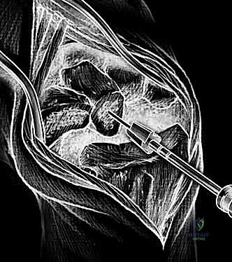
The procedure begins with precise fluoroscopic localization. A 2 to 3 cm midline incision is made directly centered over the target interspace. The lumbodorsal fascia is incised unilaterally, preserving the midline interspinous ligaments to maintain posterior tension band biomechanics. Subperiosteal dissection is meticulously performed using a Cobb elevator, elevating the paraspinal musculature off the spinous process and the hemilamina. A self-retaining retractor, such as a Taylor, McCulloch, or specialized tubular retractor system, is deployed to maintain exposure.
Under the operating microscope, a partial hemilaminotomy is performed. Using a high-speed matchstick burr or Kerrison rongeurs, the inferior edge of the superior lamina and the superior edge of the inferior lamina are resected to widen the interlaminar window. The ligamentum flavum is identified, carefully detached from its bony insertions using a curved curette, and excised to expose the underlying epidural space. Once the epidural fat is gently mobilized, the traversing nerve root is identified. It is imperative to identify the lateral border of the root. A Love nerve root retractor is utilized to gently retract the neural elements medially, exposing the underlying prominent disc herniation.
If the herniated fragment is subligamentous (contained by the posterior longitudinal ligament), a cruciate or box annulotomy is performed using a #11 blade on a long handle. Pituitary rongeurs are then introduced to extract the extruded nucleus pulposus. The surgeon must exercise extreme caution: over-aggressive exploration of the disc space can lead to penetration of the anterior annulus, risking catastrophic, often fatal, injury to the great vessels (aorta, inferior vena cava, or common iliac vessels). Instruments should never be advanced blindly beyond the pre-measured depth of the disc space (typically 30 mm). Following complete fragment removal, a Penfield dissector is passed along the nerve root into the neural foramen to ensure absolute freedom from residual compression. The wound is irrigated copiously with antibiotic saline, and the fascia is closed tightly with interrupted non-absorbable sutures to prevent postoperative muscle herniation.
Cervical Disc Operative Principles
Cervical disc herniations typically present with severe radiculopathy (e.g., C6 or C7 nerve root distribution) or insidious myelopathy if the spinal cord is globally compressed. The Anterior Cervical Discectomy and Fusion (ACDF) remains the workhorse procedure. Utilizing a standard Smith-Robinson anterolateral approach, the surgeon accesses the anterior spine by dissecting between the carotid sheath laterally and the visceral axis (trachea/esophagus) medially. This approach allows direct, complete decompression of the neural elements, resection of the posterior longitudinal ligament, and restoration of disc height and cervical lordosis via the placement of an interbody structural graft and an anterior cervical plate. In carefully selected younger patients without significant facet arthropathy, instability, or severe spondylosis, Cervical Disc Arthroplasty (CDA) serves as an excellent alternative to ACDF, designed to preserve segmental motion and theoretically mitigate the long-term risk of adjacent segment disease.
Thoracic Disc Operative Principles
Thoracic disc herniations are anatomically rare but clinically potentially devastating due to the inherently narrow thoracic spinal canal and the tenuous watershed blood supply to the thoracic spinal cord. Patients may present with axial back pain, band-like radicular chest pain, or progressive spastic myelopathy. A critical surgical absolute: posterior laminectomy alone is strictly contraindicated for central thoracic disc herniations. The required medial retraction of the thoracic spinal cord to access a central disc invariably leads to catastrophic spinal cord injury and paraplegia. Therefore, operative approaches must be anterior (transthoracic/thoracotomy) or posterolateral (costotransversectomy or transpedicular approach). These complex approaches allow the surgeon to drill away the posterior vertebral body and pull the calcified disc fragment ventrally, completely avoiding any manipulation of the delicate spinal cord.
Complications, Incidence Rates, and Salvage Management
Despite meticulous surgical technique and rigorous pre-operative planning, complications in spinal surgery are an inherent risk. A comprehensive understanding of these adverse events, their incidence, and precise salvage protocols is mandatory for the operating surgeon. Informed consent must explicitly detail these risks to manage patient expectations and establish a foundation of trust.
Incidental durotomy (dural tear) is the most frequent intraoperative complication, occurring in approximately 3% to 5% of primary lumbar microdiscectomies, with the incidence rising dramatically in revision surgeries due to the presence of dense epidural fibrosis. When a dural tear occurs, it must be recognized immediately intraoperatively. The gold standard of management is primary, watertight repair using 4-0 or 5-0 nonabsorbable sutures (e.g., Prolene or Nurolon) in a running or interrupted fashion. This repair is frequently augmented with a dural sealant (hydrogel) or an autologous free fat graft harvested from the subcutaneous tissue. Postoperatively, the patient may be kept on flat bed rest for 24 to 48 hours to decrease hydrostatic cerebrospinal fluid (CSF) pressure and prevent the formation of a pseudomeningocele or a cutaneous CSF fistula.
Recurrent disc herniation is a frustrating reality, occurring in 5% to 10% of patients following an initially successful microdiscectomy. It typically presents with an acute recurrence of the exact pre-operative radicular pain following a pain-free interval. Diagnosis is confirmed via a contrast-enhanced MRI, where gadolinium enhancement differentiates vascularized epidural scar tissue from an avascular recurrent disc fragment. Repeat microdiscectomy is often successful, though the surgical dissection is significantly more hazardous due to the loss of normal anatomical planes.
Postoperative discitis is a rare but highly morbid complication, occurring in less than 1% of cases. It classically presents with severe, unrelenting, mechanical back pain occurring 2 to 4 weeks postoperatively, often without systemic signs of infection like fever. Diagnosis is confirmed via MRI (showing endplate edema and disc space enhancement) and markedly elevated inflammatory markers (Erythrocyte Sedimentation Rate and C-Reactive Protein). Treatment requires image-guided aspiration for culture, followed by prolonged (6-12 weeks) targeted intravenous antibiotics. In cases of progressive deformity, neurological deficit, or failure of medical management, radical surgical debridement and interbody fusion are required.
| Surgical Complication | Estimated Incidence | Salvage Management & Protocol |
|---|---|---|
| Incidental Durotomy | 3% - 5% (Primary); >10% (Revision) | Primary watertight suture repair (4-0/5-0 Nurolon); Dural sealant; Bed rest 24-48 hours; Subarachnoid drain if refractory. |
| Recurrent Disc Herniation | 5% - 10% | Contrast MRI to differentiate scar vs. disc. Revision microdiscectomy. Consider fusion if massive bony resection is required. |
| Postoperative Discitis | < 1% | MRI with contrast; CRP/ESR monitoring; CT-guided biopsy; 6-12 weeks IV antibiotics. Surgical debridement/fusion if unstable. |
| Great Vessel Injury | < 0.05% | Immediate packing of disc space; Emergent vascular surgery consultation; Laparotomy for direct vessel repair. High mortality rate. |
| Nerve Root Injury | 1% - 2% | Intraoperative recognition: avoid excessive retraction. Postoperative: High-dose steroids (controversial), gabapentinoids, aggressive physical therapy. |
Phased Post-Operative Rehabilitation Protocols
The paradigm of postoperative rehabilitation following intervertebral disc surgery has shifted radically over the past two decades. The historical approach of prolonged bed rest has been universally discarded, as it promotes muscular atrophy, deep vein thrombosis, and psychological deconditioning. Modern spinal surgery emphasizes immediate, controlled, early mobilization to optimize tissue healing and restore functional biomechanics.
Phase I: Immediate Postoperative and Tissue Healing (Weeks 0-2)
Patients undergoing standard lumbar microdiscectomy are typically mobilized on the day of surgery and discharged within 24 hours. The primary goal of Phase I is the protection of the surgical site and the mitigation of acute inflammatory pain. Patients are instructed in strict "BLT" precautions: absolutely no Bending, Lifting (greater than 10 pounds), or Twisting of the lumbar spine. A progressive daily walking program is immediately instituted, as walking provides rhythmic, low-impact loading to the spine, which facilitates disc nutrition and prevents nerve root adherence to the surgical bed. Sitting is generally limited to 30-45 minute intervals to avoid excessive axial loading on the newly decompressed annulus.
Phase II: Early Stabilization and Neural Mobilization (Weeks 3-6)
As the acute inflammatory phase subsides and the annular defect begins to scar, the focus shifts to restoring neuromuscular control. Formal physical therapy is initiated. The cornerstone of this phase is lumbar and pelvic core stabilization. Exercises focus on the transverse abdominis, multifidus, and pelvic floor musculature, teaching the patient to maintain a neutral spine during dynamic movements. Gentle neural mobilization techniques (nerve gliding) are introduced to ensure the traversing nerve root moves freely within the neural foramen and does not become tethered by developing epidural fibrosis. Passive modalities (ultrasound, electrical stimulation) are of limited value and are generally discouraged in favor of active rehabilitation.
Phase III: Advanced Strengthening and Return to Function (Weeks 6-12+)
The final phase of rehabilitation is highly individualized, dictated by the patient's occupational demands and athletic aspirations. Isometric strengthening progresses to dynamic load management. Patients undergo work hardening programs if they are returning to heavy manual labor. Gradual return to high-impact sports (e.g., running, contact sports) is generally permitted between 3 to 6 months, strictly contingent upon complete symptom resolution, full restoration of motor strength, and demonstrated core competency. The patient is educated that while the herniation has been removed, the underlying degenerative disc disease remains, necessitating a lifelong commitment to spinal hygiene and core maintenance.
Summary of Landmark Literature and Clinical Guidelines
The evidence-based management of intervertebral disc disorders is anchored by several monumental clinical trials and long-term observational studies that every practicing orthopedic surgeon must intimately understand. These landmark papers guide clinical decision-making and provide the statistical foundation for patient counseling.
The Spine Patient Outcomes Research Trial (SPORT), led by Weinstein et al., is arguably the most influential modern study evaluating the efficacy of surgical versus non-operative treatment for lumbar disc herniation. A massive, multi-center prospective cohort and randomized trial, SPORT demonstrated that patients who underwent standard open discectomy experienced significantly greater improvement in pain, function, and satisfaction compared to those treated non-operatively, with these surgical advantages maintained at 4-year and 8-year follow-ups. However, SPORT also highlighted the favorable natural history of the disease: patients in the non-operative cohort also demonstrated significant improvement over time, validating a trial of conservative care for non-emergent cases.
Historically, Weber's classic 1983 prospective randomized study provided the foundational data on the long-term outcomes of disc herniation. Weber randomized patients with confirmed disc herniations to either surgical discectomy or conservative care. At the 1-year mark, the surgical group showed statistically superior outcomes. However, at the 4-year and 10-year follow-up intervals, there was no statistically significant difference in clinical outcomes between the two groups. This seminal finding underscores the concept that surgery rapidly accelerates the relief of radicular pain, but over a decade, the natural history of disc resorption and neural accommodation often matches surgical intervention.
As previously noted, the work by Boos et al. fundamentally altered the interpretation of advanced imaging. By performing MRIs on asymptomatic volunteers and comparing them to symptomatic patients, Boos demonstrated an exceptionally high prevalence of disc herniations and degenerative changes in individuals with absolutely no back or leg pain. This study cemented the clinical maxim that surgeons must "treat the patient, not the MRI," and firmly established the role of psychosocial profiling in surgical patient selection.
Future advancements in the treatment of disc disorders must pivot aggressively from merely developing new mechanical implants to better defining and targeting the underlying pathophysiological processes. Biomolecular therapies, mesenchymal stem cell injections, and viral-vector gene therapy targeting the aggrecan and MMP-3 pathways hold immense promise for actually regenerating the intervertebral disc. This represents a monumental paradigm shift from mechanical salvage (decompression and fusion) to biological restoration. Until reliable criteria and FDA-approved protocols for these biological diagnoses are established, the orthopedic surgeon must rely on meticulous clinical evaluation, strict adherence to evidence-based surgical indications, and flawless operative technique to optimize patient outcomes.
This academic synthesis is based on established protocols from Hutaifortho's Operative Orthopaedics and has been medically reviewed by Prof. Dr. Mohammed Hutaif, Consultant Orthopedic & Spine Surgeon. It is designed to assist orthopedic residents, fellows, and practicing surgeons in surgical preparation and board reviews (AAOS, FRCS, Arab Board).
