Master Radiation Safety in Orthopedic Imaging & Special Studies
Key Takeaway
This topic focuses on Master Radiation Safety in Orthopedic Imaging & Special Studies, Imaging and special studies include diagnostic procedures like fluoroscopy and nuclear medicine bone scans, often using Technetium Tc 99m to detect conditions such as fractures, avascular necrosis, or osteomyelitis. Radiation safety is paramount, involving minimizing exposure time, using collimation, protective shielding, and maximizing distance from the x-ray source for patient and clinician well-being.
As an academic orthopedic surgeon and medical educator, the imperative for comprehensive understanding and meticulous application of radiation safety principles in orthopedic imaging and special studies cannot be overstated. Our daily practice, spanning from initial diagnostic evaluations to complex intraoperative guidance, inherently involves exposure to ionizing radiation for both patients and the surgical team. Mastering radiation safety is not merely a regulatory obligation; it is a fundamental ethical responsibility rooted in the ALARA principle—As Low As Reasonably Achievable—to minimize potential harm while optimizing diagnostic and therapeutic outcomes.
This review systematically addresses the foundational physics, biological effects, clinical applications, and practical strategies for minimizing radiation exposure within the orthopedic domain. It is tailored for orthopedic surgeons, residents, and medical students, aiming to elevate clinical acumen and foster a pervasive culture of radiation safety throughout our specialty.
Introduction and Epidemiology
The indispensable role of imaging in contemporary orthopedic practice necessitates a profound understanding of radiation safety. From plain radiographs guiding initial fracture assessment to fluoroscopically-guided complex spinal instrumentation and nuclear medicine scans elucidating subtle pathology, ionizing radiation is a ubiquitous tool. However, its benefits are balanced against the inherent risks of cellular damage and the potential for deterministic and stochastic health effects.
The cumulative nature of radiation exposure, particularly for orthopedic surgeons who routinely operate under fluoroscopic guidance, underscores the critical need for robust safety protocols. Epidemiological data highlight that orthopedic surgeons receive some of the highest occupational radiation doses among medical specialists, second only to interventional radiologists and cardiologists. This persistent exposure, even at low doses, contributes to a lifelong risk profile. Therefore, embracing and rigorously applying ALARA principles is not just a recommendation but a cornerstone of responsible surgical practice and a commitment to the well-being of both our patients and ourselves. This review aims to equip practitioners with the knowledge and practical strategies to navigate this essential aspect of orthopedic care effectively.
Principles of Radiation Physics and Biological Effects
Understanding the fundamental principles of radiation physics and its interaction with biological tissues is paramount for effective radiation safety. Without this foundation, efforts to minimize exposure remain mechanistic rather than principle-driven.
Ionizing Radiation Fundamentals
Ionizing radiation, including X-rays and gamma rays, possesses sufficient energy to remove electrons from atoms, leading to ionization. This process is the basis of both its diagnostic utility and its potential for biological harm. X-rays, used in conventional radiography and fluoroscopy, are generated electrically when high-speed electrons strike a metal target. Gamma rays, originating from the nucleus of radioactive atoms, are used in nuclear medicine studies.
The interaction of X-rays with tissue primarily occurs through two mechanisms: the photoelectric effect and Compton scattering. The photoelectric effect predominates at lower X-ray energies and in denser materials (e.g., bone), where the X-ray photon is completely absorbed, contributing significantly to image contrast and patient dose. Compton scattering, more prevalent at higher X-ray energies and in less dense tissues, involves the partial transfer of X-ray photon energy to an electron, resulting in a scattered photon. This scattered radiation is the primary source of occupational exposure for the surgical team, degrading image quality and increasing dose.
Units of radiation measurement are critical for quantifying exposure and risk. The Roentgen (R) measures exposure in air. The Rad (Radiation Absorbed Dose) or Gray (Gy, 1 Gy = 100 rad) quantifies the energy absorbed per unit mass of tissue. The Sievert (Sv, 1 Sv = 100 rem) accounts for the biological effectiveness of different types of radiation and is used to express effective dose and equivalent dose, which are more relevant for assessing biological risk. For X-rays, 1 Gy is approximately equal to 1 Sv.
Radiation Biology and Health Effects
Ionizing radiation induces cellular damage primarily through two mechanisms: direct action and indirect action. Direct action occurs when radiation directly strikes and damages critical cellular macromolecules, particularly DNA. Indirect action, more common (approximately 70% of damage), involves radiation interacting with water molecules to produce highly reactive free radicals, which then chemically damage DNA and other cellular components.
The biological consequences of radiation exposure are categorized into deterministic and stochastic effects:
- Deterministic Effects: These effects have a threshold dose below which they do not occur, and their severity increases with increasing dose above the threshold. They typically manifest relatively quickly (days to weeks) after acute high-dose exposure. Examples relevant to orthopedic practice include skin erythema, desquamation, epilation, and cataracts. Surgical staff, particularly those involved in prolonged or complex fluoroscopic cases, are at risk for cumulative deterministic effects, especially on the skin and ocular lens.
- Stochastic Effects: These effects do not have a dose threshold; any amount of radiation is presumed to carry some probability of inducing them. The probability of occurrence increases with dose, but the severity is independent of dose. Stochastic effects are typically long-term and include radiation-induced cancers (e.g., leukemia, thyroid, breast, and other solid tumors) and heritable (genetic) effects. For both patients and staff, these effects represent the primary long-term concern, emphasizing the ALARA principle. Factors influencing the biological effect include the total dose, dose rate (e.g., acute vs. protracted exposure), and tissue radiosensitivity (e.g., rapidly dividing cells, pediatric tissues, and the developing fetus are highly radiosensitive).
Indications for Advanced Orthopedic Imaging Modalities
The appropriate selection and judicious use of advanced orthopedic imaging modalities are critical for diagnostic accuracy and patient management, while concurrently informing radiation safety strategies. Each modality, with its unique advantages and radiation profile, must be considered within the clinical context.
Fluoroscopy Applications in Orthopedic Surgery
Fluoroscopy, a dynamic X-ray imaging technique, provides real-time visualization, making it invaluable for intraoperative guidance. Its widespread use, however, underscores the need for constant vigilance regarding radiation exposure.
Common orthopedic applications include:
- Fracture Reduction and Fixation: Fluoroscopy is essential for confirming satisfactory reduction and precise hardware placement in a multitude of fractures, including those of long bones, pelvis, and spine. It allows for immediate feedback during manipulation and pin/screw insertion.
- Joint Aspiration and Injection: Accurate placement of needles for diagnostic aspirations (e.g., septic arthritis) or therapeutic injections (e.g., pain management, visco-supplementation) into joints or bursae is significantly enhanced by fluoroscopic guidance.
- Vertebroplasty and Kyphoplasty: These spinal procedures rely entirely on fluoroscopic guidance for safe access and cement delivery into vertebral bodies.
- Intramedullary Nailing: Achieving optimal entry points, nail insertion, and distal locking in long bone fractures is routinely performed under fluoroscopic control.
- Guidance for Biopsy: Precise localization of bone or soft tissue lesions for biopsy, particularly deep-seated or small lesions, often requires fluoroscopic or CT guidance.
- Pediatric Orthopedics: While highly effective, fluoroscopy in pediatric patients demands extreme caution due to their increased radiosensitivity and longer life expectancy for stochastic effects to manifest. Every fluoroscopic case, regardless of patient age or procedure complexity, should involve careful consideration of radiation safety protocols from the outset.
Nuclear Medicine Imaging Indications
Nuclear medicine studies involve the administration of radiopharmaceuticals that concentrate in specific tissues or organs, emitting gamma rays detected by external cameras. These studies provide physiological information often not obtainable with anatomical imaging.
Bone Scintigraphy Technetium Tc 99m
Bone scans utilizing Technetium (Tc) 99m-labeled phosphate complexes are a cornerstone of nuclear orthopedic imaging. The mechanism relies on the radiopharmaceutical's ability to reflect increased blood flow and osteoblastic activity (metabolism). Once administered, the Tc 99m phosphate complexes are rapidly absorbed onto the hydroxyapatite crystals in bone, particularly in areas of increased bone turnover.
Clinical utility of Tc 99m bone scans is broad:
- Subtle or Stress Fractures: Early detection of occult fractures, particularly stress fractures not visible on plain radiographs, where increased osteoblastic activity precedes structural changes.
- Avascular Necrosis (AVN): Characteristically, early AVN demonstrates hypoperfusion and decreased uptake (a "cold" spot) due to ischemic bone. In the reparative phase, increased uptake (a "hot" spot) is observed as bone attempts to heal.
- Osteomyelitis: Bone scans are highly sensitive for detecting osteomyelitis due to increased blood flow and bone turnover associated with infection. However, specificity can be an issue as other conditions (e.g., trauma, tumor) also cause increased uptake. Therefore, bone scans are often performed in conjunction with more specific agents like gallium citrate (Ga 67) or indium (In 111) labeled white blood cell scans to differentiate infection from inflammation or trauma.
- Total Hip Arthroplasty (THA) and Total Knee Arthroplasty (TKA) Loosening: Bone scans can help differentiate between aseptic loosening and periprosthetic infection. Aseptic loosening typically shows diffuse, moderate uptake around the component. Periprosthetic infection often presents with more intense and focal uptake, especially in the three-phase bone scan.
- Tumor Staging: Identification of bone metastases from primary malignancies.
Both whole-body views and more detailed, localized (pinhole) views are possible, allowing for comprehensive skeletal assessment or focused evaluation of specific areas of concern.
Other Nuclear Modalities
Beyond standard bone scans, other nuclear medicine techniques provide specialized insights:
- PET-CT (Positron Emission Tomography-Computed Tomography): Often using 18F-fluorodeoxyglucose (FDG), PET-CT is invaluable for detecting metabolically active processes. In orthopedics, it's increasingly used for diagnosing prosthetic joint infections, evaluating musculoskeletal tumors, and identifying occult inflammatory conditions.
- White Blood Cell Scans: Indium-111 or Technetium-99m labeled white blood cell scans are highly specific for infection, particularly useful in differentiating infection from other causes of increased uptake on bone scans (e.g., in differentiating osteomyelitis from Charcot arthropathy or aseptic prosthetic loosening).
- Gallium-67 Scans: Ga-67 citrate accumulates in areas of inflammation and infection, often used as an adjunct to bone scans for infection localization, though largely superseded by WBC scans in many settings.
Table of Imaging Modality Indications
| Imaging Modality | Primary Orthopedic Indications | Radiation Dose Considerations |
|---|---|---|
| Plain Radiography | Initial assessment of fractures, dislocations, degenerative changes, tumor screening, follow-up of healing and hardware. | Low dose per image, but cumulative. ALARA still applies to minimize repeat imaging and optimize views. |
| Fluoroscopy Standard C-Arm | Intraoperative fracture reduction and fixation (long bones, pelvis, spine), joint injections, biopsy guidance, vertebroplasty/kyphoplasty, arthrography. | Variable, potentially high dose depending on procedure complexity, duration, and operator technique. Significant scatter radiation to staff. |
| Mini C-Arm | Distal extremity fracture reduction and fixation (hand, wrist, foot, ankle), small joint injections, foreign body removal. | Significantly lower dose than standard C-arm due to smaller field of view and lower energy output. Preferred for appropriate indications. |
| Computed Tomography (CT) | Complex fracture patterns (intra-articular, comminuted), periarticular fractures, spinal trauma, tumor staging and characterization, surgical planning, assessment of non-union/malunion, infection localization. | Moderate to high dose. Doses vary by body region, slice thickness, scan length, and number of passes. Iterative reconstruction techniques help reduce dose. |
| Nuclear Bone Scintigraphy (Tc 99m) | Early detection of stress fractures, subtle fractures, avascular necrosis, osteomyelitis, tumor screening/staging (metastases), evaluation of prosthetic loosening (THA/TKA). | Moderate internal dose (internal radionuclide). Dependent on tracer activity, half-life, and excretion. |
| PET-CT | Oncologic staging and restaging, assessment of metabolic activity in tumors, diagnosis of prosthetic joint infection, localization of occult infections, assessment of inflammatory arthropathies. | High dose (combination of CT and radionuclide exposure). Reserved for specific diagnostic challenges where high sensitivity and specificity are required. |
| Dual-Energy X-ray Absorptiometry (DXA) | Bone mineral density assessment for diagnosis and monitoring of osteoporosis and osteopenia, fracture risk assessment. | Very low dose, specialized application for bone densitometry. |
Pre Operative Planning and Intraoperative Radiation Safety Protocols
Effective radiation safety begins well before the first incision, with meticulous preoperative planning and the establishment of clear intraoperative protocols. This proactive approach minimizes unforeseen challenges and ensures a safer environment for all.
Patient Specific Risk Assessment
A thorough patient evaluation is critical to tailor radiation exposure to individual needs.
- Age and Reproductive Status: Younger patients and children are inherently more susceptible to radiation-induced stochastic effects due to their longer life expectancy and greater proportion of rapidly dividing cells. For women of childbearing age, pregnancy screening (e.g., serum β-hCG test) is mandatory before elective procedures involving significant radiation exposure. If pregnancy is confirmed, the procedure should be postponed if possible, or alternative imaging modalities (e.g., MRI, ultrasound) considered. If fluoroscopy is unavoidable, strict shielding and dose minimization techniques are paramount.
- Prior Radiation Exposure History: A detailed history of previous diagnostic and therapeutic radiation exposures (e.g., multiple CT scans, prior fluoroscopically guided procedures, radiation therapy) should be documented. While there isn't a strict "lifetime limit," awareness of cumulative dose helps inform the risk-benefit discussion for additional studies.
- Anticipated Procedure Complexity and Duration: Procedures with high anticipated fluoroscopy times (e.g., complex pelvic or spinal trauma, deformity correction) require even more rigorous planning and team briefings on dose reduction strategies.
Equipment Selection and Preparation
The choice of imaging equipment and its meticulous preparation significantly influence intraoperative radiation dose.
- Choice Between Standard C-Arm and Mini C-Arm: The original seed content rightly highlights that utilizing a mini C-arm whenever feasible is associated with minimal radiation exposure compared to a large C-arm. Mini C-arms are ideal for distal extremity procedures (hand, wrist, foot, ankle) where a smaller field of view is sufficient, offering lower X-ray output and significantly reduced scatter. For larger body parts, the standard C-arm remains necessary, but its use dictates heightened safety measures.
- Ensuring Proper Function: Before any case, the fluoroscopy unit's proper function, including collimators, automatic dose rate control, and dose reduction features (e.g., pulsed fluoroscopy, last image hold), must be verified. Regular preventative maintenance and calibration by qualified personnel are essential.
- Availability of Lead Shielding: All necessary lead shielding (aprons, thyroid collars, leaded eyewear for staff, and gonadal/breast/thyroid shields for patients) must be readily available, in good condition, and positioned correctly.
Team Education and Role Assignment
A well-informed and coordinated surgical team is fundamental to minimizing radiation exposure.
- Review of ALARA Principles: A brief pre-procedure huddle to review the ALARA principles and specific dose reduction strategies pertinent to the upcoming case is highly beneficial.
- Designation of Roles: Clearly designate individuals responsible for controlling the C-arm, monitoring fluoroscopy time, using collimation, and ensuring protective shielding is correctly worn by all present. The surgeon should maintain control of the C-arm foot pedal to prevent inadvertent exposures.
- Communication: Foster an environment where any team member can voice concerns regarding radiation safety, promoting a collective commitment to minimizing exposure.
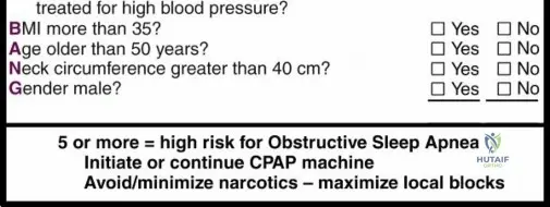
- While directly unrelated to radiation safety, comprehensive preoperative patient evaluation is paramount for any surgical procedure. This includes screening for conditions like obstructive sleep apnea, as illustrated by the STOP-BANG questionnaire. Such assessments contribute to overall patient safety, complementing specific radiation safety measures by ensuring the patient is optimally prepared for surgery and potential complications are anticipated and managed.

Detailed Intraoperative Radiation Exposure Minimization Techniques
The core of practical radiation safety lies in the systematic application of techniques that minimize exposure during the procedure. The "Time, Distance, Shielding" paradigm, central to the ALARA principle, guides these efforts.
Time Distance Shielding ALARA Principles
Time
Minimizing exposure time is the most direct way to reduce total radiation dose. The seed content correctly identifies "minimizing exposure time" as a primary factor.
- Pulsed Fluoroscopy: Utilize pulsed fluoroscopy modes (e.g., 7.5 or 15 pulses per second) instead of continuous fluoroscopy whenever clinically appropriate. This can reduce dose by 50-75% with minimal impact on image quality for most orthopedic procedures.
- Last-Image Hold (LIH): Maximize the use of the LIH feature. Once a satisfactory image is captured, hold it on the screen for reference rather than continuous live fluoroscopy.
- Pre-procedure Mental Rehearsal: Mentally walk through the steps requiring fluoroscopy, anticipating required views and instrument positions, to reduce "search" time.
- Communication with Team: Clear and concise communication with the radiographer and surgical team ensures images are taken only when needed, and the C-arm is optimized for the desired view quickly.
- Surgeon Control: The surgeon should retain control of the C-arm foot pedal. This ensures that fluoroscopy is activated only when directly needed by the operating surgeon, preventing unnecessary exposures by assistants or scrub technicians.
Distance
The inverse square law dictates that radiation intensity decreases rapidly with increasing distance from the source. Doubling the distance from the X-ray source reduces exposure by a factor of four. The seed content rightly emphasizes "maximizing the distance between the surgeon and the radiation beam."
- Standing Away from Source and Patient: Surgeons and staff should stand as far as practically possible from the X-ray tube and the patient, especially during active fluoroscopy. Scatter radiation, the primary source of occupational exposure, is most intense near the patient's body.
- C-Arm Positioning: The image intensifier (detector) should be as close to the patient as possible, while the X-ray tube should be as far from the patient as possible. Positioning the X-ray tube below the patient (if feasible) directs the primary beam and most scatter away from the surgeon's upper body and eyes, which are closer to the image intensifier. The seed content mentions "Positioning the extremity closer to the x-source" increases exposure; conversely, positioning the detector closer to the extremity and the source further away (below the table if possible) is the optimal strategy.


Illustrations 1 and 2 depict optimal C-arm positioning techniques to minimize radiation exposure to the surgical team, emphasizing the principles of time, distance, and shielding. These typically demonstrate positioning the X-ray tube below the patient and the image intensifier as close as possible, thereby reducing scatter to the operating surgeon. - Rotation of Staff: If prolonged cases are anticipated, rotating staff can distribute cumulative doses, though this should not replace direct dose reduction strategies.
Shielding
Protective shielding creates a physical barrier to absorb radiation. The seed content explicitly notes "use of protective shielding."
- Lead Aprons: All personnel in the fluoroscopy suite must wear lead aprons (typically 0.25 mm to 0.5 mm lead equivalent). Aprons should be properly fitted and regularly inspected for cracks or damage.
- Thyroid Shields: The thyroid gland is highly radiosensitive. Thyroid collars must be worn to protect against scatter radiation.
- Leaded Eyewear: The ocular lens is susceptible to deterministic effects (cataracts). Leaded glasses with side shields are crucial, especially for the primary operator.
- Patient Shielding: Gonadal and breast shields should be used on patients whenever possible, provided they do not obscure the region of interest or interfere with the procedure.
- Mobile Lead Barriers: Mobile lead shields or drapes can be strategically positioned between the X-ray source/patient and the operating team to provide additional protection against scatter.
Optimized Fluoroscopic Technique
Beyond Time, Distance, and Shielding, specific fluoroscopic techniques further contribute to dose reduction.
- Collimation: "Using collimation to manipulate the x-ray beam" is a critical factor. Collimation restricts the X-ray beam to the smallest necessary field of view. This reduces patient dose by limiting the irradiated tissue volume, significantly decreases scatter radiation (thus reducing staff dose), and improves image quality by reducing scattered photons reaching the detector. Always collimate down to the area of interest.
- Image Intensifier and X-ray Source Positioning: As noted, position the image intensifier as close to the patient as possible (reduces dose, improves image quality due to less scatter reaching detector) and the X-ray tube as far below the patient as practical.
- Magnification and Zoom: While useful for detail, magnification modes generally increase the X-ray dose to the patient as the automatic dose rate control increases output to maintain image brightness. Use magnification judiciously and only when necessary.
- Last-Image Hold and Digital Subtraction Angiography: These advanced features reduce the need for live fluoroscopy by providing static reference images or allowing visualization of contrast flow without continuous radiation.
Specific Modality Considerations
- Mini C-Arm vs Standard C-Arm: The seed content highlights the difference in radiation exposure between the two. The mini C-arm, suitable for extremities, operates at lower kilovoltage (kVp) and milliamperage (mA), resulting in significantly less radiation and scatter. Its use is encouraged "whenever feasible (associated with minimal radiation exposure)." Conversely, "use of large C-arm rather than mini C-arm" is associated with increased radiation exposure.
- 3D Imaging and Navigation: Advanced intraoperative 3D imaging (e.g., O-arm, cone-beam CT C-arms) provides superior anatomical detail and can reduce the need for multiple 2D fluoroscopic shots. While a single 3D scan delivers a higher dose than a single 2D shot, the overall cumulative dose for complex procedures may be lower by improving accuracy and reducing the need for repeat fluoroscopy or postoperative CT scans. Navigation systems further enhance precision, potentially minimizing the number of images required.
Radiation-Related Complications and Management
Understanding the potential complications of radiation exposure is essential for patient counseling, occupational health, and effective risk management. These complications span both deterministic and stochastic effects.
Deterministic Effects
Deterministic effects are characterized by a threshold dose and increased severity with higher doses.
- Skin Injury (Radiation Dermatitis): Acute high-dose exposure, particularly from prolonged fluoroscopy where the same skin area is irradiated, can lead to erythema, desquamation, ulceration, and even necrosis. The threshold for transient erythema is around 2 Gy, with more severe reactions occurring at higher doses. In orthopedic surgery, this is rare but can occur in extremely long, complex cases (e.g., complex spine or pelvic trauma) without proper beam rotation.
- Cataracts: The ocular lens is particularly sensitive to cumulative radiation, with a threshold for detectable lens opacities around 0.5-2 Sv. Interventionalists, including orthopedic surgeons, have an elevated risk of developing posterior subcapsular cataracts due to chronic exposure to scatter radiation to the head and neck if not adequately protected with leaded eyewear.
- Sterility: High doses of radiation to the gonads can cause temporary or permanent sterility. While extremely rare in orthopedic imaging, proper gonadal shielding is a standard precaution, especially for younger patients.
Stochastic Effects
Stochastic effects, primarily cancer induction and genetic mutations, are the major long-term concerns, with no presumed threshold dose.
- Increased Cancer Risk: The most significant stochastic effect is the increased risk of developing radiation-induced cancers. This includes leukemia, thyroid cancer (especially in children), breast cancer, and other solid tumors. The risk is cumulative over a lifetime, emphasizing that every exposure, no matter how small, adds to the overall probability. This risk is proportionally higher for younger patients and for highly exposed occupational workers.
- Genetic Effects: While demonstrable in animal models, evidence for significant radiation-induced heritable genetic defects in humans at diagnostic doses is limited. Nonetheless, the theoretical risk underscores the importance of gonadal shielding and minimizing exposure to reproductive organs.
- Teratogenic Effects: In utero exposure to ionizing radiation can lead to congenital malformations, growth restriction, and intellectual disability. The risk is highly dependent on the gestational age and dose. The first trimester is particularly vulnerable. This necessitates rigorous pregnancy screening and, if radiation is unavoidable, meticulous shielding and careful dose estimation for pregnant patients.
Monitoring and Dose Tracking
Effective management of radiation risks relies on robust monitoring systems.
- Personal Dosimetry: All personnel routinely exposed to ionizing radiation (e.g., orthopedic surgeons, residents, technologists) are required to wear personal dosimeters. These include thermoluminescent dosimeters (TLDs) or optically stimulated luminescence (OSL) dosimeters, typically worn on the collar outside the lead apron (to measure head/neck exposure) and sometimes under the apron (to estimate whole-body dose). These are read monthly or quarterly.
- Regular Monitoring and Reporting: Institutions must maintain records of individual and departmental dose levels. Elevated readings should trigger immediate investigation and counseling, potentially leading to adjustments in practice or additional training.
- Dose Reference Levels (DRLs): Establishing and adhering to institutional and national DRLs for common procedures helps optimize protocols and identify outliers where doses might be unnecessarily high.
- Incident Reporting: Any suspected overexposure or equipment malfunction leading to unintended radiation exposure must be promptly reported and investigated to prevent recurrence.
Management of Accidental Overexposure
Should an accidental overexposure occur, a structured management plan is critical.
- Medical Evaluation and Follow-up: The exposed individual must undergo medical evaluation, potentially including specialized blood tests or consultations with radiation oncologists or geneticists, depending on the estimated dose. Long-term follow-up may be necessary to monitor for potential health effects.
- Counseling and Risk Communication: Clear, empathetic, and evidence-based counseling must be provided regarding the estimated dose, potential risks, and available support.
- Modification of Practice: The circumstances leading to the overexposure must be thoroughly analyzed, and practice patterns or equipment protocols modified to prevent similar incidents in the future. This may involve retraining, equipment replacement, or procedural changes.
Table of Radiation Related Complications
| Complication Type | Description | Incidence | Management/Prevention Strategy |
|---|---|---|---|
| Deterministic Effects | (Threshold dose, severity increases with dose) | ||
| Radiation Dermatitis | Skin erythema, desquamation, epilation, ulceration, or necrosis from direct, high cumulative beam exposure. | Rare in typical orthopedic fluoroscopy; higher in prolonged, complex interventional cases without beam rotation. | Strict adherence to ALARA (time, distance, shielding), beam angulation changes, awareness of cumulative skin dose (fluoroscopy dose meters). Symptomatic treatment. |
| Radiation-Induced Cataracts | Lens opacification (posterior subcapsular) from cumulative exposure to scatter radiation, particularly to the head/neck region. | Increased risk in interventionalists without consistent leaded eye protection over time. | Consistent use of leaded eyewear, optimal C-arm positioning to direct scatter away from the head/eyes, minimizing head exposure. |
| Sterility (Gonadal) | Temporary or permanent impairment of reproductive function due to high direct gonadal radiation doses. | Extremely rare in orthopedic imaging; typically requires very high doses. | Routine use of gonadal shielding for patients, especially those of reproductive age. Minimizing exposure time and field size. |
| Stochastic Effects | (No threshold, probability increases with dose) | ||
| Radiation-Induced Cancer | Increased lifetime risk of various cancers (e.g., leukemia, thyroid, breast, solid tumors) with cumulative ionizing radiation exposure. | Small but statistically demonstrable increase in risk over general population for both patients and staff. | Strict ALARA adherence, justification for every exposure, patient and staff shielding, personal dosimetry, dose management systems. Long-term surveillance. |
| Genetic Effects | Damage to germ cells potentially leading to hereditary mutations in offspring. | Theoretical risk; not conclusively demonstrated in humans at diagnostic doses. | Avoidance of unnecessary radiation to reproductive organs, particularly in younger patients. |
| Teratogenic Effects | Fetal abnormalities, growth restriction, intellectual disability, or spontaneous abortion from in-utero exposure. | Dose- and gestational age-dependent; critical thresholds exist. | Rigorous pregnancy screening. Avoidance of elective studies in pregnant patients. Urgent procedures with meticulous abdominal/pelvic shielding; alternative imaging (US, MRI) where possible. |
| Operational Concerns | (Factors affecting safe practice) | ||
| Equipment Malfunction | Over-radiation or image quality degradation due to C-arm errors, collimator issues, or dose rate control failure. | Infrequent but potentially critical. | Regular equipment calibration and preventative maintenance. Pre-use checks of all safety features. Immediate reporting of any anomalies to biomedical engineering. |
| Staff Non-Compliance | Failure to adhere to safety protocols (e.g., not wearing PPE, ignoring distance rules, unnecessary fluoroscopy). | Variable, depends on training, awareness, and institutional safety culture. | Continuous education, regular safety audits, robust institutional safety protocols, strong leadership by example from senior surgeons. |
Post Exposure Monitoring and Long-term Surveillance
Effective radiation safety extends beyond the intraoperative period, encompassing a robust system of post-exposure monitoring and long-term surveillance for both occupational workers and patients. This continuous oversight ensures accountability, informs future practice, and safeguards health.
Personal Dosimetry Programs
Personal dosimetry programs are a cornerstone of occupational radiation protection.
- Requirement for Personnel: All orthopedic surgeons, residents, medical students, and other personnel who may receive an occupational dose likely to exceed 10% of the annual occupational dose limit (typically 50 mSv per year for whole body) are required to wear personal dosimeters.
- Types of Dosimeters: Thermoluminescent dosimeters (TLDs) and optically stimulated luminescence (OSL) dosimeters are the most common. They measure cumulative dose over a specified period.
- Monitoring Periods and Reporting: Dosimeters are typically exchanged and processed monthly or quarterly. Detailed reports are generated, providing individual cumulative dose information. These reports are crucial for tracking exposure trends and ensuring compliance with regulatory limits.
- Interpretation and Investigation: Any readings that approach or exceed established action levels or regulatory limits must trigger an immediate investigation. This involves reviewing the individual's work activities, fluoroscopy logs, and adherence to safety protocols. Corrective actions may include re-training, equipment review, or reassignment of duties.
Medical Surveillance for Radiation Workers
While routine medical examinations are generally not mandated for radiation workers at diagnostic exposure levels, a nuanced approach to surveillance is prudent.
- Periodic Health Examinations: Institutions should consider periodic health examinations for long-term radiation workers, with an emphasis on identifying potential deterministic effects, such as ocular lens opacities (e.g., annual ophthalmologic exams for interventionalists).
- Counseling: Regular counseling sessions regarding cumulative dose, long-term risks, and the importance of continued adherence to ALARA principles are valuable. This reinforces a culture of safety and addresses individual concerns.
Patient Dose Tracking
Tracking patient radiation exposure is becoming increasingly feasible and important with advancements in electronic health records and dose management software.
- Cumulative Patient Dose: Establishing a system to track patient radiation exposure from all modalities (plain films, fluoroscopy, CT, nuclear medicine) over time allows for a comprehensive understanding of a patient's total dose. This is particularly relevant for pediatric patients or those requiring multiple imaging studies.
- Dose Management Software: Integration of dose management software with imaging equipment (PACS, RIS) automatically records and monitors patient doses. This data can be used to set and refine diagnostic reference levels (DRLs), optimize imaging protocols, and flag potentially high-dose examinations for review.
- Communication: Providing patients with a record of their radiation exposure, especially when referring them to other specialties or institutions, can prevent unnecessary repeat examinations and facilitate informed decision-making.
Institutional Review and Audit
A robust institutional framework for radiation safety, including regular review and audit, is essential for continuous improvement.
- Safety Audits: Regular audits of fluoroscopy suites, CT scanners, and nuclear medicine departments by medical physicists or radiation safety officers ensure equipment is functioning correctly, protocols are followed, and shielding is adequate.
- Review of Incident Reports: Thorough review of all incident reports and "near misses" related to radiation exposure provides valuable learning opportunities and helps refine safety protocols.
- Continuous Improvement Programs: Radiation safety is not static. Institutions should implement continuous quality improvement programs that regularly evaluate new technologies, update protocols based on current literature and guidelines, and provide ongoing education to staff. This proactive approach ensures that the highest standards of radiation protection are consistently maintained.
Summary of Key Radiation Safety Literature and Guidelines
Adherence to established guidelines and a continuous awareness of evolving literature are paramount for maintaining best practices in radiation safety within orthopedic surgery. These guidelines are developed by expert bodies to protect both patients and healthcare professionals.
International and National Regulatory Bodies
Several authoritative bodies provide the foundational principles and regulatory frameworks for radiation protection:
- International Commission on Radiological Protection (ICRP): The ICRP provides the global scientific basis for radiation protection. Its core recommendations, embodied in principles like justification (each exposure must have a net benefit), optimization (ALARA principle), and dose limitation (for occupational exposure), serve as the benchmark for national regulations worldwide. The ICRP has specifically addressed medical exposure and occupational exposure.
- National Council on Radiation Protection and Measurements (NCRP): In the United States, the NCRP is a non-profit organization that provides independent scientific advice and recommendations on radiation protection. Its reports offer detailed guidance on dose limits, shielding design, and operational procedures, often influencing federal and state regulations (e.g., for maximum permissible doses for occupational workers).
- Local State and Federal Regulations: Compliance with specific local, state, and federal regulations (e.g., those enforced by the Nuclear Regulatory Commission (NRC) for radioactive materials, or state health departments for X-ray equipment licensing) is mandatory. These regulations often translate ICRP and NCRP recommendations into enforceable legal requirements.
Professional Society Recommendations
Orthopedic and radiological professional societies actively contribute to radiation safety through position statements, guidelines, and educational initiatives tailored to their specific disciplines:
- American Academy of Orthopaedic Surgeons (AAOS) Guidelines: The AAOS regularly publishes clinical practice guidelines and position statements that incorporate radiation safety considerations relevant to orthopedic procedures. These often provide practical recommendations for fluoroscopy use, patient shielding, and occupational protection.
- Radiological Society of North America (RSNA) and American College of Radiology (ACR) Statements: These societies are at the forefront of promoting radiation safety in medical imaging. Their "Image Gently" (for pediatric imaging) and "Image Wisely" (for adult imaging) campaigns advocate for judicious imaging practices, emphasizing dose reduction techniques and appropriate modality selection. Their guidelines often provide detailed technical specifications and best practices for various imaging modalities.
- Specific Subspecialty Societies: Spine societies, trauma associations, and other subspecialty groups within orthopedics often issue specific recommendations addressing radiation safety pertinent to their highly specialized procedures (e.g., C-arm positioning for spinal fusion, radiation protection in pelvic trauma surgery).
Best Practices and Emerging Technologies
The field of radiation safety is continuously evolving with new research and technological advancements:
- Review of Recent Studies on Dose Reduction Techniques: Ongoing research consistently identifies new methods for dose reduction. This includes optimizing fluoroscopy protocols (e.g., frame rates, pulse width), utilizing iterative reconstruction algorithms in CT to significantly lower doses while maintaining diagnostic image quality, and exploring the dose-saving potential of newer C-arm technologies.
- Impact of 3D Imaging and Robotic Assistance: The integration of intraoperative 3D imaging (e.g., O-arm, robotic-assisted navigation) is transforming surgical precision. While these technologies involve an initial radiation dose, they often reduce the need for repetitive 2D fluoroscopic images, potentially lowering the overall cumulative dose and reducing operative time. Studies continue to evaluate the net radiation burden and benefits of these advanced systems.
- Future Directions in Radiation Protection Research: Research continues into areas such as personalized dosimetry, advanced radiation detection technologies, novel shielding materials, and a deeper understanding of low-dose radiation biological effects.
- Culture of Safety and Continuous Education: Ultimately, mastering radiation safety is a dynamic process that requires a pervasive "culture of safety" within every orthopedic practice. This involves continuous education for all staff, regular reinforcement of ALARA principles, active participation in quality improvement initiatives, and a commitment from leadership to prioritize radiation protection. Staying abreast of the latest literature and guidelines is not merely academic; it is a critical component of providing the safest and most effective care in orthopedic surgery.
