Arthroscopic Lysis of Adhesions, Osteoarthritis Debridement, and Complication Management
Key Takeaway
Arthroscopic lysis of adhesions is a highly effective intervention for post-traumatic or postoperative knee stiffness. This comprehensive guide details the Sprague technique for systematic adhesiolysis, indications for osteoarthritis debridement, and evidence-based management of knee arthroscopy complications. Key protocols include managing dropped anterior cruciate ligament grafts, mitigating arthrofibrosis, and addressing postoperative intra-articular infections to optimize patient outcomes and restore joint kinematics.
ARTHROSCOPIC LYSIS AND EXCISION OF ADHESIONS
Postoperative or post-traumatic arthrofibrosis of the knee presents a formidable challenge to the orthopaedic surgeon. The proliferation of dense, intra-articular fibrous scar tissue can severely restrict patellofemoral kinematics and tibiofemoral tracking, leading to profound functional deficits. The systematic arthroscopic lysis of adhesions, originally popularized by Sprague, remains the gold standard for restoring joint mobility while minimizing the iatrogenic trauma associated with open arthrolysis.
Indications and Patient Selection
Arthroscopic adhesiolysis is indicated for patients with refractory knee stiffness that has failed to respond to an aggressive, well-structured physical therapy regimen. Common etiologies include prior anterior cruciate ligament (ACL) reconstruction, prolonged immobilization, intra-articular fractures, and total knee arthroplasty (TKA).
Surgical Warning: Careful preoperative assessment is mandatory. Differentiate between true intra-articular adhesions and extra-articular contractures (e.g., quadriceps scarring or capsular contracture). Arthroscopic lysis will not resolve extra-articular tethering.
Preoperative Planning and Biomechanics
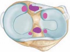
Normal knee flexion requires the patella to glide distally and the anterior interval (the space between the infrapatellar fat pad and the anterior tibia) to remain supple. Adhesions in the suprapatellar pouch restrict patellar excursion, while scarring in the anterior interval and intercondylar notch limits terminal extension. The surgical goal is to systematically release these tethering points without damaging the underlying chondral surfaces or native ligamentous structures.
The Sprague Technique: Step-by-Step
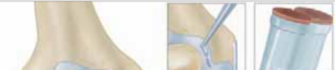
1. Initial Portal Placement and Blind Dissection
* Establish standard anterolateral and anteromedial portals.
* Insert an arthroscopic sheath equipped with a blunt trocar. Do not use a sharp trocar, as the distorted anatomy and obliterated joint spaces dramatically increase the risk of iatrogenic chondral injury.
* Carefully pass the blunt trocar beneath the patella and direct it superiorly into the suprapatellar pouch.
* Utilize a sweeping motion with the blunt trocar to mechanically disrupt the initial layer of adhesions within the suprapatellar pouch and the medial and lateral gutters.
2. Arthroscopic Inspection and Debridement
* Insert the arthroscope and establish visualization. In cases of dense arthrofibrosis, the patellofemoral joint itself is often spared, though the surrounding recesses are obliterated.
* Begin systematic motorized débridement in the peripatellar region, utilizing a shaver or radiofrequency ablation wand, and extend the resection outward.
* Once the suprapatellar pouch has been adequately restored, insert an inflow cannula through a superior portal to maintain joint distension and improve visualization.
3. Gutter and Notch Clearance
* Continue the dissection inferiorly into the medial and lateral gutters, clearing the capsular recesses.
* Proceed into the medial and lateral tibiofemoral compartments.
* Address the intercondylar notch. Proliferation of fibrous tissue (often termed a "Cyclops lesion" if localized anteriorly post-ACLR) is frequently present within the notch and anterior interval. This must be meticulously excised, as it acts as a mechanical block to terminal extension.
* Cruciate Protection: Exercise extreme caution when navigating the notch to avoid inadvertent damage to the native cruciate ligaments or a previously placed graft.
4. Adjunctive Procedures and Manipulation
* If patellar mobility remains restricted after comprehensive arthroscopic release, a lateral retinacular release may be indicated to restore normal patellar tilt and glide.
* Following systematic lysis, perform a gentle manipulation under anesthesia (MUA).
* If further adhesions are audibly or palpably disrupted during manipulation, re-introduce the arthroscope to débride the newly freed fibrous bands and ensure hemostasis.
Surgical Pitfall: Avoid aggressive, forceful manipulation. The osteopenic bone frequently seen in chronically stiff knees is highly susceptible to iatrogenic supracondylar femur fractures or patellar tendon avulsions during excessive manipulation.
Postoperative Protocol
The success of arthroscopic adhesiolysis is heavily dependent on the postoperative rehabilitation protocol.
* Analgesia: Continuous epidural anesthesia or an indwelling adductor canal catheter is highly recommended, maintained for 2 to 3 days postoperatively to ensure pain-free early mobilization.
* Drainage: Thoroughly irrigate the joint, insert an intra-articular suction drain (removed at 48 hours), and apply a bulky compressive dressing to mitigate hemarthrosis.
* Mobilization: The patient must be placed in a continuous passive motion (CPM) machine immediately in the recovery room. Aggressive, supervised physical therapy begins on postoperative day one.
DÉBRIDEMENT OF OSTEOARTHROSIS AND ABRASION ARTHROPLASTY
The role of arthroscopy in the management of degenerative arthritis of the knee is highly nuanced. While it is not a cure for osteoarthritis (OA), targeted arthroscopic intervention can provide significant symptomatic relief in carefully selected patients.
Indications and Patient Selection
The most favorable outcomes are observed in patients presenting with relatively acute mechanical symptoms superimposed on mild-to-moderate degenerative changes.
Appropriate Indications:
* Resection of mobile, unstable flaps of degenerative menisci.
* Excision of excessive, impinging synovial fronds.
* Removal of loose bodies.
* Chondroplasty of unstable, severe chondromalacic flaps.
Contraindications:
* Patients with chronic, progressive, generalized knee pain.
* Advanced degenerative changes (e.g., bone-on-bone articulation evident on standing 45-degree posteroanterior flexion radiographs).
* Malalignment (varus/valgus) without concurrent corrective osteotomy.
Techniques for Chondral Defects
For isolated, full-thickness chondral defects (Grade IV chondromalacia) measuring 1 cm² or less, marrow stimulation techniques can be employed to promote the formation of a fibrocartilage repair tissue.
- Abrasion Arthroplasty and Drilling: These older techniques involve mechanically removing the calcified cartilage layer to expose the subchondral vascularity.
- Microfracture: The current standard for marrow stimulation. Using specialized awls, multiple perforations are made into the subchondral bone plate, spaced 3-4 mm apart. This stimulates a vascular response, releasing mesenchymal stem cells and marrow elements into the defect to form a "super clot."
- Verification: The success of the marrow stimulation must be verified intraoperatively. Deflate the tourniquet and reduce intra-articular fluid pressure; the surgeon should observe distinct fat droplets and blood emanating from the microfracture holes.
Clinical Pearl: Microfracture techniques, osteochondral autograft transfers (OATS), and autologous chondrocyte implantation (ACI) are strictly contraindicated in patients with diffuse, extensive degenerative arthritis. These procedures require healthy surrounding cartilage shoulders to contain the repair tissue.
Postoperative Rehabilitation for Marrow Stimulation
Healing of the fibrocartilage clot requires a delicate balance of protection and stimulation.
* Weight Bearing: Strict protective weight-bearing (touch-down only) for 6 weeks.
* Motion: Constant passive motion is critical for molding the regenerating fibrocartilage. Historically, CPM for 6 to 8 hours a day for 6 weeks was recommended, though modern literature questions the cost-benefit ratio of extended CPM use compared to active-assisted range of motion exercises.
COMPLICATIONS ASSOCIATED WITH KNEE ARTHROSCOPY
Despite being minimally invasive, knee arthroscopy carries a distinct complication profile. Large epidemiological series from the late 1980s (encompassing over 380,000 cases) established a baseline overall complication rate of less than 2%. Modern literature confirms that this rate remains relatively constant, though the complexity of the procedures has increased.
Epidemiology of Complications
Reported complications include:
* Infection (Septic arthritis)
* Hemarthrosis
* Vascular injury (e.g., popliteal artery injury during posterior horn meniscal work)
* Nerve injury (Saphenous, Peroneal)
* Deep vein thrombosis (DVT) and thrombophlebitis
* Arthrofibrosis (Stiff knee)
* Complex Regional Pain Syndrome (CRPS / RSD)
Complication rates correlate directly with procedural complexity. For example, isolated medial meniscectomy carries a complication rate of approximately 1.78%, whereas meniscal repairs and ACL reconstructions carry rates approaching 2.4% and 2.0%, respectively.
Arthrofibrosis and Surgical Timing
Arthrofibrosis is a devastating complication, particularly following ACL reconstruction. The incidence of postoperative stiffness increases significantly when ligamentous reconstruction is combined with meniscal repair or peripheral ligamentous repair (e.g., medial collateral ligament repair on the femoral side).
Mitigation Strategies:
* Surgical Timing: Early surgical intervention for ACL injuries—before the patient has regained normal muscular tone, resolved their effusion, and achieved full range of motion—is a primary risk factor for arthrofibrosis.
* The "Calm Knee": Allowing the knee to "calm" (resolution of acute inflammatory phase, restoration of terminal extension) before proceeding with reconstruction has been definitively shown to decrease postoperative stiffness.
Neurovascular Injuries
Nerve injuries remain a persistent risk, particularly during complex repairs.
* Saphenous Nerve: The infrapatellar branch of the saphenous nerve is highly vulnerable during the harvest of bone-patellar tendon-bone (BTB) or hamstring autografts, as well as during medial meniscal repairs (inside-out techniques).
* Peroneal Nerve: At risk during lateral meniscal repairs, particularly when placing sutures in the posterior horn, or during posterolateral corner (PLC) reconstructions.
Infection and the Contaminated Graft Protocol
The incidence of infection following ACL reconstruction is slightly increased when performed in conjunction with meniscal repair, likely due to increased surgical time, additional exposure, and the potential for joint contamination during the passing and retrieving of needles.
Management of the Dropped Graft
A dropped autograft is a highly stressful intraoperative event. The surgeon must decide whether to harvest a different graft (autograft or allograft) or attempt to sterilize and salvage the contaminated tissue.
Evidence-based protocols support the salvage of dropped grafts. Molina et al. demonstrated the efficacy of various sterilization solutions. Based on their findings and subsequent literature, the following protocol is recommended for a dropped graft:
- Immediate Retrieval: Retrieve the graft immediately from the floor.
- Mechanical Wash: Rinse the graft thoroughly using sterile technique with a large volume of sterile saline (e.g., pulse lavage or vigorous agitation).
- Chlorhexidine Soak: Soak the graft in a 4% chlorhexidine gluconate solution. While 90 seconds is the minimum, a 10-minute soak is highly recommended.
- Antibiotic Soak: Transfer the graft to a solution containing neomycin and polymyxin B (e.g., 40 mg neomycin and polymyxin in 1000 mL sterile saline) for an additional 10 minutes.
- Final Rinse: Rinse the graft thoroughly with sterile saline before implantation to remove cytotoxic residual chemicals.
Clinical Pearl: In a survey of 196 sports-trained surgeons regarding ACL graft contamination, 75% of those who experienced a dropped graft proceeded with cleansing and reconstruction using the salvaged graft. Zero infections were reported in this cohort, validating the safety of rigorous sterilization protocols.
Management of Postoperative Septic Arthritis
When a postoperative knee infection occurs, aggressive and immediate intervention is required to protect the articular cartilage from chondrolysis.
* Surgical Intervention: Early, thorough arthroscopic irrigation and débridement (I&D) are mandatory.
* Repeat I&D: A repeat irrigation and débridement should be performed at 48 to 72 hours if clinical symptoms, synovial fluid analysis, or inflammatory markers have not significantly improved.
* Graft Retention: The ACL graft can and should be left in place, provided that its fixation is secure and there is no extensive macroscopic deterioration or necrosis of the graft tissue at the time of the initial I&D.
* Antibiotic Therapy: Intravenous antibiotics (tailored to culture sensitivities) are generally prescribed for 2 to 3 weeks, followed by targeted oral antibiotics to complete a comprehensive 6-week course.
Complex Regional Pain Syndrome (CRPS)
Historically referred to as Reflex Sympathetic Dystrophy (RSD), CRPS is a poorly understood, debilitating neuroinflammatory condition characterized by disproportionate pain, hyperalgesia, skin color changes, and temperature asymmetry.
While surgical control over the onset of CRPS is limited, risk mitigation includes:
* Careful patient selection and preoperative psychological screening.
* Minimizing tourniquet time and overall operative duration.
* Aggressive, early postoperative physical therapy to maintain neural mobility and joint kinematics.
* Note on Graft Tension: Early literature hypothesized that overtightening of the ACL graft might induce CRPS or result in failure of graft maturation; however, modern biomechanical and clinical studies have largely refuted this conclusion, emphasizing instead the importance of anatomic tunnel placement and appropriate tensioning at specific flexion angles.
You Might Also Like