PIP Joint Dislocations: Comprehensive Guide to Anatomy, Biomechanics & Management
Key Takeaway
PIP joint dislocations are common hand injuries, often from sports. These finger joint dislocations involve disruption to static stabilizers like the volar plate and collateral ligaments, and dynamic tendons. Biomechanics differ: dorsal (hyperextension, volar plate rupture) is most common, while palmar, lateral, and rotatory types result from distinct forces.
Introduction & Epidemiology
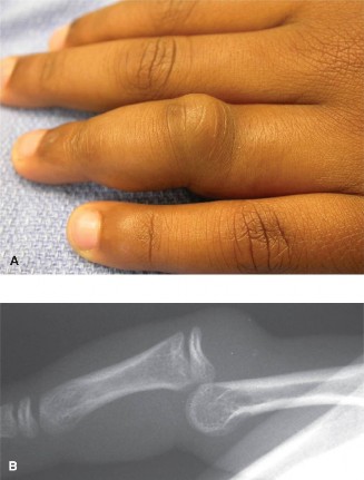
Dislocations of the proximal interphalangeal (PIP) joint are among the most common hand injuries, representing a significant proportion of all joint dislocations. These injuries are frequently encountered in athletic activities, particularly contact sports suchs as football, basketball, and rugby, as well as in falls onto an outstretched hand. The PIP joint is a ginglymus (hinge) joint, crucial for hand function and precision grip, making its stable and congruent restoration paramount for long-term functional outcomes. While often perceived as minor injuries, undertreatment or inadequate management can lead to significant morbidity, including chronic pain, stiffness, instability, and post-traumatic arthritis, severely impacting occupational and daily living activities.
Epidemiologically, PIP joint dislocations are most prevalent in the active adult population. Dorsal dislocations, resulting from hyperextension and axial loading, account for the vast majority (approximately 75-80%) of these injuries. Less common are palmar (volar) dislocations, which typically result from axial loading combined with hyperflexion, and rotatory dislocations, which involve a more complex mechanism of axial load and torsional forces. Lateral dislocations are rare in isolation but often occur in conjunction with dorsal or palmar patterns, indicative of severe collateral ligamentous disruption. Understanding the precise mechanism of injury is critical for anticipating associated soft tissue and osseous pathology.
Surgical Anatomy & Biomechanics
The PIP joint is a diarthrodial hinge joint formed by the head of the proximal phalanx and the base of the middle phalanx. Its stability is primarily conferred by a complex arrangement of static and dynamic stabilizers.
Static Stabilizers
- Volar Plate: A thick, fibrocartilaginous structure on the palmar aspect, firmly attached to the base of the middle phalanx and less firmly to the neck of the proximal phalanx (the check-rein ligaments). It prevents hyperextension and resists dorsal translation of the middle phalanx. Its rupture or avulsion is a common feature of dorsal PIP dislocations.
-
Collateral Ligaments:
Comprising proper and accessory collateral ligaments on both radial and ulnar sides.
- Proper Collateral Ligaments (PCLs): Cord-like structures originating from the head of the proximal phalanx and inserting into the volar-lateral aspect of the base of the middle phalanx. They are taut in flexion and relaxed in extension, resisting varus and valgus stresses.
- Accessory Collateral Ligaments (ACLs): Fan-shaped ligaments originating dorsal to the PCLs and inserting into the volar plate. They are taut in extension.
- Joint Capsule: Surrounds the entire joint, reinforcing the volar plate and collateral ligaments.
- Articular Congruity: The trochlear surface of the proximal phalanx head articulates with the biconcave base of the middle phalanx. The bicondylar configuration of the proximal phalanx, with its central groove and medial/lateral condyles, interlocks with the central ridge and medial/lateral depressions of the middle phalanx base, providing inherent stability, particularly in flexion.
Dynamic Stabilizers
- Extensor Mechanism: Comprises the central slip, lateral bands, and sagittal bands. The central slip inserts onto the dorsal base of the middle phalanx, primarily extending the PIP joint. The lateral bands contribute to PIP extension via the terminal tendon.
- Flexor Digitorum Superficialis (FDS) and Flexor Digitorum Profundus (FDP) Tendons: Traverse the joint volarly, providing dynamic flexion stability.
Biomechanics of Dislocation
- Dorsal Dislocation: The most common type. Typically results from hyperextension and axial load, forcing the middle phalanx dorsally relative to the proximal phalanx. The primary injury involves rupture or avulsion of the volar plate from its insertion on the middle phalanx. Often, the check-rein ligaments remain attached, causing the volar plate to displace proximally and potentially interpose in the joint. Severe dorsal dislocations can also involve rupture of one or both collateral ligaments and, in extreme cases, avulsion fractures of the middle phalanx base (dorsal lip fracture).
- Palmar (Volar) Dislocation: Less common, typically due to axial compression with hyperflexion. The central slip of the extensor mechanism is frequently involved, either avulsed from its insertion or sustaining a buttonhole rupture, allowing the lateral bands to displace volarly. This can lead to a boutonnière deformity if untreated. One or both collateral ligaments may also be ruptured.
- Lateral Dislocation: Usually associated with significant varus or valgus stress, leading to rupture of the contralateral collateral ligament. Often seen in combination with dorsal or palmar patterns, indicating complete disruption of the volar plate.
- Rotatory Dislocation: A rare and complex injury where the phalanx rotates around its longitudinal axis. It often involves impaction of the condyle of the proximal phalanx into the joint capsule or interposition of soft tissues (e.g., collateral ligament, volar plate) preventing reduction.
Understanding these anatomical relationships and biomechanical principles is critical for accurate diagnosis, reduction, and operative planning, particularly in addressing persistent instability or irreducibility.
Indications & Contraindications
Management of PIP joint dislocations spans a spectrum from conservative care for stable, reducible injuries to complex surgical reconstruction for irreducible, unstable, or chronic cases. The decision-making process is guided by the type of dislocation, associated osseous and ligamentous injuries, joint stability post-reduction, patient factors, and functional demands.
Indications for Operative vs. Non-Operative Management
| Indication Type | Non-Operative Management | Operative Management |
|---|---|---|
| Dorsal Dislocation | * Simple Dorsal Dislocation: Closed reduction is successful, and the joint is stable through a functional arc of motion (e.g., 30-70 degrees flexion). | * Irreducible Dislocation: Due to incarcerated volar plate, collateral ligament, or osteochondral fragment. |
| * Stable Dorsal Dislocation with Volar Plate Avulsion Fracture: If the fragment is small (<20-30% of articular surface), non-displaced, and joint is stable post-reduction. | * Unstable Dorsal Dislocation: Recurrent subluxation/dislocation after closed reduction, suggesting significant volar plate and/or collateral ligament disruption. | |
| * Large Volar Plate Avulsion Fracture: >20-30% of articular surface, displaced, or involving a significant portion of the collateral ligament insertion, leading to instability. | ||
| * Pilon-type Fracture of Middle Phalanx Base: Comminuted intra-articular fracture requiring open reduction and internal fixation (ORIF) and potential bone grafting. | ||
| Palmar Dislocation | * Stable Palmar Dislocation: Closed reduction successful, joint stable without central slip disruption. | * Irreducible Dislocation: Due to incarcerated collateral ligament or central slip, or buttonhole rupture. |
| * Central Slip Avulsion: If minimal displacement and joint stability maintained with dynamic splinting. | * Unstable Palmar Dislocation: Persistent instability or extensor lag >30 degrees after closed reduction, indicating significant central slip disruption. | |
| * Chronic Boutonnière Deformity: Established deformity requiring central slip repair/reconstruction. | ||
| Lateral Dislocation | * Isolated Lateral Dislocation: Closed reduction successful, stable with collateral ligament repair/reconstruction. | * Irreducible Lateral Dislocation: Due to interposition of soft tissue (e.g., collateral ligament). |
| * Unstable Lateral Dislocation: Post-reduction instability requiring collateral ligament repair or reconstruction. | ||
| Fracture-Dislocations | * Small, Non-displaced Avulsion Fractures: Stable after reduction. | * Large or Displaced Intra-articular Fractures: Especially those involving a significant portion of the articular surface or creating joint incongruity. |
| * Rotatory Dislocation with Irreducibility: Due to soft tissue interposition or bone impaction. | ||
| Chronic Instability | N/A | * Chronic PIP Joint Instability: Following failed conservative management or acute surgical repair, requiring ligamentous reconstruction or arthroplasty in severe cases. |
Contraindications to Operative Management
Absolute contraindications are few and generally relate to the patient's overall health status or local factors.
*
Acute Infection:
Active infection in the surgical field.
*
Severe Comorbidities:
Medical instability precluding safe anesthesia and surgery.
*
Poor Skin Quality:
Compromised skin envelope (e.g., severe degloving injury, crush injury, extensive scar tissue) that would hinder wound healing or increase infection risk.
*
Unrealistic Patient Expectations:
While not a strict contraindication, understanding patient goals and educating them on realistic outcomes is crucial.
*
Severe Osteopenia/Osteoporosis:
May preclude adequate fixation with standard techniques, necessitating alternative strategies.
Relative contraindications include poor patient compliance with post-operative rehabilitation, which is paramount for successful outcomes.
Pre-Operative Planning & Patient Positioning
Thorough pre-operative planning is essential for predictable outcomes in PIP joint surgery. This involves a detailed assessment of the injury, careful selection of the surgical approach, and meticulous preparation.
Pre-Operative Planning
-
Clinical Assessment:
- History: Mechanism of injury, timing, previous hand injuries, dominant hand, occupational/recreational demands.
- Physical Examination: Careful assessment of joint stability, range of motion (active and passive), neurovascular status, associated soft tissue injuries, and presence of irreducible deformity. Assessment of post-reduction stability is critical if closed reduction was attempted.
-
Imaging Studies:
- Standard Radiographs: AP, lateral, and oblique views of the involved digit are mandatory. A true lateral view is crucial to assess joint congruity and fragment displacement, especially for fracture-dislocations. Traction radiographs can be helpful for assessing irreducibility or the size of volar plate avulsion fragments.
- Stress Radiographs: In cases of suspected subtle instability, stress radiographs (varus/valgus, hyperextension) can quantify joint laxity, though these are typically performed intra-operatively after nerve block.
- Computed Tomography (CT) Scan: Indicated for complex fracture-dislocations, particularly when assessing articular comminution, fragment size, displacement, and orientation of intra-articular fragments (e.g., pilon-type fractures, large volar lip fractures). It aids in surgical planning for plate and screw fixation or bone grafting.
- Magnetic Resonance Imaging (MRI): Less commonly used in acute settings but can be valuable for assessing purely ligamentous injuries, especially chronic ones, or to rule out occult soft tissue interposition if reduction is persistently difficult and radiographs are unremarkable.
- Anesthesia Consultation: Standard evaluation for general, regional (axillary block), or local anesthesia with sedation. Tourniquet use is routine for bloodless field.
- Surgical Strategy: Based on clinical and imaging findings, determine the specific approach, reduction technique (closed vs. open), fixation method (K-wires, mini-screws, mini-plates, external fixator), and any requirements for bone grafting or ligament reconstruction. Plan for potential need for implant removal in the future.
Patient Positioning
- Supine Position: The patient is positioned supine on the operating table.
- Hand Table: The involved upper extremity is placed on a dedicated hand table, allowing for comfortable and stable positioning.
- Arm Preparation: The arm is prepped and draped from the shoulder to the fingertips to allow for sterile access to the entire limb, including potential donor sites for grafts if required.
- Tourniquet Application: A pneumatic tourniquet is applied to the upper arm (preferred) or forearm. This provides a bloodless field, which is essential for visualizing small structures and precise dissection. Tourniquet pressure should be individualized to the patient, typically 250-300 mmHg for upper arm, or 50-70 mmHg above systolic pressure.
- Magnification: Surgical loupes (2.5x to 4.5x magnification) are routinely used to enhance visualization of delicate structures. A microscope may be considered for highly complex microvascular repairs or nerve repairs, though less common for primary PIP joint dislocations.
Detailed Surgical Approach / Technique
The choice of surgical approach depends on the type of dislocation, associated fractures, and soft tissue damage. Common approaches include dorsal, volar (midaxial or palmar zigzag), and sometimes lateral incisions.
1. Dorsal Dislocation with Volar Plate Avulsion or Ligament Disruption (Dorsal Approach)
This approach is often favored for large dorsal lip fractures of the middle phalanx and certain irreducible dorsal dislocations where dorsal interposition is suspected, or if dorsal extensor repair is necessary.
- Incision: A dorsal longitudinal or chevron incision centered over the PIP joint. Alternatively, a Y-shaped or lazy-S incision can be employed, particularly if extensor mechanism repair is anticipated. Meticulous care is taken to avoid injury to the dorsal digital nerves and veins.
-
Dissection:
- Carefully incise the skin and subcutaneous tissue. Identify and protect the dorsal digital nerves and veins, retracting them radially/ularly.
- Incise the extensor mechanism longitudinally over the central slip, or through one of the lateral bands. If a central slip avulsion is suspected, the incision should be planned to facilitate its repair.
- Retract the extensor mechanism to expose the dorsal aspect of the PIP joint capsule.
- If a large dorsal lip fracture is present, the fragment will be visible. The joint may be dislocated dorsally or subluxated.
-
Reduction and Fixation:
- Clear any interposed soft tissue (e.g., scar tissue, periosteum) from the fracture site and articular surface.
- Reduce the dorsal lip fracture fragment anatomically. This often requires gentle traction and manipulation.
-
Fixation options for dorsal lip fractures:
- K-wires: Small diameter K-wires (0.028" or 0.035") can be used for temporary or definitive fixation. If using definitive K-wires, they are typically inserted across the fracture site and into the middle phalanx, then potentially retrograde into the proximal phalanx or out the dorsum of the digit.
- Mini-screws: For larger fragments, one or two 1.0mm or 1.3mm lag screws can provide stable compression. Care must be taken to avoid over-drilling or stripping the fragile bone.
- Suture anchors: For pure ligamentous avulsions without a significant bone fragment, small suture anchors can be used to reattach the central slip or collateral ligaments to the bone.
- After fixation, assess joint stability through a full range of motion. If unstable, consider additional fixation (e.g., transarticular K-wire in 30 degrees of flexion) or ligament repair.
- Closure: Repair the extensor mechanism (central slip or lateral band) with non-absorbable sutures. Close the skin with fine sutures.
2. Dorsal Dislocation with Volar Plate Avulsion (Volar Approach)
This approach is preferred for most dorsal dislocations with volar plate injuries, especially those with large volar plate avulsion fractures or irreducible dislocations due to volar plate interposition.
- Incision: A Bruner zigzag incision across the PIP joint on the palmar aspect provides excellent exposure while minimizing flexion contracture risk. The apex of the distal flap should be distal to the PIP joint crease.
-
Dissection:
- Elevate skin flaps carefully, protecting the neurovascular bundles at the apices of the flaps. These bundles lie volar to the collateral ligaments, passing along the midaxial line.
- Identify the flexor tendon sheath. Incise the sheath longitudinally, usually between A2 and A3 pulleys, or along one side, to expose the flexor tendons. Retract the flexor tendons to one side.
- The volar plate and accessory collateral ligaments will be visible. In a dorsal dislocation, the volar plate is usually avulsed from the middle phalanx base and may be displaced proximally, blocking reduction.
-
Reduction and Fixation:
- Clear any interposed tissue from the joint. This may involve excising small, comminuted fragments of the volar plate or carefully dissecting it away if it's blocking reduction.
- Perform gentle closed reduction if possible, or open reduction by applying longitudinal traction and gentle flexion while pushing the middle phalanx head volarly.
-
Volar Plate Repair/Reconstruction:
- Suture Anchors: If the volar plate is avulsed from the middle phalanx base, drill a pilot hole (1.0mm) into the base of the middle phalanx, insert a mini suture anchor, and use the sutures to reattach the volar plate to its anatomical insertion site.
- Pull-out Wires: For larger, intact volar plate avulsions, pull-out wires can be used. Drill tunnels through the middle phalanx from dorsal to volar, pass sutures through the volar plate, and pull them through the tunnels to tie over a dorsal button.
- Lag Screws: For large osteochondral fragments of the middle phalanx volar lip, direct fixation with one or two 1.0mm or 1.3mm lag screws can be performed. This requires careful contouring of the fragment and precise drilling to avoid joint penetration.
- Transarticular K-wire: After reduction and volar plate repair, temporarily stabilize the joint with a transarticular K-wire in 30-40 degrees of flexion, passing from the dorsal middle phalanx into the proximal phalanx, ensuring the wire does not impinge on repaired structures. This provides additional stability during early healing.
- Collateral Ligament Repair: If associated collateral ligament instability is noted, small suture anchors or direct repair with fine non-absorbable sutures can be performed to reattach the torn ligament to its bony origin or insertion.
- Closure: Repair the flexor tendon sheath if it was opened significantly. Close subcutaneous tissues and skin meticulously with fine sutures.
3. Palmar (Volar) Dislocation (Dorsal Approach)
Palmar dislocations often involve central slip rupture or avulsion. A dorsal approach is preferred.
- Incision: Dorsal longitudinal or chevron incision over the PIP joint.
-
Dissection:
- Carefully elevate skin and subcutaneous tissue, protecting neurovascular structures.
- Identify the extensor mechanism. A central slip avulsion or "buttonhole" tear will be evident, with the lateral bands displaced volarly.
-
Reduction and Fixation:
- Reduce the dislocation. This may require gentle manipulation and reduction of the lateral bands.
- Central Slip Repair: Reattach the central slip to the dorsal base of the middle phalanx using a mini suture anchor or drill holes with pull-out sutures. If the central slip has a "buttonhole" rupture, repair it primarily with non-absorbable sutures.
- Collateral Ligament Repair: Assess collateral ligament stability. If disrupted, repair with suture anchors or direct sutures.
- Transarticular K-wire: Stabilize the joint with a transarticular K-wire in full extension (0 degrees) or slight flexion (e.g., 10-20 degrees) to protect the central slip repair.
- Closure: Close the extensor mechanism, subcutaneous tissue, and skin.
4. Irreducible Rotatory Dislocation
These dislocations are often irreducible by closed means due to interposition of the collateral ligament or volar plate.
- Approach: Typically a midaxial incision, as it provides good access to the collateral ligaments and joint capsule. A dorsal or volar approach can also be used depending on the suspected interposed structure.
- Dissection: Follow standard dissection to expose the joint capsule and collateral ligaments. Identify the interposed structure.
- Reduction: Gently de-rotate the digit while applying longitudinal traction. It may be necessary to release the interposed structure (e.g., collateral ligament) temporarily, reduce the joint, and then repair the released structure.
- Fixation: Often, a stable reduction can be achieved without internal fixation, but a transarticular K-wire may be placed for temporary stabilization in a functional position (e.g., 30 degrees flexion) for 3-4 weeks. If a collateral ligament was released and repaired, assess stability and consider K-wire fixation if significant laxity persists.
- Closure: Standard layered closure.
General Surgical Principles
- Magnification: Always use surgical loupes (2.5x to 4.5x) for precise dissection and repair.
- Delicate Tissue Handling: Minimize retraction and avoid crush injury to soft tissues, especially the joint capsule and articular cartilage.
- Anatomical Reduction: Strive for anatomical reduction of all bony fragments and soft tissue structures to restore joint congruity and stability.
- Stable Fixation: Ensure fixation is sufficiently stable to allow for early controlled motion, which is crucial for preventing stiffness.
- Tourniquet Time: Be mindful of tourniquet time, typically aiming for under 90 minutes.
- Irrigation: Copious irrigation of the joint and wound to remove debris and reduce infection risk.
Complications & Management
Despite meticulous surgical technique, complications can arise following PIP joint dislocation and surgical intervention. Anticipation and early recognition are key to effective management.
Common Complications and Salvage Strategies
| Complication | Incidence | Pathophysiology / Presentation | Management / Salvage Strategy |
|---|---|---|---|
| Stiffness / Loss of Motion | Very Common (>50%) | Post-traumatic fibrosis, prolonged immobilization, heterotopic ossification, articular incongruity. Pain, limited ROM (esp. flexion). |
*
Prevention:
Early controlled active and passive ROM, dynamic splinting.
* Non-operative: Hand therapy, static progressive or dynamic splinting, steroid injections. * Operative: Capsulectomy, tenolysis, arthrolysis. Arthroplasty (e.g., silicone, pyrocarbon) or arthrodesis for severe, refractory cases. |
| Recurrent Instability / Dislocation | 5-15% | Inadequate repair, progressive ligamentous laxity, malunion/nonunion of avulsion fracture, persistent articular incongruity. |
*
Non-operative:
Splinting, activity modification.
* Operative: Revision ligament repair/reconstruction (e.g., using palmaris longus or FDS tendon graft), revision fracture fixation, hemi-hamate arthroplasty for large volar lip defects. Fusion as salvage. |
| Post-Traumatic Arthritis (PTA) | 10-30% (long-term) | Articular cartilage damage at time of injury, persistent joint incongruity, chronic instability. Pain, crepitus, stiffness, swelling. |
*
Non-operative:
Activity modification, NSAIDs, steroid injections, splinting.
* Operative: Joint debridement, osteophyte excision. Salvage procedures include arthroplasty (silicone, pyrocarbon) or arthrodesis for end-stage arthritis. |
| Infection | Rare (1-5%) | Contamination during surgery, hematogenous spread, poor wound care. Pain, redness, swelling, purulent discharge, fever. |
*
Prevention:
Strict aseptic technique, prophylactic antibiotics.
* Management: Oral or IV antibiotics (guided by culture), incision and drainage, debridement, removal of hardware if present and stable reduction can be maintained. Joint irrigation. |
| Extensor Lag / Boutonnière Deformity | 5-10% (palmar dislocations) | Inadequate central slip repair, persistent lateral band volar subluxation, scarring. PIP flexion contracture, DIP hyperextension. |
*
Prevention:
Meticulous central slip repair, appropriate post-op splinting (PIP extension).
* Non-operative: Dynamic splinting (Boutonnière splint), hand therapy. * Operative: Central slip reconstruction, lateral band repositioning, tenolysis, release of secondary contractures. |
| Neurovascular Injury | <1% | Direct trauma during injury or iatrogenic during surgical dissection (digital nerves/arteries). Numbness, tingling, pallor, coldness. |
*
Prevention:
Meticulous surgical technique, proper retraction, anatomical knowledge.
* Management: Immediate exploration and repair (microsurgical for nerves/arteries). Nerve grafting for significant defects. |
| Hardware-Related Complications | Variable | K-wire migration/breakage, screw loosening/breakage, soft tissue irritation from prominent hardware. Pain, erythema, loss of fixation. |
*
Prevention:
Proper implant selection and placement.
* Management: Hardware removal if symptomatic or causing infection. Revision fixation if stability is compromised. |
| CRPS (Complex Regional Pain Syndrome) | Rare (<5%) | Sympathetic nervous system dysfunction, often after trauma/surgery. Severe pain, swelling, skin changes, allodynia, stiffness. |
*
Prevention:
Early mobilization, pain control, vitamin C supplementation post-injury.
* Management: Aggressive multimodal therapy: physical/occupational therapy, regional nerve blocks, sympathetic blocks, medications (gabapentin, tricyclics), psychological support. Refer to pain specialist. |
| Nonunion/Malunion | Rare (fracture-dislocations) | Inadequate fixation, poor bone quality, excessive motion, infection. Deformity, pain, instability. |
*
Non-operative:
Immobilization (for nonunion if stable), bracing.
* Operative: Revision ORIF with bone grafting, corrective osteotomy for malunion. Arthroplasty or arthrodesis as salvage for severe cases. |
Post-Operative Rehabilitation Protocols
Post-operative rehabilitation is as critical as the surgical intervention itself in achieving optimal functional outcomes for PIP joint dislocations. Protocols vary based on the specific injury, surgical technique, and intra-operative stability, but generally emphasize early controlled motion to prevent stiffness while protecting repairs.
General Principles
- Protection vs. Mobilization: A delicate balance must be struck between protecting the surgical repair and initiating early motion to prevent joint stiffness, which is the most common complication.
- Individualization: Protocols are guidelines and must be tailored to the individual patient's injury, stability of fixation, healing response, and compliance.
- Close Collaboration: A strong working relationship between the surgeon and a specialized hand therapist is paramount.
Phase 1: Immobilization/Early Protection (Weeks 0-3/4)
- Goal: Protect surgical repair, minimize swelling and pain, initiate passive motion within protected range.
-
Splinting:
- Dorsal Dislocation (Volar Plate Repair): Often immobilized in a dorsal blocking splint or custom orthosis, allowing PIP flexion but blocking full extension (e.g., 20-30 degrees short of full extension) to protect the volar plate. This protects against recurrent dorsal subluxation.
- Palmar Dislocation (Central Slip Repair): Immobilized in full PIP extension (0 degrees) using a dorsal static splint or custom orthosis, allowing DIP flexion and MCP joint motion. This protects the central slip repair.
- Collateral Ligament Repair: Often immobilized in 30-40 degrees of PIP flexion using a dorsal splint, allowing limited flexion/extension within a stable arc.
- Fracture-Dislocations: Immobilization depends on fracture stability and fixation. Often a dorsal blocking splint or similar, allowing limited protected motion.
-
Exercises:
- Passive Range of Motion (PROM): Gentle, pain-free PROM of the PIP joint within the protected arc of motion defined by the splint or K-wire (if present).
- Active Range of Motion (AROM): Begin gentle AROM of adjacent joints (DIP, MCP, wrist, elbow, shoulder) to prevent stiffness elsewhere.
- Tendon Glides: Active gentle FDP and FDS glides, avoiding stress on the PIP joint.
- Swelling Management: Elevation, gentle massage, compression gloves.
- Pain Management: Analgesics as prescribed.
Phase 2: Controlled Active Motion (Weeks 3/4 - 6/8)
- Goal: Gradually increase active and passive range of motion, improve tendon gliding, begin gentle strengthening.
-
Splinting:
- Continue night splinting as per Phase 1 to protect the repair and prevent contractures.
- During the day, transition to a dynamic splint (e.g., dynamic extension splint for flexion contractures, dynamic flexion splint for extension lag) or progressive static splints as needed, under therapist guidance.
- Buddy taping the injured finger to an adjacent finger can provide protection and support for certain injuries.
-
Exercises:
- AROM: Gradually increase active flexion and extension of the PIP joint. Emphasis on gentle, controlled movements.
- PROM: Continued gentle PROM to regain full range.
- Blocking Exercises: Isolate motion to the PIP joint by blocking adjacent joints (e.g., block MCP to enhance FDS excursion).
- Gentle Strengthening: Initiate isometric exercises for grip and pinch, progressing to light resistance with therapeutic putty or soft balls.
- Scar Management: Begin scar massage and silicone sheeting to minimize scar adhesion and hypertrophy.
Phase 3: Advanced Strengthening & Functional Return (Weeks 6/8 - 12+)
- Goal: Restore full strength, endurance, and functional use of the hand. Return to activity.
- Splinting: Discontinue day splinting. Night splinting may continue for contracture prevention as needed.
-
Exercises:
- Progressive Strengthening: Advance strengthening exercises with increased resistance (theraputty, hand grippers, weights).
- Functional Activities: Incorporate activities that simulate daily living and work tasks.
- Proprioception/Coordination: Dexterity drills, fine motor tasks.
- Return to Activity: Gradual return to light activities and sports, with appropriate bracing or taping as advised. High-impact or contact sports may require 3-6 months or more before full return.
Key Considerations
- Stiffness: The most common and challenging complication. Aggressive, yet controlled, therapy is crucial. Dynamic and static progressive splinting are vital tools.
- Patient Education: Patients must be thoroughly educated on the importance of adherence to the rehabilitation program and the potential for prolonged recovery.
- K-wire Removal: If K-wires were used for temporary stabilization, they are typically removed at 3-4 weeks post-operatively, after which more aggressive ROM can be initiated, provided clinical stability is confirmed.
Summary of Key Literature / Guidelines
The management of PIP joint dislocations has evolved considerably, with increasing emphasis on anatomical reduction, stable fixation, and early mobilization. Several seminal articles and consensus guidelines inform current surgical practice.
-
Volar Plate Injuries and Dorsal Dislocations:
- Eaton et al. (1971) & Green (1990): These early works established the classification of dorsal PIP dislocations based on stability and the role of the volar plate. Eaton emphasized the concept of "stable dorsal blocking" for conservative management. Green detailed the anatomy and biomechanics, highlighting the irreducibility caused by volar plate interposition.
- Kiefhaber and Stern (1998): Provided a comprehensive review of PIP joint injuries, emphasizing the importance of anatomical reduction of avulsion fractures and stable fixation to prevent post-traumatic arthritis.
- Rayan (2004): Described the surgical technique for large volar plate avulsion fractures, advocating for open reduction and internal fixation with small screws or pull-out sutures for fragments involving >30% of the articular surface to restore joint congruity and stability.
- Chen and Jupiter (2015): A contemporary review reinforcing the indications for surgical intervention, particularly for unstable or irreducible dislocations and large fracture fragments.
-
Palmar Dislocation and Boutonnière Deformity:
- D.A. Harris (1975) & S.L. Lee (1997): Detailed the pathomechanics of palmar dislocations and their association with central slip injuries, leading to boutonnière deformity. Emphasized the need for early recognition and repair of the central slip.
- Zancolli (1991): His work on reconstruction of chronic boutonnière deformities provides a foundation for complex reconstructive procedures when primary repair fails.
-
Fracture-Dislocations:
- Hastings and Burke (1994): Highlighted the challenges in managing complex PIP fracture-dislocations, especially pilon-type fractures of the middle phalanx base. They described various techniques including internal fixation, external fixation, and traction methods, emphasizing the need for articular reconstruction.
- Richard and Jupiter (2012): Reviewed current concepts in the treatment of intra-articular fractures of the PIP joint, including the use of modern mini-fragment plates and screws, as well as joint distraction techniques, for restoring articular surface and achieving stable fixation.
- Alp et al. (2018): Explored the role of hemi-hamate autograft arthroplasty for large volar lip fracture-dislocations of the PIP joint, a technique gaining traction for difficult cases involving significant bone loss and instability. This technique provides a new articular surface and stabilizes the joint.
-
Rehabilitation:
- Cannon et al. (2005) & P.R. Schreck (2010): Numerous hand therapy literature underscores the critical role of early, controlled motion protocols. Protocols for dorsal blocking splinting, dynamic splinting, and progressive ROM exercises are well-established for preventing stiffness and promoting optimal recovery. The importance of specific protocols tailored to the type of injury and surgical repair is a recurring theme.
Current Guidelines and Consensus
- American Society for Surgery of the Hand (ASSH) and American Academy of Orthopaedic Surgeons (AAOS): While not providing specific CPGs solely for PIP dislocations, their educational materials and position statements align with the principles of anatomical reduction, stable fixation for unstable injuries, and early rehabilitation.
- Evidence-Based Practice: The trend in literature supports surgical intervention for unstable or irreducible PIP dislocations and those with significant intra-articular fractures (>20-30% of articular surface involved, or significantly displaced). The choice of surgical technique (e.g., volar plate repair, screw fixation, hemi-hamate arthroplasty) depends on the specific injury pattern and surgeon expertise.
- Focus on Outcomes: Long-term studies consistently show that anatomical reduction, joint stability, and adherence to rehabilitation protocols are the most critical factors determining functional outcomes, including range of motion, grip strength, and pain levels. The prevention of post-traumatic arthritis and chronic stiffness remains a primary goal.
